Keywords
Bone Marrow Cytology, Forensic Pathology, Postmortem Interval (PMI)
This article is included in the Health Services gateway.
Bone Marrow Cytology, Forensic Pathology, Postmortem Interval (PMI)
Thanatology, derived from the Greek word thanatos (meaning “death”), refers to the scientific study of death and its processes. It represents an interdisciplinary field encompassing significant elements of Anthropology, Pathology, Biochemistry, and Entomology.1
A forensic autopsy is a vital process that helps shed light on the circumstances of deaths that are suspicious, sudden, or legally disputed. It plays a crucial role in uncovering the truth and providing answers for families, investigators, and the judicial system. For forensic pathologists, the primary goals include establishing the identity of the deceased, determining the cause of death, and, importantly, estimating the post-mortem interval (PMI), often referred to as Time Since Death (TSD).2
Traditionally, Time since death (TSD) is calculated by looking at external physical changes such as body cooling (algor mortis), settling of blood (livor mortis), stiffening of muscles (rigor mortis), and the process of decomposition (putrefaction). Although these methods have been widely used, they are susceptible to numerous external influences, which complicates the precise determination of the time of death.3
Owing to these limitations, forensic practitioners have shifted attention to histological examination and blood-based biochemical assays, which often yield more reliable estimates of the TSD than external findings alone.4
Bone marrow has some unique advantages when it comes to postmortem studies, due to its protected anatomical location and slower rate of decomposition compared to soft tissues. This protected setting helps preserve the blood-forming (hematopoietic) cells inside, making bone marrow a valuable resource for understanding what happened after death.5
After death, the cells in the bone marrow start to break down in a predictable way, offering important clues for estimating the time since death. Once life stops, natural self-digesting (autolytic) processes start, disrupting the delicate balance of cellular homeostasis. The cytoplasm starts to undergo series of progressive morphological alterations, from early vacuolization to eventual dissolution, providing a sequential framework that can be correlated with elapsed time since death.6
Understanding the morphological changes of cytoplasmic degeneration in bone marrow cells allows forensic experts to approximate PMI with improved accuracy, especially in the early postmortem period. This study aims to examine these cytoplasmic changes in bone marrow after death and assess their value as reliable biological markers for determining the time since death.
This observational study was designed as a descriptive, cross-sectional investigation conducted from June 2023 to November 2024 in the Department of Forensic Medicine at a tertiary care hospital in Mangalore, India.
Based on a previous study by Biradar G et al. (2016),7 which reported a standard deviation (σ) of 75.66, and using an acceptable margin of error (d) of 13.5 with a 99% confidence interval (Z = 2.58), the minimum calculated sample size was determined to be 43.
The inclusion criteria for this study were cases with a documented time of death based on inquest or hospital records, bodies stored in cold chambers prior to autopsy, no history of hepatological malignancies or bone marrow disorders, and cases where informed consent and to publish clinical details and images was obtained from the legal next of kin of the deceased individuals. The exclusion criteria included cases with a history of hypothermia, those involving burns, neoplasms, or malnutrition, fractures of the sternum, significant pre-mortem blood loss, unknown or disputed time of death, exposure to extreme environmental conditions such as immersion, extreme heat, or freezing temperatures and individuals who had undergone chemotherapy, radiotherapy, or bone marrow transplantation before death.
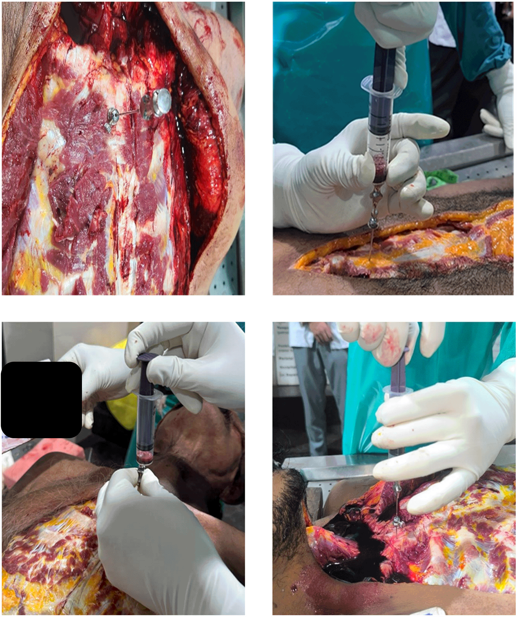
Sample collection was carried out by first making an I-shaped incision according to standard medico-legal autopsy protocols, carefully exposing the sternum to avoid injury to major vessels. Bone marrow was then aspirated from the junction of the first and second parts of the sternum using a Salah bone marrow aspiration needle attached to either a 10 ml or 50 ml syringe, depending on the resistance ( Figure 1). The aspirated samples were promptly transferred into EDTA tubes to prevent clotting. For smear preparation and staining, the samples were placed on frosted slides using a sterile pipette and spread evenly to create uniform smears. Multiple smears were prepared for thorough analysis. These slides were air-dried at room temperature, fixed with absolute alcohol, and subsequently stained using Leishman stain (Romanowsky technique) to demonstrate cytoplasmic details. During microscopic examination, the stained slides were evaluated under an electronic compound microscope, with particular attention given to cytoplasmic changes such as membrane breaks, vacuolation, and eventual dissolution.
The scoring system used to assess cellular changes in the bone marrow was adopted from the study by Biradar G, SatishBabu BS, Bakkannavar S, Pradeep Kumar G, Shaila B. Estimation of time since death from cytoplasm changes of bone marrow cells (2016)”.7
This study was conducted after obtaining approval from the Institutional Ethics Committee, K. S. Hegde Medical Academy, Nitte (Deemed to be University), Mangalore (Approval No: INST.EC/EC/131/2023, REG. NO: EC/NEW/INST/2022/KA/0174, dated 15th May 2023).
Consent to publish: Written informed consent to publish clinical details and images was obtained from the legal next of kin of the deceased individuals who are included in this study. Consent was granted to the authors by the next of kin, and all efforts were made to anonymize data and images.
The time of death issued by the treating physician was collected from the hospital file.
Data entry and coding were done in Microsoft Excel.
Descriptive analysis such as frequencies, percentages, mean and standard deviation was done. Statistical analysis was performed using SPSS version 25. The Analysis of Variance (ANOVA) test was conducted to determine whether there were statistically significant differences among the means of multiple groups. p<0.05 considered as statistically significant.
A total of 43 cases were included in this study, with a clear predominance of males (79.1%) compared to females (20.9%) ( Table 2).
| Gender | N | % |
|---|---|---|
| Female | 9 | 20.9 |
| Male | 34 | 79.1 |
| Total | 43 | 100.0 |
The mean age of the cases was 45.67 years with a standard deviation of 16.21. The mean time difference was 12.74 with a standard deviation of 6.02 ( Table 3).
| N | Minimum | Maximum | Mean | Std. Deviation | |
|---|---|---|---|---|---|
| Age | 43 | 17.0 | 77.0 | 45.674 | 16.2095 |
| Time difference | 43 | 3 | 26 | 12.74 | 6.022 |
Overall, road traffic accidents (51.2%) were the most common cause of death among the cases, followed by fall from height (20.90%), poisoning(14%) and the least common was sudden death (13.90%).
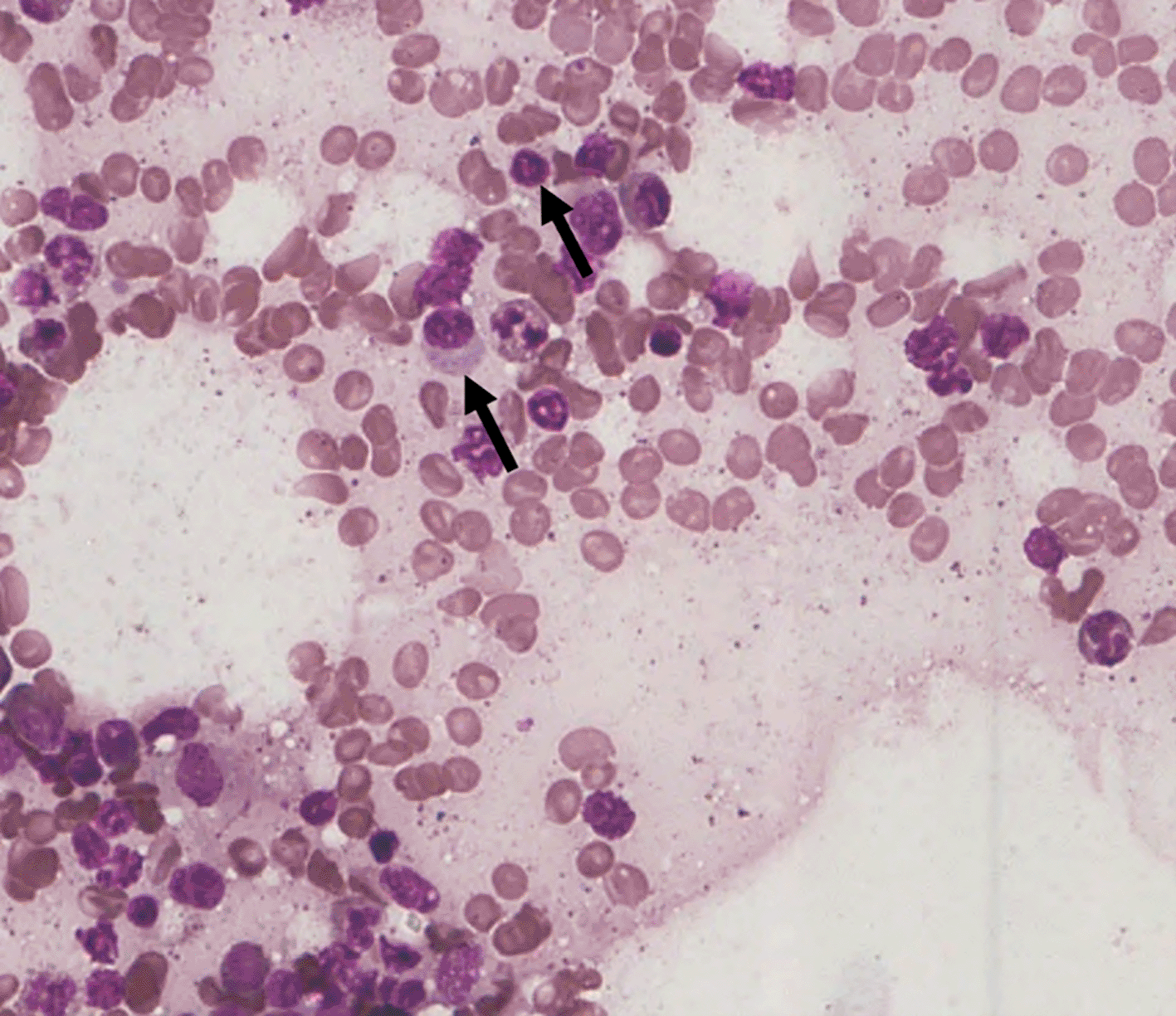
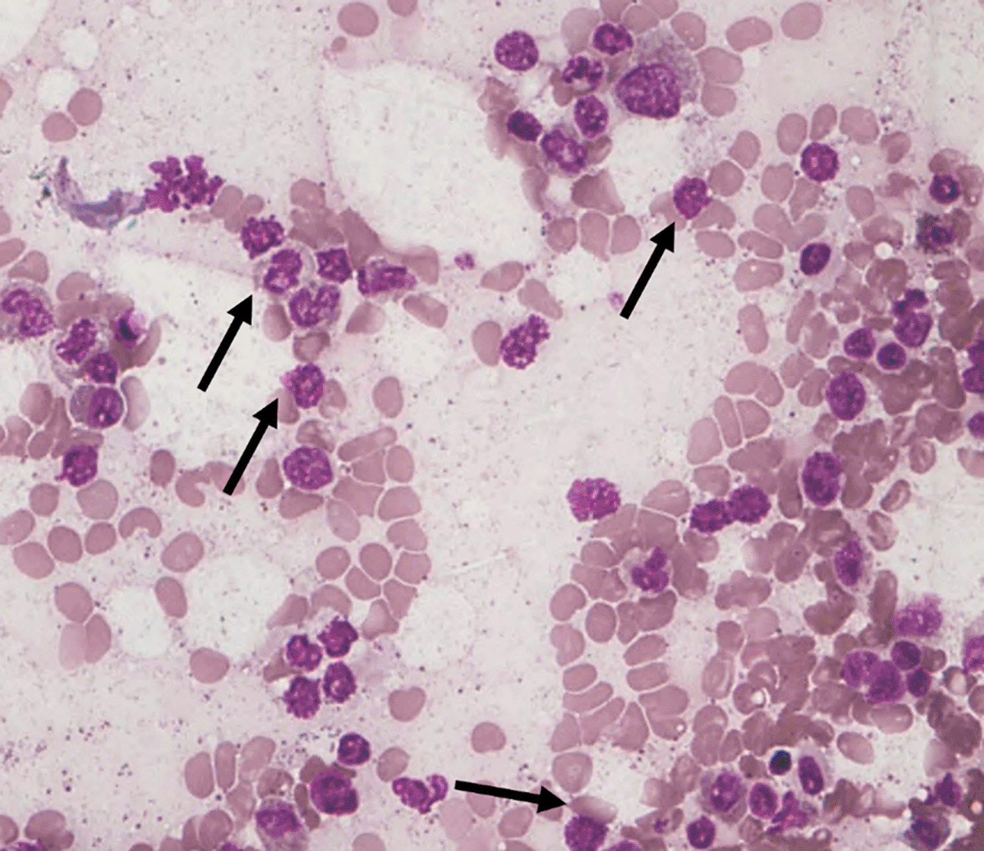
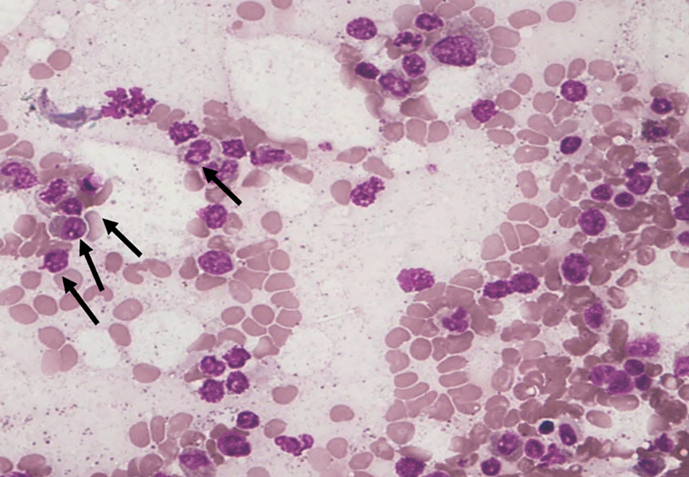
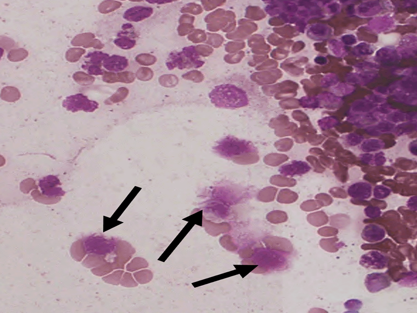
Among the 43 cases studied, cytoplasmic alterations in the bone marrow cells were graded based on the established scoring system. As shown in Table 4. The majority of cases (51.1%) exhibited mild cytoplasmic changes (S1) ( Figure 3). A smaller proportion (25.6%) demonstrated moderate changes (S2) ( Figure 4), while extensive cytoplasmic dissolution (S4) ( Figure 5) was observed in 14.0% of the cases. Notably, 9.3% of the cases (4 out of 43) showed no observable cytoplasmic alterations (S0) ( Figure 2). No cases demonstrated large vacuolation (S3 stage), hence this category is not represented in the figures.
| Cytoplasm changes | Frequency | Percent |
|---|---|---|
| S0 | 4 | 9.3 |
| S1 | 22 | 51.1 |
| S2 | 11 | 25.6 |
| S4 | 6 | 14.0 |
| Total | 43 | 100.0 |
An analysis was performed to examine the relationship between the degree of cytoplasmic changes and the time difference (used as an estimate of PMI), as presented in Table 5.
• The four cases classified under S0 (no cytoplasmic changes) had the shortest mean time difference, averaging 8.50 ± 6.56 hours, with a range from 3 to 18 hours.
• The 21 cases with mild cytoplasmic changes (S1) had a mean time difference of 14.09 ± 6.34 hours, ranging from 6 to 26 hours.
• The 11 cases showing moderate changes (S2) displayed a mean of 10.48 ± 4.31 hours, spanning 5 to 17 hours.
• The 6 cases with extensive cytoplasmic changes (S4) had the longest mean time difference, recorded at 14.97 ± 5.84 hours, with values ranging from 7 to 22 hours.
(p<0.05**).
| Cytoplasmic changes | N | Mean | Std. Deviation (p) | Minimum | Maximum |
|---|---|---|---|---|---|
| S0 | 4 | 8.50 | 6.557 | 3 | 18 |
| S1 | 22 | 14.09 | 6.344 | 6 | 26 |
| S2 | 11 | 10.48 | 4.307 | 5 | 17 |
| S4 | 6 | 14.97 | 5.849 | 7 | 22 |
| Total | 43 | 12.74 | 6.022 | 3 | 26 |
While a trend suggesting increasing cytoplasmic changes with longer post-mortem intervals was noted, variability was observed across categories, and no statistically significant correlation was found between time difference and cytoplasm changes (p>0.05).
The estimation of the post-mortem interval (PMI) remains a cornerstone of forensic practice, guiding both legal and investigative processes. While traditional markers such as rigor mortis, hypostasis, and core body temperature are routinely employed, their reliability diminishes under certain circumstances, including when bodies have been refrigerated or environmental conditions vary significantly. In this context, microscopic evaluation of internal tissues, particularly bone marrow, the primary hematopoietic site, is relatively protected and decomposes more slowly than most soft tissues, so its cytoplasmic and nuclear autolytic changes provide a sequential, biologically plausible framework for PMI estimation.8
Tattoli et al. (2014) emphasised the slower autolysis of bone marrow compared to other tissues, highlighting its potential utility in PMI estimation, especially in the early post-mortem period when traditional markers may yield equivocal results. Although our study did not find a statistically significant correlation, the observable pattern of progressive cytoplasmic changes supports this notion, reinforcing the potential role of bone marrow cytology as a complementary tool in forensic time since death estimation.9
Biradar et al. (2016) observed the onset of cytoplasmic vacuolation at 5–7 hours postmortem, with advanced lysis beyond 16 hours.7 Our study aligns with this timeline, showing mild cytoplasmic changes (S1) in over half the cases (51.1%) and extensive dissolution (S4) most commonly beyond 14–15 hours.
Similarly, Babu et al. (2015) documented progressive vacuolation and cytoplasmic breakdown with increasing PMI, findings echoed in the present work.10 More recently, Sakr et al. (2024) described minimal cytoplasmic changes at 6 hours, with substantial loss of cellular detail by 10–12 hours and necrosis beyond 18 hours.11 The present results concur, showing that while early PMI cases exhibited intact or mildly altered cytoplasm, progressive vacuolation and dissolution became evident after 12 hours.
Michalova et al. (2011) further demonstrated that hematopoietic stem cells remained viable for 2–12 hours within the intact femur, with marrow cell suspensions preserving viability for up to two days at 37°C and four days at 4°C.12 Although our study did not examine stem cell–specific survival, the general cytoplasmic changes we observed beginning at 6–12 hours and advancing by 20 hours are consistent with these findings. Taken together, this highlights the influence of storage conditions, particularly refrigeration, in delaying marrow cell autolysis and thereby extending the preservation window for cytoplasmic morphology.
Although a general trend of increasing cytoplasmic alteration with longer PMIs was observed, statistical correlation was not significant. This variability is likely attributable to confounding factors such as environmental conditions, cold storage, cause of death, and individual biological differences, all of which influence the rate of cytoplasmic autolysis. Nevertheless, the consistency of trends across multiple studies reinforces the potential of cytoplasmic changes as supportive markers for PMI estimation, particularly in the early postmortem period when external indicators are unreliable.
This study demonstrates a gradual progression of cytoplasmic autolysis in sternal bone marrow cells with increasing post-mortem intervals, from early vacuolation to complete dissolution. Although no statistically significant correlation with PMI was established, likely due to biological variability, environmental influences, and a modest sample size, the observed trends highlight bone marrow cytology as a useful adjunct for PMI estimation. Its simplicity, cost-effectiveness, and relative resistance of bone marrow to decomposition, cytoplasmic evaluation can strengthen forensic assessments when interpreted alongside other morphological, biochemical, and circumstantial evidence to strengthen accuracy and medico-legal reliability.
The study was limited by a small sample size, a single-centre setting, and restriction to sternal marrow, which may limit generalizability. Variability in refrigeration, environmental factors, and individual biological differences also influenced cytoplasmic autolysis. Additionally, interobserver subjectivity in smear interpretation may have introduced variability.
Future studies should adopt standardised scoring systems, larger and multicentric samples, and comparative analyses with other organs. Incorporating molecular tests, enzyme assays, and immunohistochemistry may improve sensitivity and precision. Such refinements could enhance the reliability of bone marrow cytology as a supportive tool for postmortem interval estimation.
| Views | Downloads | |
|---|---|---|
| F1000Research | - | - |
|
PubMed Central
Data from PMC are received and updated monthly.
|
- | - |
Provide sufficient details of any financial or non-financial competing interests to enable users to assess whether your comments might lead a reasonable person to question your impartiality. Consider the following examples, but note that this is not an exhaustive list:
Sign up for content alerts and receive a weekly or monthly email with all newly published articles
Already registered? Sign in
The email address should be the one you originally registered with F1000.
You registered with F1000 via Google, so we cannot reset your password.
To sign in, please click here.
If you still need help with your Google account password, please click here.
You registered with F1000 via Facebook, so we cannot reset your password.
To sign in, please click here.
If you still need help with your Facebook account password, please click here.
If your email address is registered with us, we will email you instructions to reset your password.
If you think you should have received this email but it has not arrived, please check your spam filters and/or contact for further assistance.
Comments on this article Comments (0)