Keywords
Radiology; Digital health; Artificial intelligence; Large language models; Workflow optimization; Medical education

This article is included in the AI in Medicine and Healthcare collection.
Radiology; Digital health; Artificial intelligence; Large language models; Workflow optimization; Medical education
From computer hardware to artificial intelligence (AI), technological advances over the past decade have profoundly transformed healthcare systems and the practice of medicine. Rapid progress in computing power, data processing, connectivity, and intelligent algorithms has reshaped diagnostic pathways, clinical decision-making, and the organization of healthcare.1–3
Among medical specialties, radiology has been particularly impacted by this technological revolution due to its intrinsic reliance on advanced digital imaging technologies. The most visible effects have been the continuous hardware and software improvements of imaging modalities such as computed tomography (CT) and magnetic resonance imaging (MRI), which constitute the core tools of radiological practice. These advancements have led to substantial benefits, including improved image quality, enhanced diagnostic accuracy, reduced radiation exposure, and increased operational efficiency.4,5
However, limiting the scope of radiological work to image interpretation alone would provide an incomplete representation of the impact that technology has had on the profession. As in other medical specialties, radiologists perform a wide range of secondary tasks that are often time-consuming and not directly related to image interpretation, which means their diagnostic expertise is not always fully leveraged. Indeed, a study demonstrated that radiologists spend only 36.4% of their working time on image interpretation, while 43.8% is dedicated to non-interpretative clinical tasks, with frequent interactions that directly influence patient care.6
These activities extend far beyond image interpretation and encompass a broad range of technical, administrative, and organizational tasks that are integral to modern radiological practice. They include, but are not limited to, examination protocolling, deciding on patient image acquisition parameters, management of clinical and imaging workflows, human-computer interaction with hospital information systems and picture archiving and communication systems (PACS), reporting logistics, communication and documentation, and collaborative work with fellow radiologists and other medical specialists. Many of these tasks are repetitive, cognitively demanding, or bureaucratic in nature, yet they critically shape efficiency, diagnostic accuracy, and patient care.7 Thus, they represent a domain in which digital technologies have substantial potential to augment radiologists’ performance and reduce their non-clinical burden.8
In this narrative review, we aim to explore the current landscape of digitally augmented radiology ( Figure 1), defined as the integration of digital technologies, including software applications, connected devices, immersive interfaces, and AI, to enhance radiologists’ education, workflow efficiency, cognitive performance, and overall clinical practice.9,10
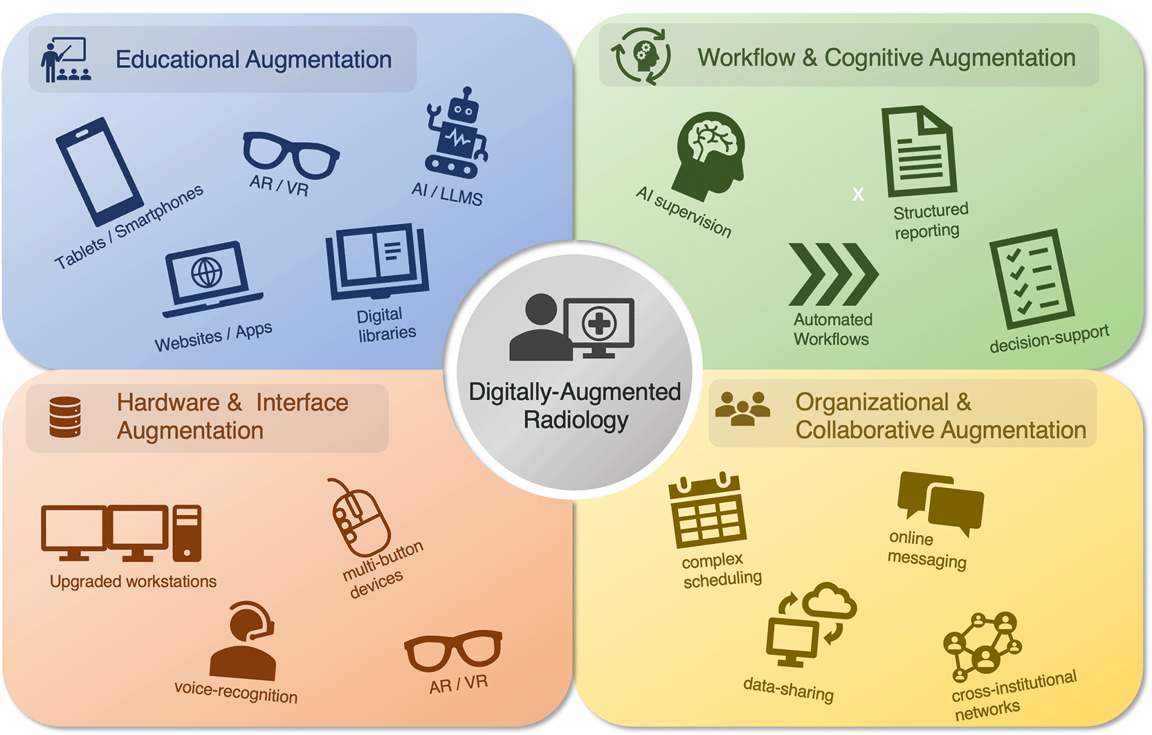
VR = Virtual Reality; AR = Augmented Reality; AI = Artificial Intelligence; LLM = Large Language Model.
The impact of these technologies is discussed across four main thematic domains: educational augmentation, workflow and cognitive augmentation, hardware and interface augmentation, and organizational and collaborative augmentation.
Each section examines traditional approaches that preceded or accompanied the early adoption of digital technologies, current practices in today’s digital era, emerging trends driven by artificial intelligence and advanced digital tools, and future perspectives in light of increasingly sophisticated AI-based solutions that, while currently perceived as futuristic, may soon become integral to routine radiological practice. Finally, the last section addresses key challenges, risks, and ethical considerations related to digital technologies, particularly AI, in radiology, highlighting limitations and areas requiring careful evaluation.
In this first section, we discuss how radiologists acquire and update their knowledge through a combination of traditional methods, current digital tools, and emerging technologies. We cover classical approaches such as textbooks, lectures, and mentorship; digital platforms including online courses, web-based resources, and mobile applications; and emerging innovations like virtual and augmented reality (VR/AR) and large language models ( Figure 2), which offer immersive and AI-assisted educational experiences.
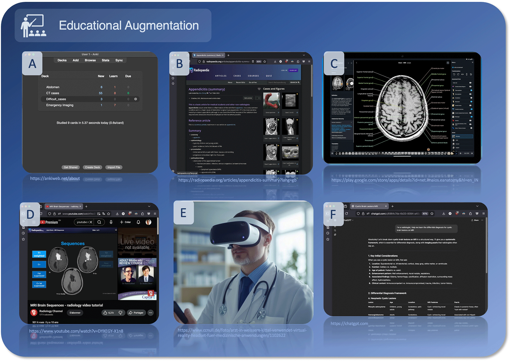
A) Anki flashcard app for image-based learning, B) Radiopaedia.org interactive cases, C) tablet-based educational apps, D) YouTube video lectures, E) VR ultrasound simulation, F) LLM chatbots (ChatGPT, DeepSeek AI) for AI-assisted learning. Screenshots courtesy of publicly available sources.
The methods of acquiring medical and radiological knowledge have traditionally relied on three pillars: textbooks, lectures, and practice. Although they make up the cornerstone of knowledge acquisition, textbooks have the disadvantage of often relying on small images which may not be of sufficient quality to accurately represent the images, which is of particular concern for a specialty like radiology where this is essential. Furthermore, though some still prefer physical books, they are often being replaced by digital versions of the literature.11
Lectures form the second pillar of knowledge acquisition, representing a major part of most medical school curriculums.12 Continuing medical education, and notably that which is available during congresses, is very often delivered in this format, making it a learning method that most radiologists will be familiar with.
The third, and arguably most important, pillar is practice, personal experience, peer-to-peer teaching and mentorship, which also account for an important way in which radiologists will continue to learn throughout their career.13
Flashcard applications are a popular tool used by medical students with the majority of first-year medical student making use of this application.14 This can be particularly useful to radiology students as this method of learning has been shown to improve image-based recall and is associated with improved exam performance and high overall satisfaction, medical students and residents.15
Web-based learning tools are another way in which radiologists acquire knowledge and follow classes. One of the most widely used of these is Radiopaedia.org, with more than 17′000 articles and more than 67′000 dynamic radiology cases, which are notable as users are encouraged to submit their own articles and edit articles which are already posted in order to grow the website and improve upon what is already written, leveraging the knowledge of the community.16 Other websites providing sources of education such as radiologyassistant.nl, as well as personal knowledgebase websites such as fahrnipedia.ch, also exist and provide valuable alternative sources of information. Other tools include online textbooks with a focus on illustrations and teaching (e.g., www.statdx.com, Elsevier), while others focus on specific topics such as learning radiological anatomy (e.g. www.imaios.com//e-anatomy , IMAIOS) and have the advantage of allowing zooming and dynamic interactions with images when compared to traditional textbooks.
Online education in the format of videos is another popular way of furthering one’s radiological education. Of the platforms that offer this service, YouTube (Alphabet, California, USA) represents a free and widely accessible method that hosts lectures and educational videos from a wide array of sources.17 Societies such as the European Society of Radiology (ESR) or the Société Francaise de Radiologie (SFR) also have online immediate libraries available to their members, though other online platforms also offer courses. Unlike traditional lectures, these have the advantage over traditional lectures in that they can be paused, rewound, and be viewed at the learners’ own pace. In parallel, scientific congresses have increasingly transitioned toward hybrid and fully virtual formats since the COVID-19 pandemic, further expanding access to continuing medical education beyond traditional in-person attendance.18
Tablets have enabled image and interaction-based learning which is particularly tailored for fields such as radiology. Many healthcare professionals use tablet computers as part of their daily work and some medical schools have even incorporated them into the curriculum.19
While they are useful for individual practice, they remain limited to teaching and do not currently have an established place in the radiological clinical workflow.20
As smartphones have increased in size and improved in performance, they have become increasingly powerful tools for education. While screen size and interaction remain limited compared with larger devices, smartphones are widely relied upon as portable tools for rapid access to the medical literature.21
The advent of AI and large language models (LLM) may represent a paradigm shift, particularly with the rapidly increasing performance of these tools which has paralleled their increased popularity.22–24 Chatbots such as ChatGPT (OpenAI, California, USA) are popular models, but even completely free models such as Deepseek (DeepSeek, Hangzhou, China) demonstrate high performance and are increasingly promising in terms of radiology education.25
As this new paradigm continues to evolve, it is increasingly probable that the medical students and, subsequently, the radiologists of tomorrow will have used LLMs throughout their higher education as a primary source of information or as an educational aid. This underlines the need to train medical professionals on both how to use the tools at their disposal and to recognize the limits and risks associated with their use.26–29
As part of this trend towards digital education, virtual reality (VR) and augmented reality (AR) are also emerging as useful tools, particularly in ultrasound education.30 In this context, the technology is increasingly able to put radiology students in virtual scenarios which allows them to perform ultrasounds and discover a wide range of pathologies, often at a far lower price than traditional phantoms, allowing for greater accessibility.
LLMs have also enabled clinicians to engage in limited application development tailored to their needs through so-called “vibe coding,” in which LLMs are used to generate applications and code based on users’ input. This opens up a wide range of possibilities, including the development of educational software specifically adapted to the needs of individual radiologists, hospitals, specific organ systems, or particular pathologies. Such tools could allow structured follow-up, feedback on previously reviewed cases, and the identification of relevant cases through PACS integration. However, due to the intrinsic limitations of vibe coding, the practical implementation of such software may be challenging.31
In this second section, we discuss how digital technologies are reshaping radiologists’ workflows and supporting their cognitive processes. We cover traditional approaches to reporting and worklist management, current tools such as structured reporting and semi-automated workflow systems, and emerging innovations including AI-assisted triage, decision support, and large language models for report generation and error reduction ( Figure 3). These developments aim to enhance efficiency, reduce cognitive burden, and improve diagnostic accuracy in increasingly complex clinical environments.
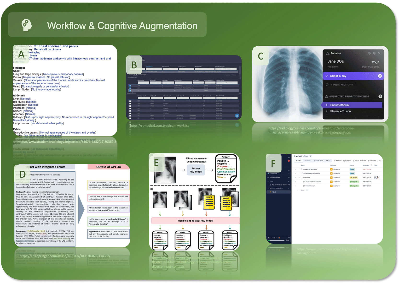
A) Structured reporting templates, B) Worklist management interfaces, C) AI-assisted triage, D) Error-check/decision support AI, E) LLM-assisted report generation, F) AI-assisted checklists and task managers. Screenshots courtesy of publicly available sources.
Radiological reports have traditionally been done in the format of free-text reporting (FTR). This entails the use of descriptive phrases and impressions, in a semi-structured fashion, with virtually unlimited freedom in describing imaging findings, but it suffers from drawbacks such as language heterogeneity, excessive variability in style and length, and reduced clarity in communication.32
Workflow optimization is important for the operational success of any organization, including a radiology department.33 Depending on multiple factors including workload, interruptions, system efficiency, and workflow integration, the need to triage studies arises with the increase in the number of exams which are requested.34
This is increasingly becoming a necessity as worklist management, exam prioritization, increased exam workload and complexity places an ever-growing burden on the cognitive capabilities of the radiologist and thus may result in lapses of concentration and subsequent errors.35
Structured reporting templates (SR) help with the organization of reports by providing a well-defined, standardized template for given radiological procedure. SR can overcome the limitations of FTR, yet some issues remain to be addressed for a more widespread adoption of SR in radiological practice.36 Limitations of SR include lack of development of individualized search patterns and failure to recognize key elements which may be missed if the radiologist only concentrates on the what is necessary to report for the given indication.37
Current systems incorporate multiple tools and plugins designed to transform simple workplace and triage systems into more advanced platforms, enabling greater organization, personalization, prioritization, and reduction of cognitive workload through rule-based or semi-automated processes.38
As the technology is developing, there is an increased potential for integrated AI-assisted models which are context-aware, integrated into the worklists and able to scan for cases with increased clinical urgency to prioritize using multimodal data as well as assist with case reporting.39–41
LLMs are emerging as promising tools for the semi-automated generation and processing of radiology reports. They appear particularly well suited for working with SR and may help address the issue of FTR by restructuring reports and standardize language, improving logical flow and clinical interpretability, and even generating patient-friendly summaries alongside clinician-focused reports.18,42 However, despite these potential benefits, significant challenges remain regarding their safe and reliable integration into routine clinical practice.43
Other applications include the use of AI to provide checklists, give useful prompts, aid in reducing errors, give feedback, provide a second look for certain lesions, detect critical errors or inconsistencies within reports and help radiologists focus on the most important aspects of an exam.44,45 However, emerging data suggest that AI use may not systematically reduce workload and has even been associated with higher rates of radiologist burnout, underscoring the need for thoughtful human–machine integration.46
Although fully integrated collaborative reading environments using AI with automatic reporting, prereading exams and automatic image flagging is still not possible, it remains within the scope of possibility that this will one day represent the future of radiology.47
In this third section, we discuss how advancements in hardware and user interfaces are transforming the radiology workspace. We cover traditional diagnostic workstations and peripherals, current high-performance multi-screen setups and remote access tools, and emerging technologies such as AI-enhanced voice recognition, eye-tracking, and virtual or augmented reality reading environments ( Figure 4). These developments aim to improve efficiency, ergonomics, and the ways radiologists interact with complex imaging data.
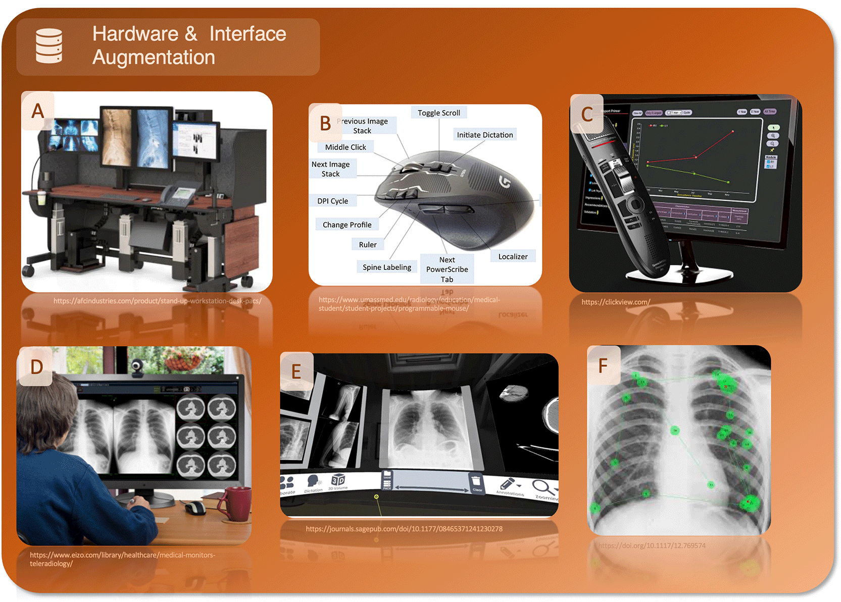
A) Multi-screen diagnostic workstation, B) Advanced peripherals (programmable mouse), C) Voice recognition software, D) Remote access/teleradiology setup, E) VR reading environment, F) Eye-tracking interfaces. Screenshots courtesy of publicly available sources.
The hardware used for radiological reporting has evolved dramatically over the past decades. Within a single generation of radiologists, the field has progressed from film-based imaging to near-complete digitization, accompanied by the development of specialized diagnostic displays, ergonomically designed workstations, and advanced software enabling image reformatting and three-dimensional visualization.48
The peripherals used by radiologists for exam interpretation and reporting have likewise evolved. Mouse and keyboard have become the primary input devices, while microphones combined with speech recognition software have largely replaced traditional dictaphones, which required manual transcription and correction by medical secretaries.49
In the current paradigm, radiologists benefit from multi-screen diagnostic workstations, high-capacity hardware capable of displaying multiple studies simultaneously, providing 3D renderings and allowing the use of other applications simultaneously for web navigation or communication with software manufacturers’ dedicated servers.50,51
As high-speed Internet access has become the norm, the possibility of remote working by accessing hospital servers is an ever-increasing possibility and the trend that has only increased after the COVID-19 pandemic.52 Furthermore, remote access to exams can allow for the possibility of getting second opinions from specialized centres.52 The peripherals used to interface with the computers have also evolved over time with it not being uncommon to see radiologists using advanced mice with multiple functions and macros in order to improve the ergonomics when interacting with the PACS, contributing to an overall increase in efficiency.53,54 Furthermore, other peripherals such as computer styluses are also gaining popularity as it becomes evident that they are practical in use cases such as image annotation.55
Alongside the adoption of microphones, voice recognition systems have also been widely adopted, allowing for real-time dictation and thus faster exam validation and publishing, speeding up the radiology workflow significantly.56
Advances in AI-based voice recognition are expanding the range and complexity of tasks and commands that can be recognized compared with traditional, non–AI-enabled systems, thereby facilitating human–computer interaction and potentially improving productivity.57 As the integration of AI into radiology remains in a relatively early stage, it is still unclear which interaction paradigms and interfaces will ultimately prove most effective in clinical practice.
Technological advances have also opened up the gateway for new ways of interacting with radiological exams, with the possibility of a VR or AR reading space being tested.58,59 However, this technology remains theoretical with regard to its application as it has not yet been made widely available. Nevertheless, as heads-up displays and AR glasses gain wider acceptance, it is possible that this technology may naturally find its way into the reading room.
Eye-tracking technology can also be leveraged to record radiologists’ search patterns, making it possible to understand the interactions with the software and images and thus optimize workflows accordingly.60
In this fourth and final section, we explore how digital tools and AI are reshaping the organization and collaboration within radiology departments. We cover traditional approaches to communication, scheduling, and staff induction, current digital practices such as cross-institutional data sharing and semi-automated planning tools, and emerging trends including instant messaging for peer-to-peer discussions and AI-driven resource allocation ( Figure 5). These innovations aim to improve efficiency, knowledge dissemination, and collaboration while reducing administrative burden and cognitive load for radiologists.
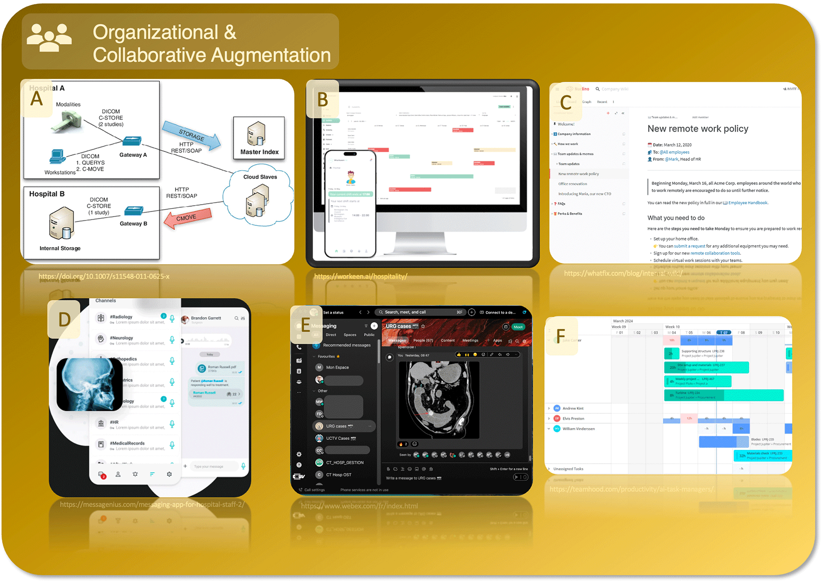
A) Cross-institutional PACS/image sharing platforms, B) AI-driven staff scheduling, C) Internal knowledge management tools, D) Instant messaging for peer-to-peer discussion, E) Collaborative case discussion platforms, F) AI-enhanced resource allocation. Screenshots courtesy of publicly available sources.
The need to be contactable at a moment’s notice is an increasingly essential part of a doctor’s life. However, this is a double-edged sword: although the adoption of telephones has undoubtedly been a boon, it has led to radiologists being increasingly interrupted, with one study reporting that 92% of calls received forced the radiologist to suspend reporting to answer them.61
Allocating shifts is a complex task that is traditionally done manually. When planning a schedule, one must consider availability, areas of competence, patient demand, as well as imaging system availability, among other requirements. This becomes even more challenging when schedules are planned several weeks or months in advance, with increased complexity when managing large teams.62 Furthermore, a variety of legal and institutional constraints must also be taken into account, which can vary at both local and national levels, representing an additional burden in terms of time and cost.
Integrating new team members can also be challenging, as local knowledge is required to adapt to the many specificities of an institution.63 Traditionally, the integration of new staff has been done organically, relying on senior members to coach others and transmit skills. However, this largely unstructured approach has many pitfalls, placing strain on both new and experienced staff, especially since this extra workload is often not formally recognized.
It is not uncommon for patients to undergo treatments and radiological examinations at multiple institutions, or for external expert opinions to be requested to review prior imaging. In such cases, it becomes necessary to transfer the patient’s files so that they can be reviewed by a physician external to the institution where the exams were performed, who otherwise would not have access to them. To address this need, it has become commonplace to request the transfer of images between institutions. This is currently accomplished through various methods, including online PACS systems as well as specially developed platforms for accessing documents across different physical locations within a defined network.64
The ability to organize shifts has improved in recent years with the development of semi-automated tools, including spreadsheets and dedicated software. However, the process remains somewhat complex and time-consuming.65,66
Staff induction has also evolved over time. Many hospitals now maintain internal tools and databases for sharing institutional knowledge, technology, and information. These measures are designed to ensure the rapid and systematic dissemination of knowledge throughout the organization.67
An emerging trend amongst physicians is to engage in large-scale peer-to-peer discussions using dedicated groups and instant messaging apps.68,69 In the increasingly fast-paced work environment, this has the advantage of allowing rapid sharing of critical information to single individuals or larger groups, facilitating information exchange and being widely appreciated by clinicians, second only to face-to-face discussions. However, it is clear that this is a legal and regulatory grey area, with many clinicians unaware of the potential repercussions of sending and storing medically sensitive data in personal devices, making education in this field critical. Nevertheless, the trend towards the use of instant messaging for rapid communication has many benefits, including flattening the hierarchical structure, showing that this method of communication merits further research to develop secure versions with appropriate user interfaces.
Cross-institutional PACS, which allow faster data sharing and enable collaboration across different hospitals, are also being developed.70 In order to avoid isolation within the hospital landscape, advanced communication systems, many of which are commercial, are used to enhance the ability of physicians to discuss cases. This development fits within the overall trend towards digitization of the hospitals, which is a pre-requisite for effective cooperation.
Resource allocation and planning are also being revolutionized by AI.71 In addition to relieving a logistical burden on the staff who are tasked with figuring out the plans, it allows those on the roster to feel that they have increased agency over their work schedule. This perceived fairness increases the feeling of wellbeing amongst staff, being preferred over manual self-rostering, alongside reducing time and cost associated with making a schedule.
AI intelligence is poised to revolutionize the knowledge databases of institutions by allowing easier access to the required information.72 By leveraging the ability of AI to extract relevant information and present it in a coherent and structured manner, it is possible to avoid searching through poorly structured databases by having access to the data which is required more easily. This can serve to reduce the cognitive workload of the searcher, though for such software to work integration within the existing systems and effective linking of the different knowledge bases is required.
Despite the considerable promise and enthusiasm surrounding digital technologies, particularly advances in AI, their integration into radiology is accompanied by substantial challenges, risks, and ethical considerations that must not be overlooked.
One of the primary challenges lies in workflow fragmentation.73 Many AI tools remain poorly integrated into PACS and hospital information systems, leading to interoperability issues between vendors and platforms. This often results in a multiplication of interfaces, additional logins, and increased clicks, ultimately contributing to alert fatigue and cognitive overload rather than efficiency gains.74 Continuous model updates and evolving regulatory requirements introduce further complexity, as software turnover may require repeated validation, retraining, and adaptation of established workflows.75
Beyond operational hurdles, several clinical risks deserve attention. Automation bias and overreliance on AI outputs may subtly influence decision-making.76 While radiologists initially maintain critical oversight, repeated exposure to automated suggestions may progressively encourage cognitive offloading and uncritical acceptance, potentially leading to deskilling and erosion of core diagnostic expertise. Furthermore, the use of “black-box” algorithms with limited explainability further challenges trust and accountability.77 Other concerns include bias in training datasets and lack of representativeness, vulnerabilities related to data privacy and limited transparency associated with proprietary (non–open-source) algorithms.78–80
Finally, the expansion of AI in radiology raises complex ethical questions. Liability remains unclear when errors occur, whether responsibility lies with the radiologist, the institution, or the vendor.81 Reimbursement models and financial incentives may influence adoption patterns, while the environmental footprint of large-scale computing infrastructures warrants consideration.82 Prior to clinical deployment, rigorous risk–benefit assessment is essential to avoid prioritizing appealing marketing features over true clinical value.
Digital technologies are reshaping radiology beyond image interpretation, enhancing education, workflow, human-computer interaction, and collaboration. Digitally-augmented radiology includes tools ranging from tablets and web-based learning platforms to VR/AR environments and AI-powered systems, including LLMs, which support learning, reduce cognitive and administrative burdens, and improve efficiency and diagnostic accuracy. While many of these technologies are already integrated into daily practice, emerging innovations promise even greater capabilities, from AI-assisted reporting to intelligent collaborative platforms. Successful adoption requires thoughtful workflow integration, awareness of limitations, and adherence to regulatory and ethical standards, paving the way for a future in which human expertise and technology work seamlessly together to advance patient care.
| Views | Downloads | |
|---|---|---|
| F1000Research | - | - |
|
PubMed Central
Data from PMC are received and updated monthly.
|
- | - |
Provide sufficient details of any financial or non-financial competing interests to enable users to assess whether your comments might lead a reasonable person to question your impartiality. Consider the following examples, but note that this is not an exhaustive list:
Sign up for content alerts and receive a weekly or monthly email with all newly published articles
Already registered? Sign in
The email address should be the one you originally registered with F1000.
You registered with F1000 via Google, so we cannot reset your password.
To sign in, please click here.
If you still need help with your Google account password, please click here.
You registered with F1000 via Facebook, so we cannot reset your password.
To sign in, please click here.
If you still need help with your Facebook account password, please click here.
If your email address is registered with us, we will email you instructions to reset your password.
If you think you should have received this email but it has not arrived, please check your spam filters and/or contact for further assistance.

Comments on this article Comments (0)