Keywords
Residues, biomaterials, nanocellulose, polysaccharide, scaffolds, nanocellulose scaffolds, biomedical applications.
This article is included in the Nanoscience & Nanotechnology gateway.
This article is included in the Plant Science gateway.
Residues, biomaterials, nanocellulose, polysaccharide, scaffolds, nanocellulose scaffolds, biomedical applications.
Bananas are among the most widely cultivated fruits globally, with an annual production of approximately 153 million tons, according to the Food and Agriculture Organization.1 This substantial yield, however, comes with a significant environmental challenge, the disposal of banana waste.2 In fact, it is estimated that about 40% of the banana plant is discarded as waste, including the peel, pseudo stems, leaves, and other by-products, resulting in millions of tons of agricultural residue each year.3,4 Traditionally, this waste has been left to decompose in landfills or used as animal feed, contributing to pollution and the inefficient use of valuable resources.5 Advancements in biopolymer production have shed light on the potential of banana waste as a raw material for sustainable alternatives, particularly in the extraction of cellulose and nanocellulose.6
Cellulose, a polysaccharide found abundantly in plant cell walls, is a renewable resource that can be processed into nanocellulose a nano-structured material with remarkable mechanical, optical, and surface properties.7 Numerous studies have demonstrated that banana peels, a key component of banana waste, can be processed to yield nanocellulose with promising properties for various industrial applications.8,9 In addition to mitigating waste disposal issues, this approach presents a sustainable pathway for producing bio-based materials, contributing to environmental conservation while adding economic value to banana cultivation.10
The use of biopolymers, particularly cellulose and its nano-derivatives, has garnered considerable attention in the field of tissue engineering due to their favorable biocompatibility, biodegradability, and structural resemblance to the extracellular matrix (ECM), which is crucial for supporting cell growth and tissue regeneration.11,12 Biopolymers derived from renewable resources like banana waste offer the potential for developing environmentally friendly alternatives to synthetic materials commonly used in biomedical applications.13 Nanocellulose stands out due to its superior mechanical properties, high surface area, and ability to be functionalized for specific biomedical uses.11 These attributes make nanocellulose an ideal candidate for fabricating scaffolds that can support the formation of new tissues, including skin, bone, and cartilage.14 Literature highlighted the versatility of nanocellulose scaffolds in promoting cellular activities such as adhesion, proliferation, and differentiation, which are critical processes in tissue regeneration.15 Furthermore, nanocellulose-based scaffolds are advantageous due to their tunable porosity, which allows for improved nutrient diffusion and waste removal in growing tissues.16
Statistical evidence suggests that the market for tissue engineering biomaterials is growing rapidly, with the global tissue engineering market expected to reach $33.8 billion by 2027, growing at a compound annual growth rate (CAGR) of 12.2%.17 This underscores the increasing interest and demand for sustainable, bio-based materials like nanocellulose in the field of regenerative medicine. Accordingly, the aim of this work was to demonstrate the feasibility of extracting nanocellulose from banana residues and its use in the fabrication of scaffolds for potential applications in tissue engineering in Ecuador, which is among the top five banana producers worldwide. By leveraging banana waste, these scaffolds not only address environmental concerns but also contribute to the advancement of regenerative medicine.
Sodium hydroxide (NaOH) (Pharmco-AAPER, Cat Number 289000000), sodium hypochlorite (NaClO) (Merck, Cat Number 425044), sulfuric acid (H2SO4) (Fisher Chemical, Cat Number A300C-212), acetic acid (Supelco, Cat Number 1.00063.2500), phosphate buffered saline (PBS) (Life Technologies, Cat Number 00–3002), dehydrated alcohol (Pharmco-AAPER, Cat Number 1110000200), chitosan (Chemsavers, Cat Number CHTS100G), and agarose (Fisher Chemical, Cat Number BP164–100). All reagents were of analytical grade.
The nanocellulose used in this study derived from commercially available banana fruits purchased from a Collection Center located in Cevallos, Tungurahua, Ecuador (geographic position 1°21 south, 78°37 west, 2894 m.a.s.l.). The study did not involve the collection or transport of regulated plant tissues. The selection criteria were based on the state of maturity (intermediate state) and physical state (no mechanical damage, fresh, no presence of fungi or bacteria). Bananas that fulfilled the criteria were purchased and stored in a refrigerator (Indurama, RI-790NE, Cuenca, Azuay, Ecuador) at 4 °C. A representative sample of the fruit was authenticated by an expert of The Misael Acosta Solís Herbarium of Ecuador (Universidad Técnica de Ambato, Ecuador) to confirm its taxonomic identity as Musa paradisiaca L.
Afterwards, banana peels were separated from the pulp and washed with distilled water to remove impurities. Clean banana peels were dried in a convection dehydrator (Kalstein, YR05265, Montpellier, Paris, France) at 50 °C for 30 h. Then, the dry residue was ground in a laboratory mill (IKA, A11, Staufen, Baden-Württemberg, Germany) and stored in sterile plastic bags. Banana residue powder was washed with distilled water at 60 °C for 4 h in a 1:10 fiber/water ratio. Finally, it was filtered and dried in the oven (Memmert, SF-30PLUS, Büchenbach, Baden-Württemberg, Germany) at 40 °C.
A mass of 25 g of banana residue powder was treated for 2 h with a 5% NaOH solution in a fiber/solution ratio of 1:20 at room temperature with vigorous stirring (Thermo Scientific, Cimarec+, Waltham, MA USA), afterwards, the solution was filtered.18 The collected insoluble banana residue was rinsed with distilled water until a neutral pH was reached in the wash water.8 Next for cellulose (CE) extraction, a bleaching process was carried out with the insoluble banana residue with a 1% NaClO solution in a fiber/solution ratio of 1:10 for 1 h at 95 °C and vigorous stirring; the bleaching was carried out twice. The obtained CE was filtered and washed with distilled water to remove any traces of NaClO solution. Finally, CE was dried at room temperature and stored in a glass container.19,20
Nanocellulose (NC) was extracted by acid hydrolysis, for that 10 g of extracted CE was treated with a 65% w/w H2SO4 solution in a fiber/H2SO4 ratio of 1:20 with constant stirring (Thermo Scientific, Cimarec+, Waltham, MA USA), for 45 minutes and a maximum of 45 °C. Cold distilled water was added to stop hydrolysis and to allow NC to precipitate. The obtained NC was centrifuged at 4000 rpm for 10 minutes and washed again until the suspension reached neutrality.19 A mechanical treatment was then carried out with the NC, for which, an ultrasound equipment (Eiwei, CD-C3, Shenzhen City, Guangdong, China) was used at 20 kHz frequency for 30 minutes. This was performed in an ice bath to avoid overheating.20,21 The suspension obtained was centrifugated and treated again with ultrasound for 30 minutes. Finally, the suspension was dried at room temperature.20
NC fibers were observed under an inverted light microscope (10x) (Euromex, DI.1053-PLPHFi, Arnhem, Guéldria, Holanda) and a scanning electron microscope (SEM) (Aspex, PSEM Express, Delmont, Pennsylvania, USA) using a voltage of 15.0 kV. To verify that the extracted product corresponded to NC, Fourier transform infrared spectroscopy (FT-IR) analysis were conducted (Perkin Elmer, L1600312 Spectrum Two, Waltham, Massachusetts, USA). Infrared data collection was carried out by scanning samples from 4000 to 500 cm−1 with a resolution of 4 cm−1.22 The PerkinElmer Spectrum Version 10.5.2 software was used for positioning the most relevant peaks present in the graph and the data was saved for further analysis.
Three types of scaffolds were fabricated ( Table 1): nanocellulose scaffold (NC-S), chitosan scaffold (CH-S) and a nanocellulose/chitosan scaffold (NC-CH). For (NC-S) and (CH-S), 1% of solutions in acetic acid 0.5 M were prepared, respectively. For (NC-CH) a solution containing 1% (NC) and 1% chitosan (CH) was prepared in acetic acid 0.5 M. Agarose 0.5% in PBS was added to previous solution as gelling agent.
| Scaffold | Nanocellulose solution in acetic acid 0.5 M | Chitosan solution in acetic acid 0.5 M | Agarose solution in PBS |
|---|---|---|---|
| (NC-S) | 1% | – | 0.5% |
| (CH-S) | – | 1% | 0.5% |
| (NC-CH) | 1% | 1% | 0.5% |
The solutions were poured into Petri dishes and refrigerated (Indurama, RI-790NE, Cuenca, Azuay, Ecuador) at 4 °C for 24 h, after that time plates were frozen (Binder, UF V 500-UL, Tuttlingen, Baden-Württemberg, Germany) at −75 °C for 24 h. The samples were then lyophilized (SP scientific, Benchtop Pro, Warminster, PA, USA) at −50 °C for 22 h. After this process, the plates were placed in the desiccator until use.
Scanning electron microscopy (SEM) (Aspex, PSEM Express, Delmont, Pennsylvania, USA) was used to analyze the morphology and pore diameter of the scaffolds. Small pieces of scaffolds were used (1 cm2). Pore size was obtained using Image-J open-source software.23,24
The porosity of the scaffold was determined by the liquid displacement method. The initial weight (W0) of the scaffolds was recorded. Then, they were immersed in dehydrated alcohol for 48 h. After this time, the scaffolds were weighed again (W1). Thus, the porosity was calculated by replacing the obtained values in Equation (1), Where (ρa) corresponds to the density of the alcohol and (ρb) to the density of the biopolymer25:
Scaffolds water absorption capacity was determined in relation to swelling, for which, the weight of the dry samples (Wdry) was recorded and then they were immersed in distilled water at room temperature for 6 h. The samples were weighed at 30 minutes intervals to obtain their wet weight (Wwet). The water absorption capacity was calculated using Equation (2)26:
Banana peel constitutes approximately 40% of the total weight of the fruit. Its main components are cellulose, hemicellulose, lignin, pectin, starch and carbohydrates.27 For the extraction, 500 g of banana peel were used and after grounding and drying a mass of 40.43 g was obtained with a 91.91% humidity ( Table 2). These values match with those reported by28 who determined values between 85–95% in banana residues.
| Weight of the starting material (g) | Final weight after drying (g) | % Moisture | |
|---|---|---|---|
| Banana residue | 500 | 40.43 | 91.91 |
Cellulose extraction continued with the banana residue powder, and after washing a reduction in weight was observed, from 40.43 g to 27.42 g of cellulose powder. The reduction can be attributed to the removal of water-soluble substances, such as sugars, phenolic compounds and impurities ( Table 2).29 Following, alkaline treatment with 5% NaOH and bleaching with 1% NaOCl, the weight decreased further, due to the removal of non-cellulosic compounds, such as lignin and hemicellulose ( Table 3). According to30 immersion of plant material in NaOH solution causes swelling of the cell walls of the fibers, inducing rupture of the outer layer and the amorphous region, which disrupts the ether and ester bonds between lignin and hemicellulose. In addition, the NaOH solution fragments the hydrogen bonds within the lignocellulosic components, making lignin and hemicellulose soluble in alkaline solutions and leading to the formation of black liquor, which is subsequently removed by washing.31 note that alkaline treatment does not completely remove lignin; therefore, NaOCl is used to remove residual lignin while serving as a bleaching agent for cellulose. NaOCl can break ether bonds within the lignin structure and improves the whiteness of the pulp.
| Treatment | Weight of the starting material (g) | Final weight (g) |
|---|---|---|
| Pretreatment | 40.43 | 27.42 |
| Alkaline treatment and bleaching | 25 | 8.40 |
| Acid hydrolysis and ultrasonication | 8.40 | 5.95 |
The acid hydrolysis and ultrasound applied to cellulose powder produced a further reduction in the mass of the treated plant material, from 8.40 g cellulose to 5.95 g nano-cellulose ( Table 3). The reduction occurred due to the elimination of non-fibrillar cellulosic compounds, residual impurities from previous treatments and losses suffered during the washing stages to achieve a neutral pH in the suspension.32 The acid hydrolysis breaks the glycosidic bonds in the amorphous region of cellulose causing degradation; the crystalline region remains intact due to its compact structure that prevents the penetration of the acid into the crystalline domains.33 During the ultrasonic process, the cavitation bubbles collapse, facilitating the formation of cellulose nanofibrils and simultaneously improving their dispersion in solvents.
The yield of nanocellulose extracted from cellulose powder was 23.8%, while the yield from the initial banana powder was 14.7%, both obtained under an acid concentration of 6% ( Table 4). In contrast,9 reported a yield of 28.1% based on the initial banana powder, using a one-pot process that combined microwave pre-treatment and oxidative hydrolysis, with lower acid concentrations ranging from 0 to 10%. According to the authors, high acid concentrations lead to the degradation of crystalline regions, thereby reducing both the efficiency of hydrolysis and the overall yield.32 noted that the yield and quality of nanocellulose are influenced by several factors, including the cellulose source, acid concentration, treatment duration, and hydrolysis temperature.
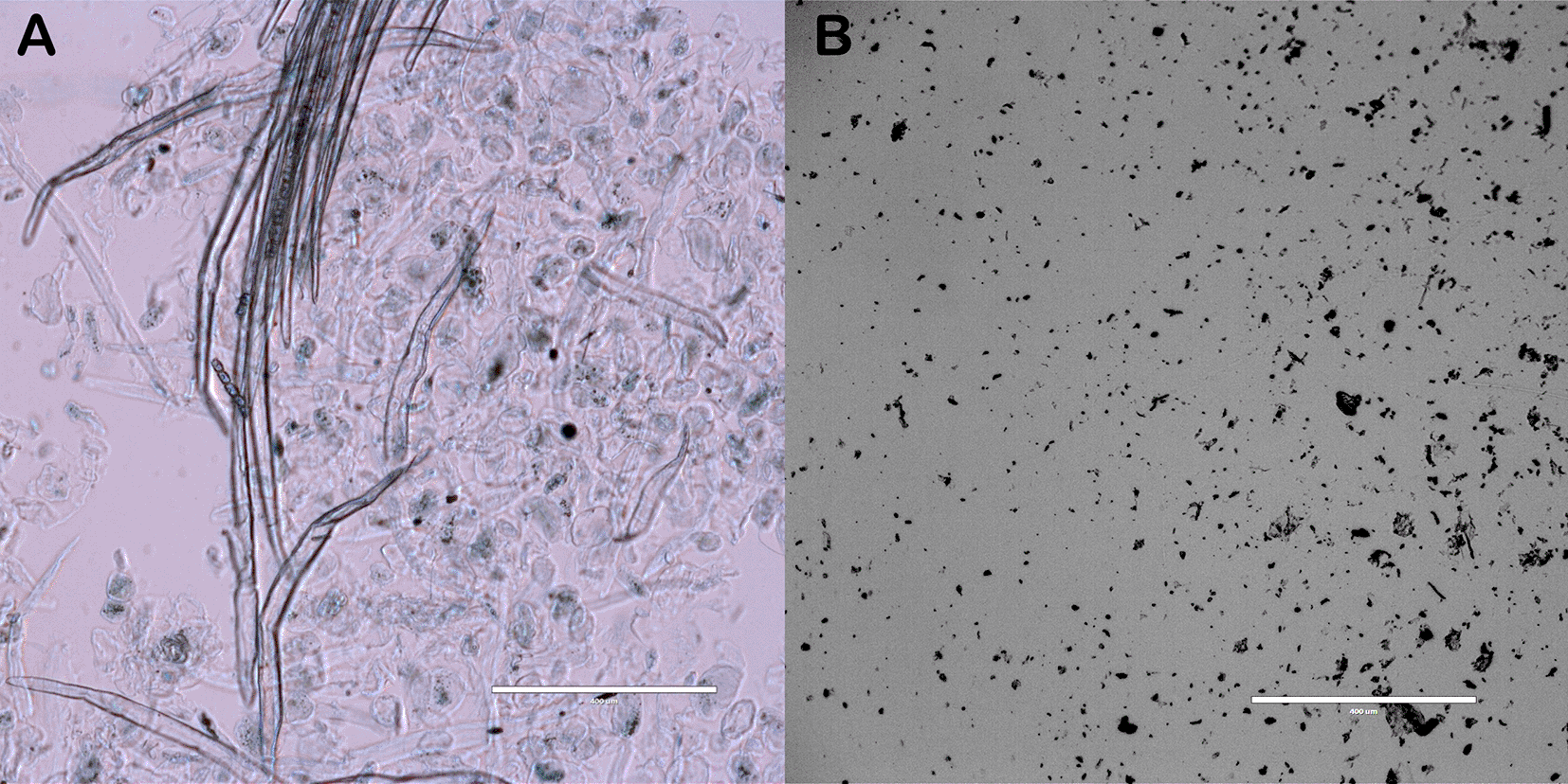
Cellulose and nanocellulose fibers were observed using an inverted light microscope equipped with a 10× objective lens. In the case of cellulose, some fibers appeared dispersed, while others remained agglomerated ( Figure 1A). During treatment with NaOH and NaClO, the lignin and hemicellulose network is weakened through hydrolysis, rendering these components soluble in the treatment solution.34 Their removal facilitates the defibrillation of fibrils; however, the presence of waxes in banana peels contributes to the retention of a compact fiber structure, thereby accounting for the observed agglomeration in certain regions. In contrast, the nanocellulose fibers appeared thinner and shorter in length ( Figure 1B). Sulfuric acid hydrolysis preferentially degrades the amorphous regions of cellulose while preserving the crystalline domains. In addition, acid hydrolysis further contributes to purification by removing residual hemicellulose and lignin not eliminated in earlier treatments, as well as waxes, oils, and fats.35 Furthermore, the application of mechanical treatment introduces high shear forces to the cellulose bundles, thereby enhancing fibril defibrillation.32
To further analyze the morphology of the extracted cellulose and nanocellulose, scanning electron microscopy (SEM) was employed. Figure 2A shows a micrograph of cellulose in which the onset of defibrillation can be observed. This suggests that the alkaline treatment effectively removed hemicellulose and lignin by hydrolyzing them into soluble forms. However, some fibers remained agglomerated due to residual non-cellulosic components, such as waxes and pectins, which act as natural adhesives.35
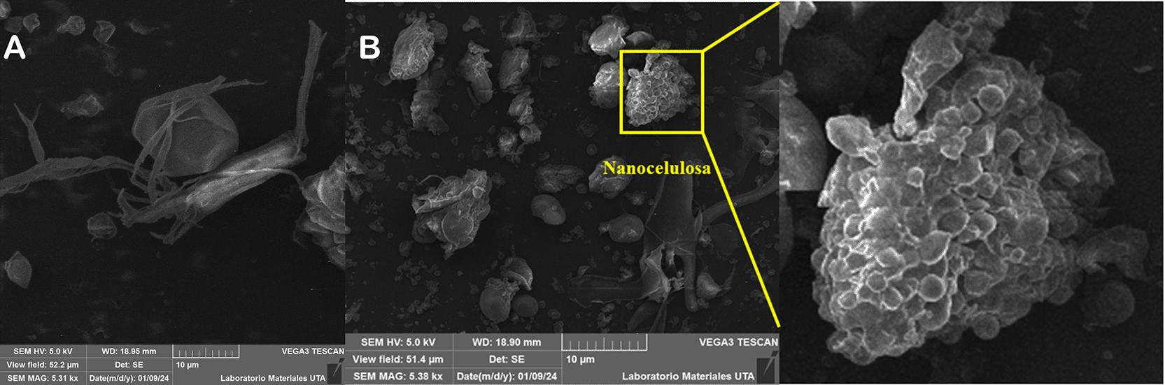
(B) Nanocellulose particles exhibiting agglomeration at 5,380×.
In contrast, nanocellulose fibers appear as agglomerated spherical structures ( Figure 2B), with decreased diameter, suggesting that non-cellulosic components have been re-moved.34 Nevertheless, upon drying, nanocellulose tends to agglomerate due to increased cohesive forces, which hinders accurate morphological visualization.18
Commercial cellulose as well as extracted cellulose and nanocellulose were analyzed by FTIR to identify their functional groups. With the use of FTIR, the infrared spectra of commercial cellulose (CE), as well as (CE) and (NC) extracted from banana residues were obtained (Figure 3). The analysis of (NC) revealed spectral similarity with both commercial (CE) and (CE) derived from banana residues, with minor variations in band intensities. According to36 this occurs because the same structure of (CE) is being analyzed, albeit on a smaller scale. These variations are due to factors such as applied treatments, particle size reduction and impurity removal.
According to37 the vibrations between 3500 and 3300 cm−1 correspond to the stretching of the -OH bond. The hydroxyl group (-OH) is evident in all three spectra at 3332.42 cm−1, 3334.47 cm−1 and 3333.97 cm−1 (Figure 3). The absorption bands between 3000 and 2800 cm−1 correspond to the stretching of the -CH bond in aliphatic compounds, including the methyl (-CH3) and methylene (-CH2) groups in (CE).38 This characteristic bond is present in all three samples.
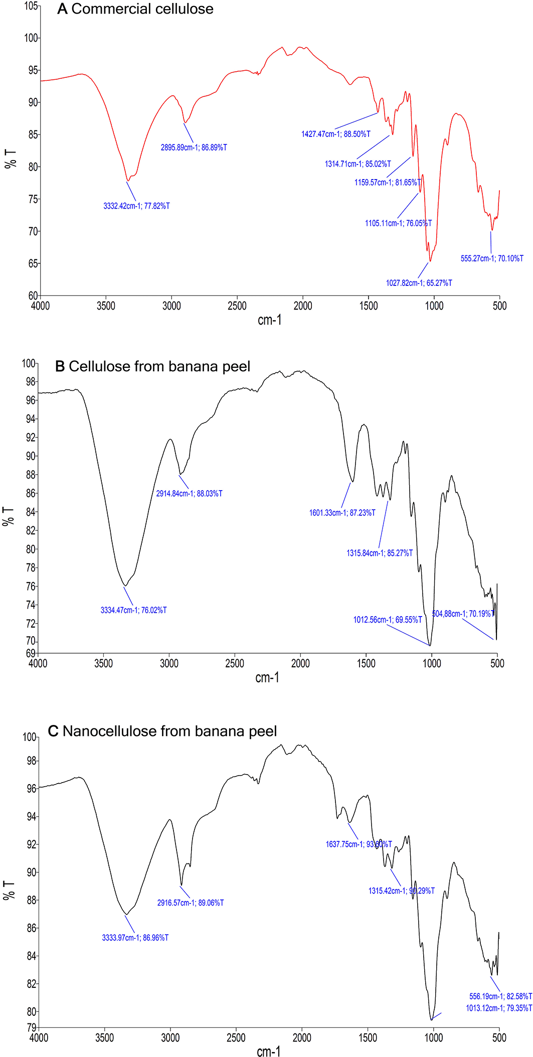
A band at approximately 1601.33 cm−1 in the spectrum of (CE) extracted from plantain residues corresponds to the stretching vibration of the C=C bond in the aromatic ring of lignin, indicating presence of residual non-cellulosic compounds.21 In contrast, the spectrum of (NC) does not show this band, suggesting that non-cellulosic residues, including lignin, were removed during acid hydrolysis. According to39 the vibrations around 1637.75 cm−1 in the (NC) spectrum are associated with the bending of the -OH bond in the adsorbed water, possibly due to residual moisture in the sample.
In addition, the absorption bands at 1314.71 cm−1, 1315.84 cm−1 and 1315.42 cm−1 in all spectra are attributed to the vibrations of -CH and -CO bonds in the methyl, methylene and aromatic rings of polysaccharides in (CE) .40 According to 35 the absorption bands at 1027.82 cm−1, 1012.56 cm−1 and 1013.12 cm−1 correspond to the stretching vibration of the C-O-C glycosidic bond, which links the glucose units to form the linear structure (CE). This characteristic wavelength is also observed in commercial (CE). In addition, bands at 555.27 cm−1, 504.88 cm−1 and 556.19 cm−1 are associated with vibrations of the C-OH and -CC bonds, which contribute to the three-dimensional framework of (CE).40 The presence of glycosidic bonds confirms the structure of (CE), as these bonds connect the anomeric carbon atoms of saccharides to form polysaccharides.
Reference 37 reported that a critical step in isolation (NC) involves the alteration of hydrogen bond structures and the cleavage of glycosidic bonds. This process reduces the availability of these bonds, altering or decreasing the presence of the corresponding absorption bands in the FT-IR spectrum. The absence of absorption bands at 1427.47 cm−1 (-CH2), 1159.57 cm−1 (C-O-C) and 1105.11 cm−1 (CO) in the (CE) and (NC) spectra extracted from plantain residues suggests that the methylene, ether and carbonyl groups at these wavelengths were not sufficiently structured to generate a detectable band. It is important to note that during the extraction process, the structure of (CE) may have been affected, resulting in changes in molecular vibrations, as sulfuric acid mainly targets the -OH and C-O-C groups.37
As reported by36 the absence of bands between 1300 and 1200 cm−1 for lignin and 1800 and 1700 cm−1 for hemicellulose indicates a partial removal of these compounds, which may not have been completely removed due to the specific conditions of the chemical treatments. By chemical and mechanical treatments, (NC) was successfully extracted from plantain residues, as confirmed by the analyses performed.
Extracted nanocellulose was used to fabricate three-dimensional (3D) structures, commonly referred to as scaffolds, through a lyophilization (freeze-drying) process. Biopolymer-based scaffolds are widely employed in tissue engineering as they can mimic the extracellular matrix, thereby supporting cell adhesion, proliferation, and tissue formation. Three scaffold types were developed: nanocellulose-only, chitosan-only, and nanocellulose/chitosan composites. These scaffolds were fabricated to evaluate the functional properties of extracted nanocellulose alone and in combination with commercially available chitosan.
Chitosan, a polysaccharide obtained through the deacetylation of chitin, is a biopolymer frequently used in scaffold fabrication due to its antimicrobial, analgesic, hemostatic, biodegradable, non-cytotoxic, and biocompatible properties. However, scaffolds composed solely of chitosan have been reported to possess limited mechanical strength and flexibility. As a result, chitosan is often combined with other biopolymers to improve the structural and mechanical performance of 3D scaffolds for tissue engineering applications.5 The incorporation of nanomaterials such as nanocellulose can significantly enhance the performance of biopolymers, as nanocellulose facilitates the formation of interconnected macro and microporous structures that promote cellular development.41 According to42 hybrid scaffolds exhibit superior mechanical strength and a more rigid network structure, which are beneficial for supporting cell growth and proliferation.
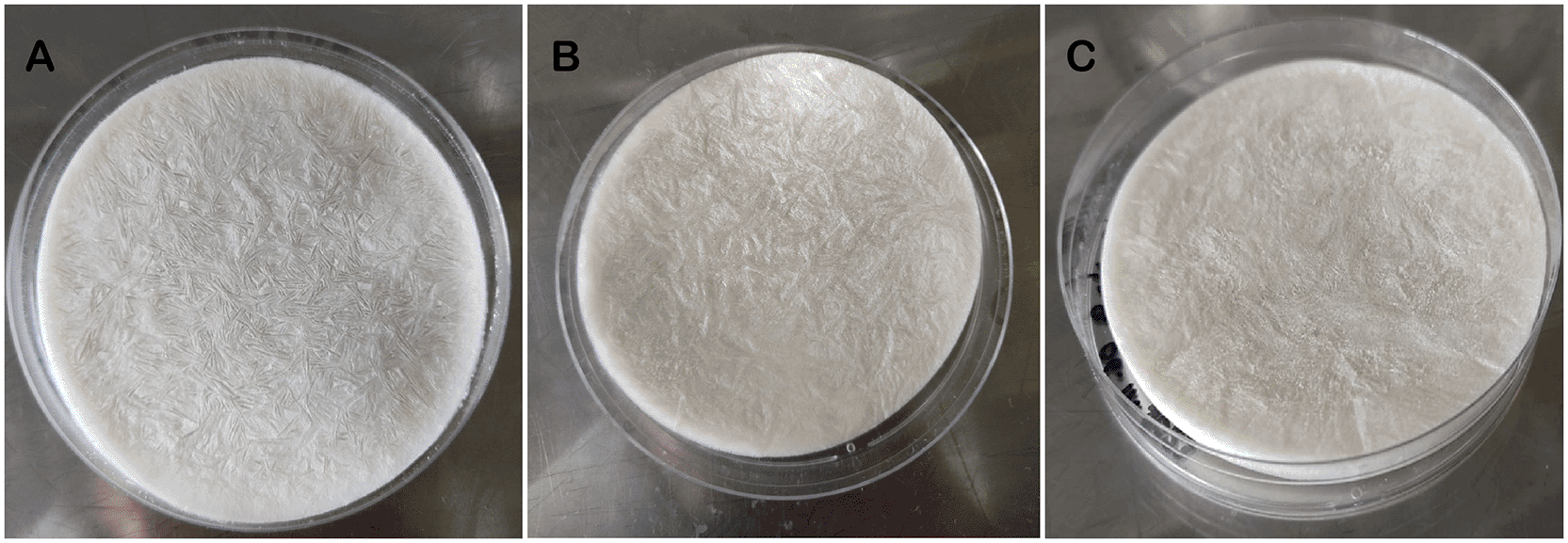
Scaffolds were fabricated in 10 cm petri dishes to analyze their visual properties. In terms of visual appearance, nanocellulose scaffolds exhibited a whitish coloration, while chitosan and nanocellulose/chitosan scaffolds showed a slightly yellowish tone ( Figure 4). Texturally, nanocellulose scaffolds were softer, whereas chitosan-based scaffolds were harder and more rigid. Notably, the nanocellulose/chitosan composite scaffolds demonstrated the greatest stiffness among the three formulations ( Figure 4C).
For optimal cell development, the architecture of 3D scaffolds must present an interconnected porous network that facilitates nutrient diffusion, cell adhesion, proliferation and migration. Porosity and pore size is a fundamental factor, because higher porosity reduces mechanical properties, such as strength and stiffness, making the material more susceptible to breakage.43 According to44 an ideal scaffold should possess an interconnected macroporous network with pore size ranging from 100 to 900 μm, allowing the passage of cells, nutrients and metabolites into the structure, promoting cell proliferation. While micropores between 2 and 20 μm enhance cell adhesion and vascularization, further promoting tissue regeneration.
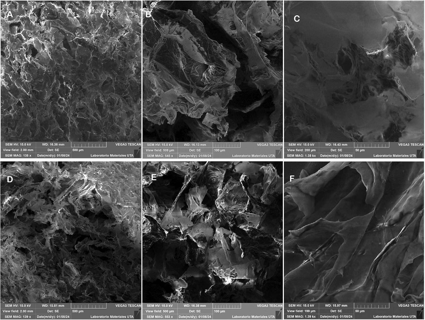
Scanning electron microscopy (SEM) analysis revealed the porous architecture of the scaffolds formed after lyophilization. All scaffolds exhibited heterogeneous porous structures with irregular surface topographies and varying degrees of pore size and interconnectivity. In nanocellulose-based scaffolds ( Figure 5), the surface displayed larger pores ( Figures 5A–C), while the interior showed smaller, more compact pores ( Figures 5D–F). These pores appeared as irregular, channel-like structures lacking defined geometry. A combination of macropores and micropores was observed, some of which were interconnected. Pore diameters ranged from 71 to 320 μm, with an average size of 133.33 ± 55.68 μm. These values were obtained from SEM micrographs captured at 553× magnification using a 100 μm scale, which provided optimal contrast for pore analysis. The observed pore morphology is consistent with previous findings by45 who reported that nanocellulose fibers exhibit limited inter fiber adhesion, thereby facilitating the formation of macropores (>100 μm). The presence of both macro and micropores, along with partial interconnectivity, is favorable for tissue engineering applications, as it may enhance nutrient diffusion, cellular infiltration, and scaffold integration with host tissue.
By contrast, chitosan scaffolds ( Figure 6) displayed smaller and more compact pores, with pore sizes ranging from 31 to 161 μm and an average of 69.92 ± 42.27 μm ( Figures 6A–F). The structure was dominated by micropores, with limited macroporosity.46 reported similar trends, where chitosan produced scaffolds with average pore sizes of ~74.5 μm.47 explained that increasing chitosan concentration raises the solution’s viscosity, restricting the formation and distribution of ice crystals during freezing and leading to reduced pore size. The dense structure of chitosan scaffolds may provide enhanced mechanical strength but may limit rapid cell infiltration.
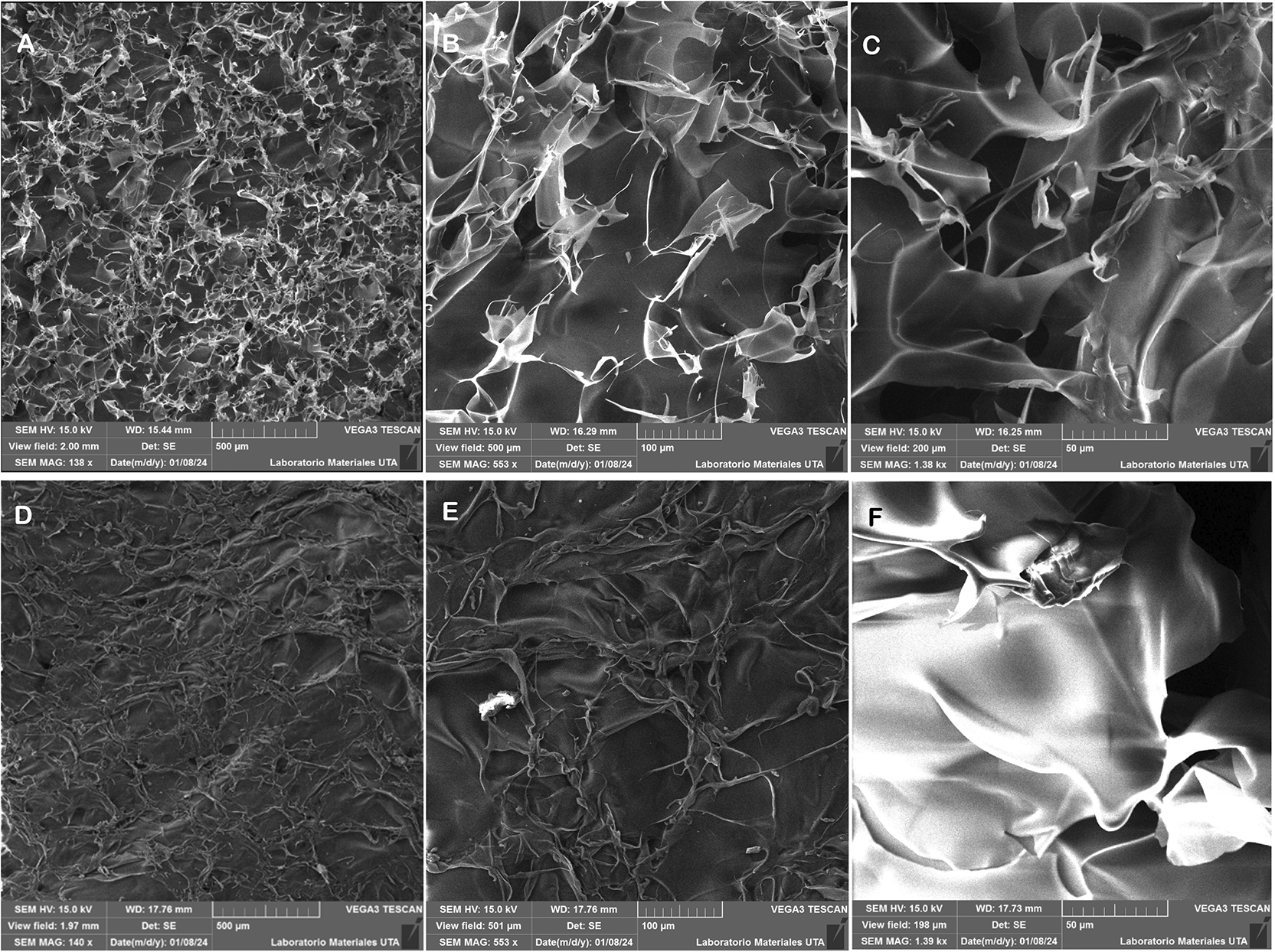
SEM micrographs showing the porosity of chitosan-based scaffolds: surface views (A, B, C) at magnifications of 138×, 553×, and 1.38 k×, respectively; and cross-sectional views (D, E, F) at 140×, 553×, and 1.39 k×, respectively.
SEM analysis of the hybrid (nanocellulose – chitosan) scaffolds ( Figure 7) showed an intermediate morphology. Pore sizes ranged from 67 to 163 μm, with an average size of 110.13 ± 33.29 μm, as determined from micrographs taken at 553× magnification with a 100 μm scale ( Figures 7A–F). The combination of nanocellulose and chitosan resulted in improved structural uniformity and more homogeneous pore distribution. According to48 the addition of nanocellulose enhances scaffold uniformity by promoting consistent pore formation, as nanofibrils crosslink with chitosan chains and enable rearrangement during freeze-drying. This leads to smaller pores than those in nanocellulose scaffolds, but larger than those in chitosan scaffolds, as also reported by.49 Furthermore, the inclusion of nanocellulose increases surface roughness, a feature that can enhance extracellular matrix (ECM) formation and support cell adhesion.
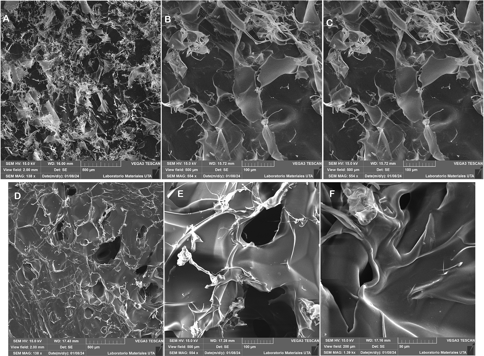
The viscosity-modifying effects of nanocellulose further contribute to the regulation of pore architecture, reinforcing the scaffold and promoting biological performance.48 The presence of macropores (>100 μm) in hybrid scaffolds may supports cell infiltration, migration, and proliferation, while smaller micropores (<50 μm) facilitate efficient nutrient exchange and waste removal.50 Moreover, as emphasized by15 pore interconnectivity is critical for effective nutrient transport and fluid exchange.
Overall, the nanocellulose scaffolds demonstrated the highest porosity with predominantly large pores, suitable for promoting cellular migration. The chitosan scaffolds displayed a denser morphology with smaller pores, offering better structural support but limited bioactivity. The hybrid scaffolds may offer a balanced profile, with moderate pore size and improved uniformity, suggesting potential advantages in both mechanical and biological performance for tissue engineering applications, such as, skin, bone and cartilage regeneration.
Reference 51 indicates that for skin tissue regeneration, scaffolds require pore sizes ranging from 20 to 120 μm to facilitate oxygen and nutrient transport. Additionally,52 report that scaffolds with pores between 45 and 106 μm are optimal for cell adhesion, whereas pores larger than 300 μm provide sufficient space for internal tissue growth, enhancing cell proliferation and vascularization. The surface area and pore size in biological scaffolds is a determining factor for cell accommodation (adhesion, survival, migration and differentiation), as well as the passage of medium and nutrients (in vitro) or blood (in vivo).
For bone regeneration, the minimum required pore size ranges from 75 to 100 μm, with an optimal range of 100 to 135 μm to facilitate proper osteoblast infiltration and migration. However, pores larger than 300 μm enhance vascularization and bone growth.53 Although osteoblasts measure between 10 and 50 μm, they preferentially colonize larger pores ranging from 100 to 200 μm, which support the mineralized bone regeneration process post-implantation.54 This pore structure also aids macrophage infiltration, bacterial clearance, and the migration of other essential cells involved in colonization, migration, and in vivo vascularization.
In arthritis, cartilage loss is a key pathological feature, and its regeneration remains highly challenging. Advances in biotechnology have led to the development of a variety of scaffolds and diverse mesenchymal stem cell (MSC) sources aimed at regenerating connective tissue, particularly cartilage. Scaffolds with pore sizes between 200 and 400 μm and an oval-to-round morphology promote osteoblast function and chondrocyte differentiation. Additionally, macropores ranging from 150 to 355 μm, when interconnected with micropores (<50 μm), enhance the internal growth of fibrocartilaginous tissue, preventing articular cartilage degeneration.55
Porosity is a fundamental property of scaffold structures, representing the percentage of void spaces within the material. It plays a crucial role in modulating cell growth and proliferation. Scaffolds generally exhibit porosity levels between 70% and 90%.56 Lower porosity offers a greater surface area for cell adhesion, thereby promoting initial cell attachment. In contrast, higher porosity, while enhancing nutrient diffusion and overall permeability, both essential for tissue regeneration, may reduce cell density and slow down proliferation.56 Thus, optimizing porosity is critical to balancing structural support and biological performance in scaffold design.
Porosity was assessed using the liquid displacement method with dehydrated alcohol, a polar solvent that does not dissolve polymeric fibers and readily penetrates scaffold pores without inducing shrinkage or swelling.57 The nanocellulose scaffolds exhibited a porosity of 89.38 ± 0.90%, chitosan scaffolds 87.72 ± 0.70%, and the hybrid scaffolds 88.94 ± 0.77% ( Figure 8). One-way ANOVA revealed no statistically significant differences in porosity among the scaffold types (p > 0.05) at the 95% confidence level. However, pairwise comparisons indicated a significant difference between nanocellulose and chitosan scaffolds.
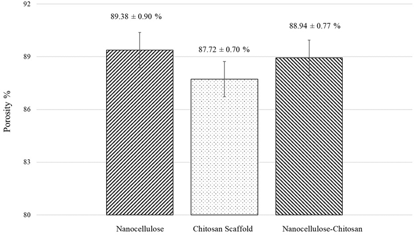
Statistical analysis showed no significant differences among the groups at a 95% confidence level.
According to58 in their study on nanocellulose materials, they reported that nano-cellulose scaffolds present a porosity of 89%. They also highlighted that the cooling rate during freeze-drying significantly influences porosity, pore alignment and mechanical properties. Similarly,45 observed that in nanocellulose scaffolds, weak adhesion between nanofibrils contributes to higher porosity. In contrast, according to a study by46 chitosan scaffolds show porosity levels between 75% and 85%. The study suggests that increasing the concentration of chitosan causes a reduction in porosity due to the higher viscosity of the solution, which promotes the formation of aggregates, which can negatively affect porosity. Although no statistically significant differences were observed in the porosity of hybrid scaffolds,45 suggested that the interconnected nanostructures in these combined scaffolds enhance the porosity. Similarly,58 in their study on nanocellulose/polyvinyl alcohol (PVA) hybrid scaffolds for skin tissue regeneration and wound healing, reported porosity levels ranging from 88% to 95%.
Scaffolds with porosity between 60% and 90% are considered ideal for oxygen and nutrient exchange, cell activity, and extracellular matrix (ECM) production during wound healing.59 Moreover, for bone tissue regeneration,54 emphasized that porosity greater than 90% is essential to support osteogenesis. It is important to note that the type of polymer used for scaffold fabrication not only influences the morphology, interconnectivity and pore size but also the porosity percentage and the mechanical properties.
Based on the results, nanocellulose scaffolds demonstrated high porosity due to the lack of fiber adhesion, while chitosan scaffolds exhibited lower porosity because of the solution’s viscosity. Finally, the hybrid scaffold displayed improved porosity due to the crosslinking and interaction between the biopolymer structures. Nevertheless, all three scaffold types exhibited porosity levels suitable for applications in skin and bone tissue regeneration.
The swelling potential of scaffolds reflects their capacity to retain water within their structure. This property not only affects the morphology and architecture of the scaffold but also influences cell growth. Adequate water absorption indicates that the scaffold can prevent the loss of essential fluids and nutrients required for cellular proliferation.60 According to59 scaffolds should provide a moist environment to prevent wound dehydration while also facilitating the removal of excess wound exudate.
The water absorption capacity was assessed by measuring the swelling behavior of the different scaffold types. Scaffold weights were recorded at 30-minute intervals over a 6-hour period following immersion in water ( Figure 9). The nanocellulose scaffold exhibited a swelling percentage of 2084 ± 352%, the chitosan scaffold reached 2524 ± 244%, and the hybrid scaffold displayed a swelling percentage of 2418 ± 221%.
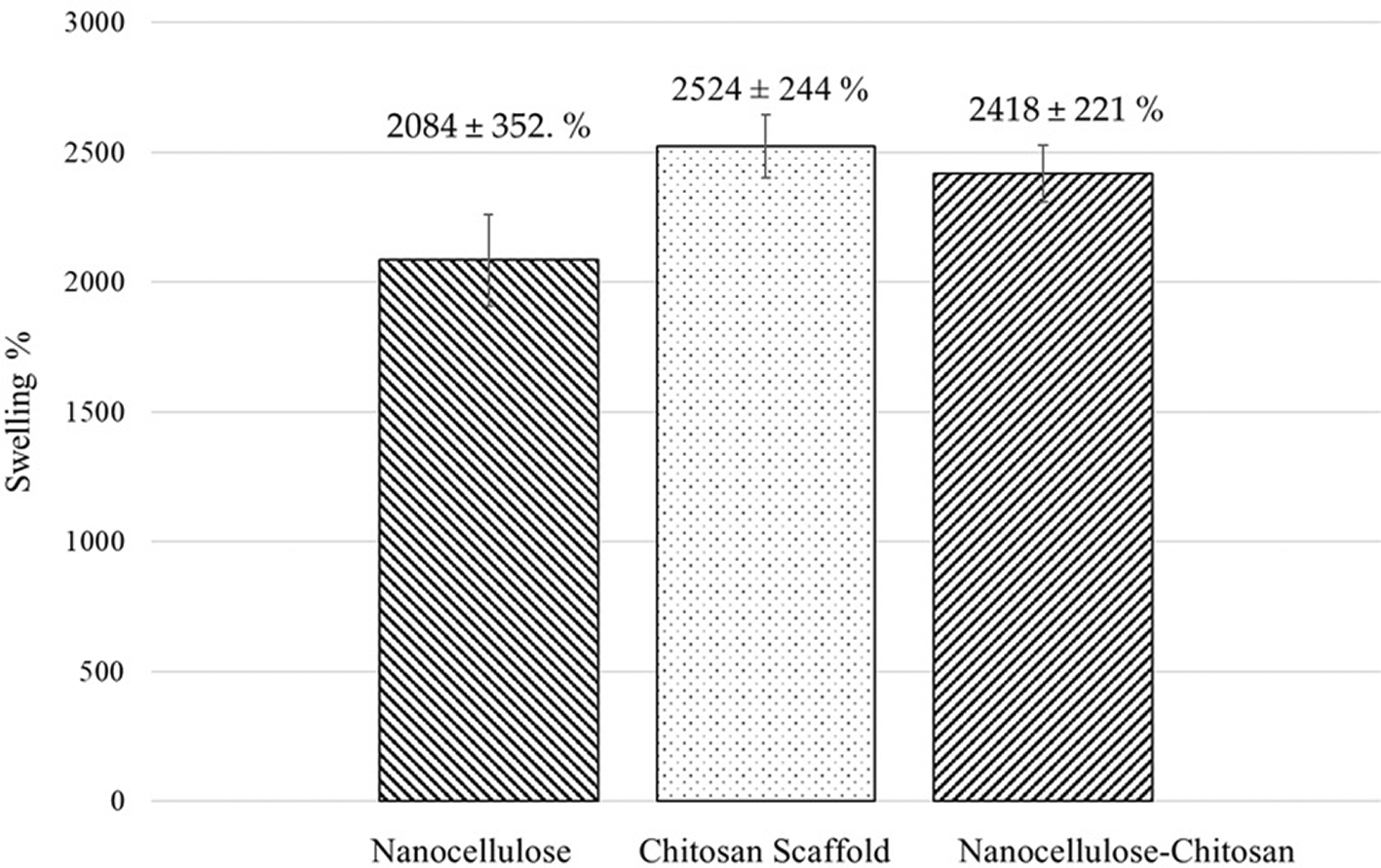
These results indicated a statistically significant difference among scaffold types at a 95% confidence level.
ANOVA analysis revealed a statistically significant difference (p-value <0.05) in the mean water absorption percentages among the different scaffold types, with a 95% confidence level. Significant differences were identified when comparing the water absorption percentages of nanocellulose with both chitosan and hybrid scaffolds. However, when comparing chitosan with hybrid scaffold, no statistically significant difference was observed.
Reference 61 reported that the swelling efficiency of nanocellulose scaffold is largely due to the availability of hydroxyl groups in the biopolymer structure. These groups form hydrogen bonds, facilitating water retention within the porous scaffold structure. Similarly,62 found that the pore size and pore distribution pattern in scaffolds significantly affect water absorption capacity. Nanocellulose disperses readily in water, forming stable suspensions, which can influence its overall absorption characteristics.
Chitosan scaffolds exhibited the highest water absorption capacity compared to the other scaffold types. This is attributed to chitosan’s structural hydroxyl (-OH) and primary amine (-NH2) groups, which enhance its affinity for water through hydrogen bonding.60,63 reported that when chitosan scaffolds are immersed in aqueous environments, the chitosan membranes swell and retain a specific volume of water absorbed within the scaffold’s three-dimensional network. For biomedical applications, chitosan scaffolds must absorb bodily fluids to support cellular transfer, while also allowing for the distribution of nutrients, metabolites, and growth factors through the extracellular matrix.
For nanocellulose – chitosan scaffolds, the combination of both biopolymers resulted in high swelling efficiency. This is due to nanocellulose’s hydrophilic nature, which acts as a bridging agent between polymer chains, improving the scaffold’s mechanical strength and promoting water absorption and retention within the structure.61 Additionally, chitosan forms hydrogen bonds with other biopolymers through its polar groups (-OH and -NH2), enhancing the scaffold’s water retention capacity.64,65 demonstrated that nanocellulose/collagen scaffolds for wound treatment exhibited swelling rates exceeding 1500%, reaching approximately 2037 ± 125% water absorption.
The water absorption capacity of scaffolds is closely linked to pore size, distribution, and interconnectivity, as well as the biopolymers’ ability to form hydrogen bonds. These factors enhance the scaffold’s affinity for water and contribute to improved water retention within its three-dimensional structure. This creates an ideal moist environment for epi-dermal cell migration and accelerates the re-epithelialization in the case of wound healing.
The valorization of organic waste, specifically banana peel residues, through the extraction of nanocellulose via acid hydrolysis and ultrasonic treatment represents a sustainable and cost-effective alternative to conventional synthetic polymers. Banana peels are an abundant agro-industrial by-product rich in cellulose, making them an ideal renewable source for nanocellulose production. Utilizing this biomass contributes to waste reduction, promotes circular economy principles, and adds value to agricultural residues that would otherwise be discarded. In the biomedical field, nanocellulose exhibits excellent properties such as biocompatibility, biodegradability, non-toxicity, and a high surface area, making it suitable for applications in tissue engineering. This study confirmed these attributes through the fabrication and characterization of lyophilized scaffolds composed exclusively of nanocellulose and nanocellulose/chitosan composites derived from banana peels. Both scaffold types demonstrated favorable porosity, water absorption capacity, and complete biodegradability. Notably, the incorporation of chitosan improved scaffold performance and structural uniformity, highlighting the potential of combining nanocellulose with other biopolymers to tailor structural and functional characteristics to specific biomedical applications. Following the extraction of plant-based nanocellulose from banana peel and its application in scaffold fabrication, analyses confirmed that the resulting scaffolds possess adequate morphological characteristics required for biomedical use. Therefore, nanocellulose-based scaffolds fabricated in this study hold great promise for tissue engineering. These materials represent viable candidates for future studies focused on evaluating cell adhesion, proliferation, and tissue regeneration, while simultaneously advancing sustainable material development.
| Views | Downloads | |
|---|---|---|
| F1000Research | - | - |
|
PubMed Central
Data from PMC are received and updated monthly.
|
- | - |
Provide sufficient details of any financial or non-financial competing interests to enable users to assess whether your comments might lead a reasonable person to question your impartiality. Consider the following examples, but note that this is not an exhaustive list:
Sign up for content alerts and receive a weekly or monthly email with all newly published articles
Already registered? Sign in
The email address should be the one you originally registered with F1000.
You registered with F1000 via Google, so we cannot reset your password.
To sign in, please click here.
If you still need help with your Google account password, please click here.
You registered with F1000 via Facebook, so we cannot reset your password.
To sign in, please click here.
If you still need help with your Facebook account password, please click here.
If your email address is registered with us, we will email you instructions to reset your password.
If you think you should have received this email but it has not arrived, please check your spam filters and/or contact for further assistance.
Comments on this article Comments (0)