Keywords
Bioprospecting; Coral Triangle; Drug discovery; Marine natural products; Secondary metabolites.
Bioprospecting; Coral Triangle; Drug discovery; Marine natural products; Secondary metabolites.
Life in the oceans has existed for approximately 3.7 billion years, nearly three times longer than life on land, resulting in a much higher level of evolutionary diversity. Of the 34 major animal phyla, only 12 are found in terrestrial environments, whereas 33 occur in marine ecosystems. Moreover, it is estimated that up to 90% of marine species remain undescribed, indicating that marine biodiversity represents an immense and largely untapped resource for scientific research and the development of innovative solutions to global challenges, particularly in the discovery of new drugs.1 Natural products derived from marine environments demonstrate substantially higher efficiency in the drug discovery process compared to those obtained from other natural sources. The success rate of developing marine-derived compounds into therapeutic applications is reported to be nearly twice as high, reflecting the significant advantage of marine chemical diversity. To date, tens of thousands of marine natural products have been successfully identified, with thousands of new compounds added each year, underscoring the vast potential of marine resources for human health applications.2 Given that many bioactive compounds represent advanced developments of previously characterized biological systems, strengthening investment in the early stages of marine biodiscovery has the potential to accelerate innovation and generate broad and sustainable social and health impacts.
The extreme conditions of marine environments, such as high salinity, elevated pressure, temperature fluctuations, and intense ecological competition, have driven marine organisms to evolve unique biosynthetic pathways, leading to the production of structurally complex secondary metabolites. These metabolites play critical roles in chemical defense, inter-organism communication, and ecological adaptation, and frequently exhibit potent and highly specific biological activities.3 In parallel, drug discovery continues to face major global challenges, particularly in combating antibiotic resistance, addressing the complexity of cancer, and responding to emerging infectious and chronic diseases.4 Although significant advances have been made in synthetic chemistry and computational drug design, the pace of discovering novel drug scaffolds has slowed over the past two decades. This situation has renewed interest in natural products as a primary source of chemical diversity for drug discovery. Of the approximately 400,000 structurally characterized natural compounds, only about 10% are derived from marine sources, highlighting the limited exploration of marine natural products and their vast potential for therapeutic development.
Within this global marine context, the Coral Triangle area is recognized as the global center of marine biodiversity and the richest region on Earth ( Figure 1).6 It encompasses Indonesia, Malaysia, Papua New Guinea, the Philippines, the Solomon Islands, and Timor-Leste. This region covers only about 1.1% of the surface of the Earth. It is home to 100,000 km2 of diverse coral reefs, which constitute one-third of the world’s coral reefs. But it has the world’s highest diversity of coral reef species, representing 76% of the world’s coral species.7
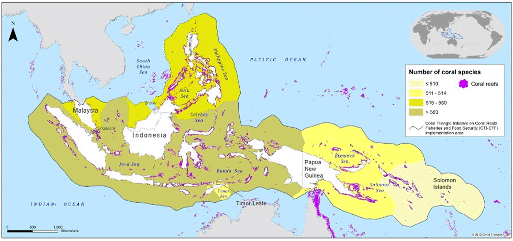
Adapted from Coral Triangle Atlas (2012).5
To enhance marine biodiversity conservation, fisheries management, and food security, countries in the Coral Triangle have collaborated through the Coral Triangle Initiative (CTI). The initiative has received significant international support and has led to improved management outcomes, including better MPA (Marine Protected Area) enforcement and increased regional management capacity for marine resource governance.
The high biodiversity of the Coral Triangle is shaped by a combination of geological setting, physical environment, ecological, and evolutionary processes. The Coral Triangle has a complex geological history, including tectonic activity and sea-level changes.8 During the Pliocene and Pleistocene periods, the Coral Triangle served as a refuge for marine species amid major environmental changes, particularly sea-level variations.9 The geological history created a variety of habitats and environmental conditions conducive to high biodiversity.
In addition to geological factors, the physical environment also contributes to its role as a biodiversity hotspot, because the region’s central position between the Indian and Pacific Oceans facilitates species accumulation and dispersal. Study found that dispersal out of the Coral Triangle may increase due to global warming.10 In addition, high rates of in situ speciation, particularly among species with limited dispersal abilities like dwarfgobies, have driven local biodiversity.8
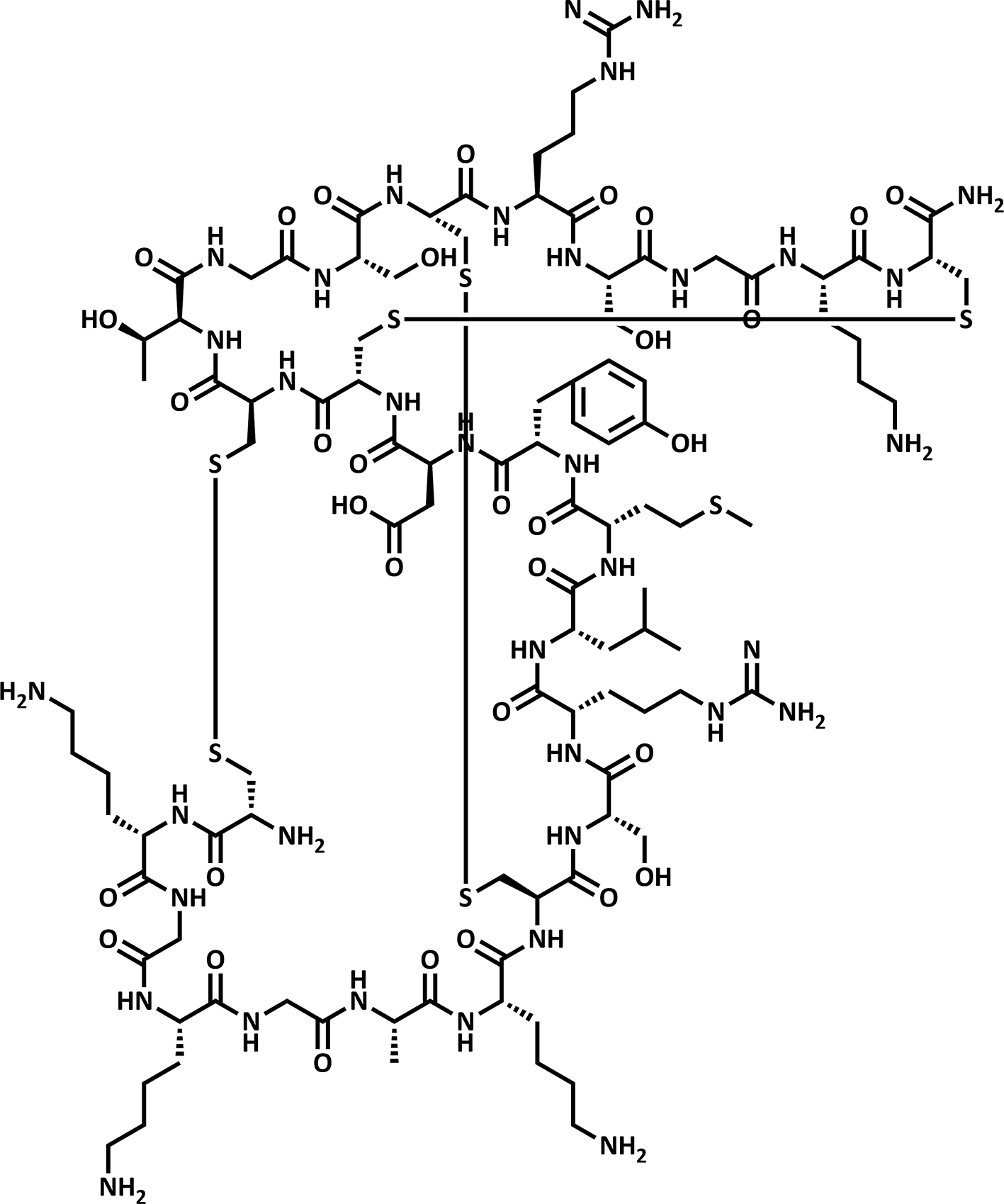
Beyond its ecological significance, the Coral Triangle holds significant potential for bioprospecting, particularly for the discovery of unique compounds for pharmaceutical and cosmetic applications. This region’s marine organisms, including sponges, corals, and mollusks, are prolific sources of bioactive metabolites with diverse biological activities. For instance, Ziconotide (Prialt®) was isolated from Pionoconus magus, a marine mollusk from the Pacific Ocean, the Philippines ( Figure 2).11 This compound comprises hundreds of peptides and hormones. Different short peptides termed conotoxins block the prey neuromuscular channel/receptors. At the same time, hormones interfere with the prey transductional signals. Based on their efficacy in managing severe chronic pain, analgesic drugs have been developed. In 2004, the FDA officially licensed Ziconotide or Prialt for intrathecal administration in the treatment of severe pain.12 The successful development of Ziconotide underscores the immense potential of marine-derived natural products in drug discovery.
Therefore, this systematic literature review aims to provide a comprehensive overview of the potential of bioactive compounds from the Coral Triangle region, analyze current research trends, and discuss prospects, while considering the ethical aspects of marine bioprospecting.
This review was conducted by combining two complementary approaches, bibliometric analysis to quantitatively assess literature to discern trends, patterns, and key research entities within a discipline, and a systematic review to qualitatively interpret and discuss scientific findings reported in the literature. This integrative approach facilitates comprehensive mapping of research trends while providing critical synthesis to identify knowledge gaps and future research directions for algae, which is commonly observed in small puddles, streams, rivers, and oceans.13 The bibliographic data were retrieved from Scopus database on December 14, 2025, using a query (see Supplementary Information) applied to titles, abstracts, and keywords. The article selection process followed the PRISMA (Preferred Reporting Items for Systematic Reviews and Meta-Analyses) workflow using predefined criteria ( Figure 3).
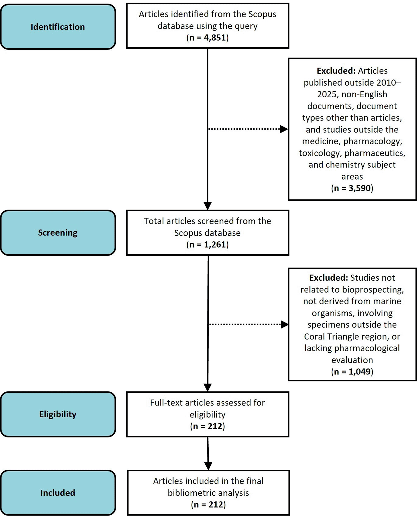
The inclusion criteria established were: (1) articles published within the period 2010–2025, (2) English-language documents, (3) research articles only, and (4) focusing on the subject area of medicine, pharmacology, toxicology, pharmaceutics, and chemistry. The initial search yielded 4,851 documents, which were subsequently manually screened to ensure the content relevance, resulting in a final dataset of 212 documents ( Figure 3). The final dataset was analyzed using Biblioshiny (Bibliometrix-R package) to evaluate annual publication trends. The relationships among organisms, compound classes, and reported bioactivities were visualized using a Sankey diagram generated in Power BI, while the geographical distribution of sampling locations was mapped using QGIS.
A systematic review was subsequently conducted to synthesize information from the selected studies, focusing on investigated organisms, identified compound classes, and associated biological activities. The primary literature sources consisted of the final dataset generated from the bibliometric analysis. To ensure comprehensive coverage of relevant studies, additional publications were identified through manual searches in Google Scholar and PubMed. The manual search employed combinations of the keywords “natural products”, “secondary metabolites”, “isolated compounds”, and “isolation”, combined with organism-specific terms (“sponges”, “corals”, “tunicates”, “echinoderms”, “mollusks”, “bacteria”, and “fungi”) and geographic descriptors (“Coral Triangle”, “Indonesia”, “Malaysia”, “Philippines”, “Papua New Guinea”, “Solomon Islands”, and “Timor-Leste”).
Despite the comprehensive overview presented in this review, several limitations should be acknowledged. The literature search was primarily conducted using the Scopus database, while additional studies were identified through manual searches in Google Scholar and PubMed. Consequently, some relevant studies indexed in other databases may not have been captured. Furthermore, only English-language publications within the 2010–2025 period were included, which may introduce language and temporal bias.
According to the data retrieved from the Scopus database, it has been ascertained that during the period 2010–2025, scholarly work about bioprospecting in the Coral Triangle comprises 212 articles. The number of publications showed a fluctuating pattern over the study period ( Figure 4a). In the early phase, publication output declined from 15 articles in 2010 to 7 articles in 2013, indicating limited research activity at the beginning of the period. A gradual recovery was observed in 2014–2015, although a slight decrease occurred again in 2016. A significant peak was recorded in 2017, with 26 publications, representing the highest output across the observed years. Following this surge, publication numbers stabilized between 15 and 16 articles annually during 2018–2021, suggesting a consolidation phase of research activity. A temporary decline was observed in 2022, followed by a moderate increase in 2023 and 2024. The slight decrease observed in 2025 may be attributed to incomplete indexing, as the data were collected on December 14, 2025. Overall, this trend indicates that the research topic has developed steadily despite annual fluctuations, with a notable surge occurring around the middle of the observation period, further confirming that the Coral Triangle region remains one of the key areas for bioprospecting research and the discovery of natural bioactive compounds.
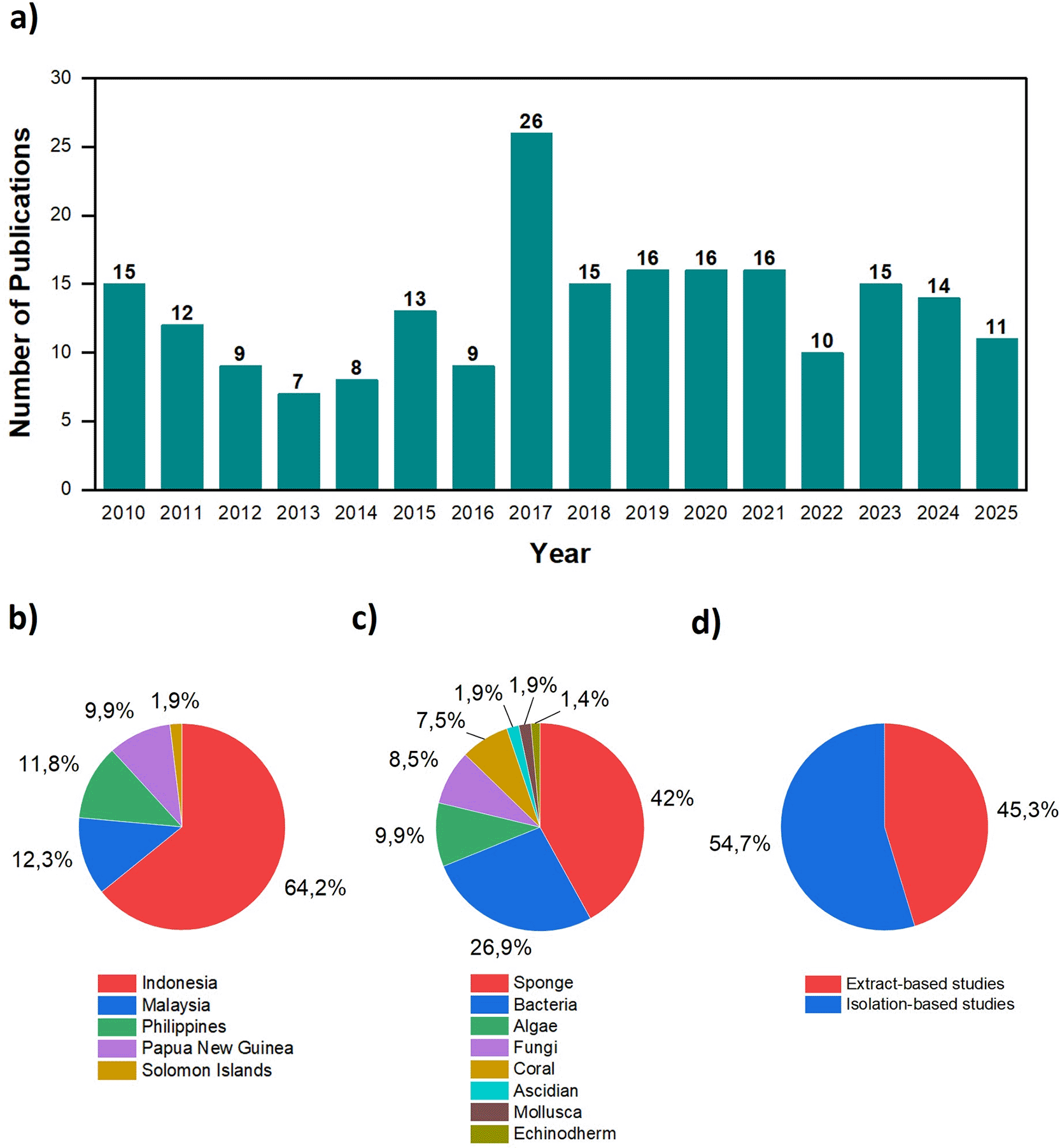
Spatial analysis reveals a significant dominance of Indonesia as the primary sampling location (64.2%), followed by Malaysia, the Philippines, Papua New Guinea, and the Solomon Islands ( Figure 4b). This dominance reinforces Indonesia’s position as both the research epicenter and the main source of specimens in the region, likely driven by its vast maritime territory and exceptionally rich biodiversity. From a taxonomic perspective, marine sponges emerge as the most prominent research subject (42%), followed by bacteria, algae, fungi, and other organisms ( Figure 4c). These findings align with global trends positioning sponges as a preferred marine source in the discovery of bioactive compounds; statistical data from 2010–2019 recorded 2,659 natural products derived from sponges, accounting for 47.2% of all compounds isolated from marine invertebrates.14 Furthermore, the distribution of methodological approaches shows that isolation-based studies (54.7%) slightly outnumber extract-based studies ( Figure 4d). This competitive ratio indicates the maturity and continuity of the research ecosystem in the region, where initial screening efforts are consistently followed by compound identification and characterization.
Spatial mapping of sampling locations indicates that bioprospecting activities within the Coral Triangle region are concentrated in several major biodiversity hotspots, particularly in the Indonesian archipelago and Papua New Guinea. A significant density of sampling locations is specifically observed in three main areas: the waters surrounding Bunaken National Park in North Sulawesi, the Spermonde Islands off the coast of South Sulawesi, and the vicinity of New Britain Island ( Figure 5). Sampling clusters are also present in the Solomon Islands, although with lower density. The overlap of sampling points from different organism groups in several locations suggests that a single area often serves as a source for multiple research target organisms, further highlighting the rich marine biodiversity of the region. Collectively, this spatial pattern confirms that bioprospecting activities are focused on major biodiversity centers, reinforcing the critical role of this region in the global discovery of marine natural products.
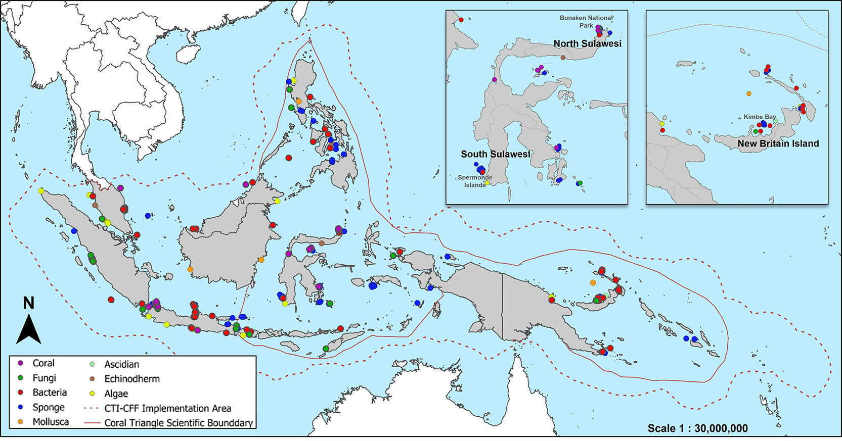
Map generated by the authors using QGIS software.
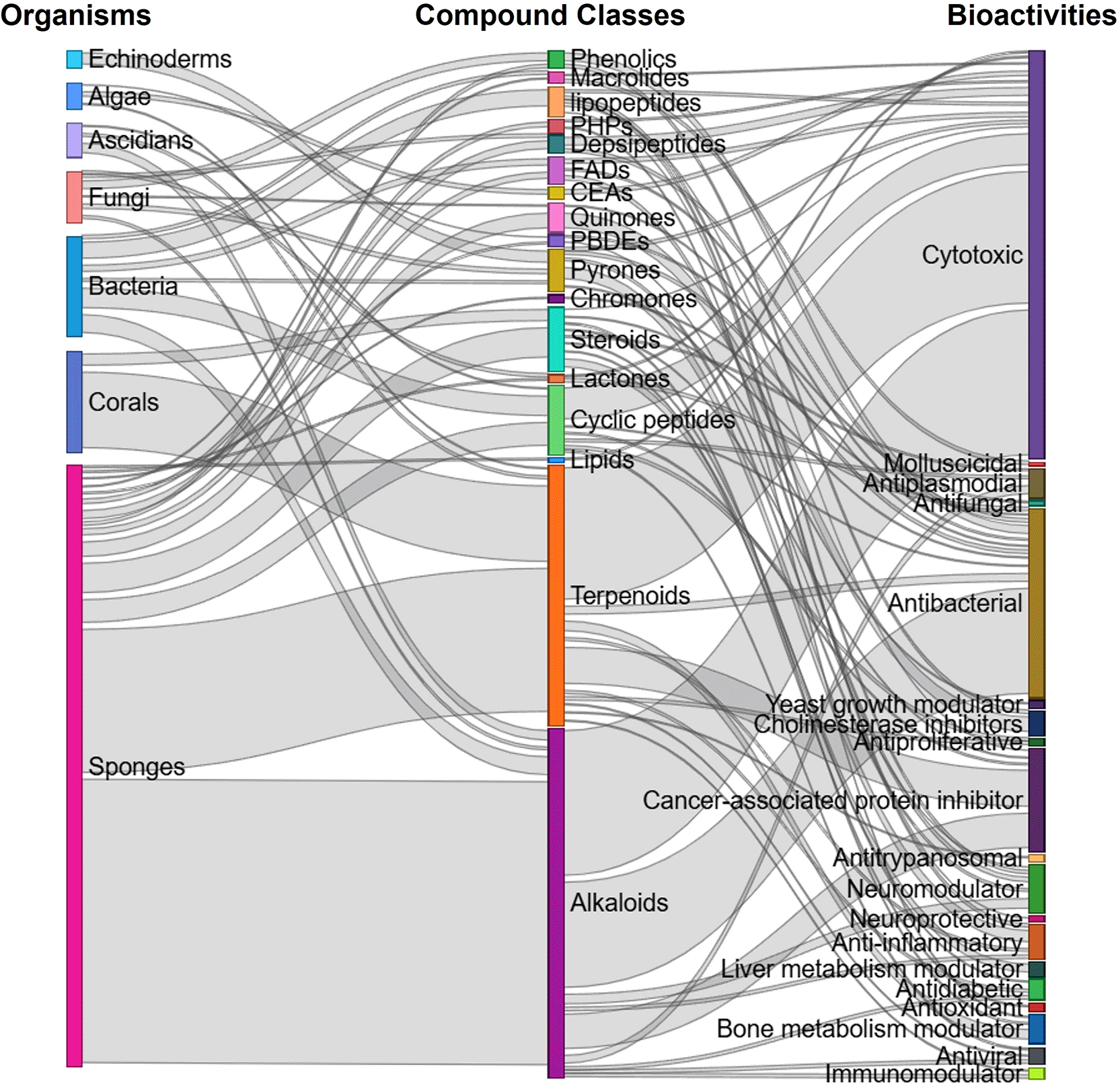
An integrative analysis using a Sankey diagram reveals a systematic relationship between organism groups, chemical compound classes, and their resulting bioactivity potentials ( Figure 6). Marine sponges emerge as the most significant contributors, particularly in the production of alkaloids and terpenoids. This flow pattern indicates that these two classes of secondary metabolites serve as the primary components underlying the diverse biological activities of marine organisms within the study region. Additionally, bacteria and corals provide consistent contributions, specifically in the production of steroids and cyclic peptides, although in smaller proportions compared to sponges. From a functional perspective, the spectrum of identified bioactivities is heavily dominated by cytotoxic activity, which is largely linked to the alkaloids and terpenoids. Beyond this cytotoxic potential—relevant for anticancer candidate development-antibacterial activity also emerges as a major bioactivity, reinforcing the critical role of marine organisms as sources of novel antibiotic candidates. Various other bioactivities, including antiviral, antidiabetic, neuromodulatory, and anti-inflammatory properties, are also detected, although with relatively lower frequencies.
The Coral Triangle is known for the richness and endemism of shallow-water reef-building corals, reef fishes, and mangrove biodiversity. The Coral Triangle includes areas of high biodiversity importance such as the southern Philippines, northeastern Malaysian Sabah, central to eastern Indonesia, eastern Papua New Guinea, and the Solomon Islands.
Coral reef ecosystems depend on a diverse assemblage of free-living and host-associated microorganisms to capture, retain, and recycle nutrients and trace elements, thereby enabling these ecosystems to be productive in the marine environment. Coral reefs are often referred to as the “rainforests of the ocean” because of their high biodiversity and ecological significance. These ecosystems provide numerous essential services, including coastal protection, fisheries, economic services, and tourism, which are vital for the livelihoods of millions of people.15 In addition to their ecological functions, reef-associated organisms employ various survival strategies, including the production of secondary metabolites that contribute to chemical defense and mediate ecological interactions. The secondary metabolites isolated from the marine products in the Coral Triangle and their bioactivity are provided in Supplementary Information.
Marine sponges are among the most abundant marine organisms in Indonesian waters. Results from the Snellius-II expedition reported the presence of approximately 830 sponge species in western Indonesia. Sponges are also recognized as highly productive hosts for diverse microorganisms. These microbial symbionts, including both bacteria and fungi, may account for 40–70% of the sponge biomass and are responsible for the production of various secondary metabolites. Especially Phylum Porifera, the dominant contributor to metabolite discovery in the Coral Triangle. Their sessile lifestyle and reliance on chemical defense mechanisms have resulted in the evolution of structurally diverse secondary metabolites, including alkaloids, terpenoids, macrolides, peptides, and polyketides.16
Consistent with global trends in marine natural product discovery, sponges from Indonesian and adjacent Coral Triangle waters have yielded an exceptional diversity of bioactive compounds spanning multiple chemical classes, including alkaloids, terpenoids, polyketides, peptides, macrolides, and brominated phenolics. The majority of reported compounds originate from demosponges (Class Demospongiae), underscoring their dominant contribution to metabolite discovery in this region. Numerous studies have demonstrated potent biological activities associated with these metabolites, most notably cytotoxic, antibacterial, antiviral, anti-inflammatory, antiplasmodial, antidiabetic, and enzyme inhibitory effects, even in extreme habitats such as cold springs and hot deserts.17 Their extreme habitats indicate their ability to adapt through structural changes in the plasma membrane and the production of protective compounds, such as polyols, sugars, and secondary metabolites.18
Various secondary metabolites identified from algae have biological activity; their chemical structure is presented in Figure 7.19–21
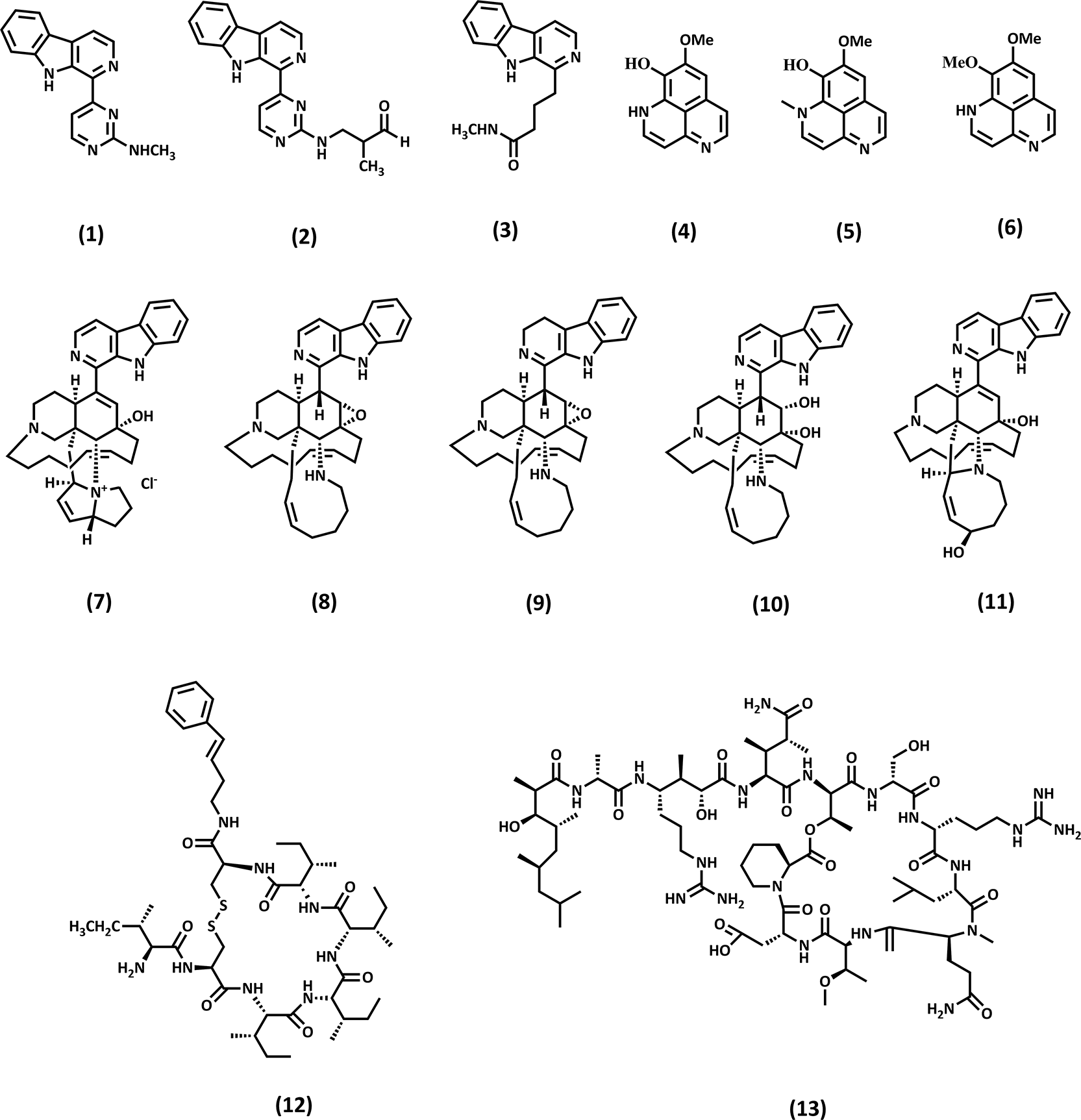
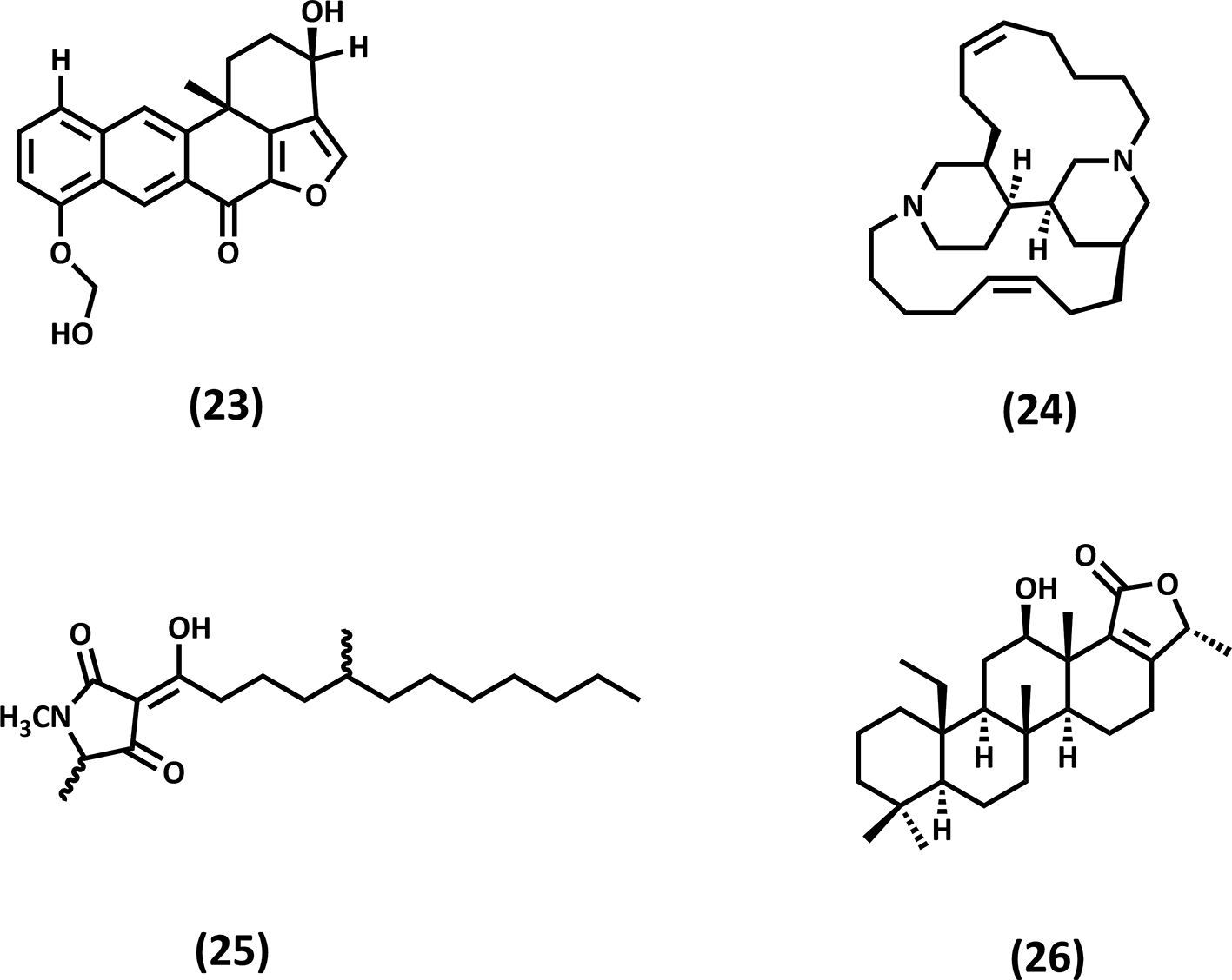
Cytotoxic and anticancer activities are among the most frequently reported bioactivities of sponge-derived metabolites from Indonesian waters; the chemical structure of the compounds is presented in Figure 7. Alkaloids such as Ingenines (1–3), Aaptamine derivatives (4–6), and Manzamines (7–11), as well as peptide and polyketide metabolites including Microcionamide (12) and Daedophamide (13), have shown strong growth inhibition against a wide range of human cancer cell lines, often at low micromolar or submicromolar concentrations. Several compounds also target specific molecular pathways relevant to cancer progression, including tyrosine kinases, ubiquitin-related enzymes, and proteasomal activity, highlighting their potential as lead structures for mechanism-driven drug development.22–24
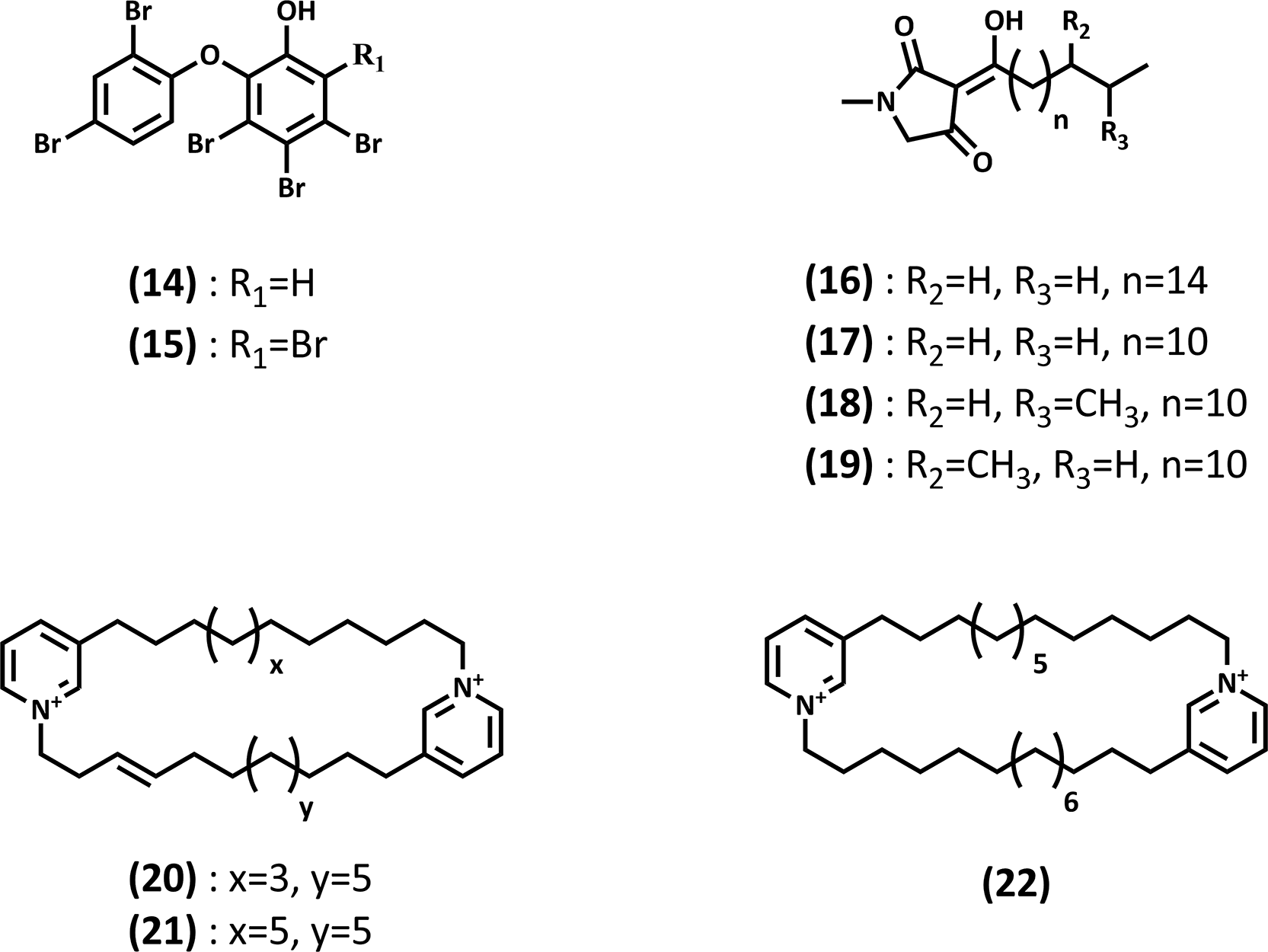
In addition to anticancer activity, sponge-derived metabolites from the Coral Triangle exhibit notable antibacterial and anti-infective properties. Brominated phenols (14–15), tetramic acid derivatives (16–18), and lipo-alkaloids (20–22) isolated from genera such as Lamellodysidea, Xestospongia, Haliclona, and Melophlus have demonstrated inhibitory activity against both Gram-positive and Gram-negative bacteria, including clinically relevant pathogens. The chemical structure is presented in Figure 8. Importantly, several compounds show activity against drug-resistant strains or interfere with bacterial virulence mechanisms, suggesting alternative therapeutic strategies beyond conventional bactericidal approaches.19,20,25,26
Apart from infectious disease applications, secondary metabolites isolated from marine sponges exhibit diverse bioactivities relevant to degenerative disorders, notably diabetes mellitus and Alzheimer’s disease, by targeting enzymes involved in insulin signaling, amyloidogenesis, and proteostasis regulation. Marine sponges from the Indo-Pacific region have yielded structurally diverse metabolites with promising activities relevant to Alzheimer’s disease (AD) and type 2 diabetes mellitus (T2DM). Xestosaprols H (23), isolated from Xestospongia sp., exhibited inhibitory activity against β-site amyloid precursor protein cleaving enzyme 1 (BACE1).19 BACE1 is a critical enzyme in amyloid β generation, and its inhibition is considered a validated therapeutic strategy for AD. The hydrophobic terpenoid scaffold may enhance binding within the catalytic pocket of BACE1, while oxygenated functionalities likely contribute to hydrogen-bond interactions, supporting enzyme inhibition. Additionally, Acanthocyclamine A (24) from Acanthostrongylophora ingens demonstrated inhibition of aftin-5–induced amyloid β-42 production at 26 μM.27 Although not directly evaluated in an Alzheimer’s disease model, suppression of Aβ-42 production suggests interference with amyloidogenic processing pathways. The nitrogen-containing heterocyclic framework may facilitate molecular interactions with enzymatic components involved in amyloid precursor protein metabolism.
Many studies have presented an association between diabetes mellitus (DM) and a clinical diagnosis of Alzheimer’s disease (AD) dementia.19–21,27 The bioactive compounds as anti-Alzheimer and antidiabetic are presented in Figure 9. Type 2 diabetes mellitus is characterized by insulin resistance and dysregulated glucose metabolism. One validated molecular target in T2DM is protein tyrosine phosphatase 1B (PTP1B), a negative regulator of insulin signaling. Melophlin C (25), a tetramic acid derivative isolated from Petrosia sp., exhibited PTP1B inhibitory activity (IC50 14.6 μM).20 The β-dicarbonyl system of tetramic acid provides strong hydrogen-bonding capacity and potential interaction with catalytic residues in the PTP1B active site. Inhibition of PTP1B enhances insulin receptor signaling, positioning tetramic acid–containing marine metabolites as promising scaffolds for antidiabetic drug development. Hyattellactones A (26) also exhibited PTP1B inhibitory activity, with an IC50 of 7.45 μM, further supporting the relevance of oxygen-rich marine metabolites in modulating insulin signaling pathways.21 Their macrocyclic lactone frameworks may enable multipoint interactions within enzyme binding pockets, contributing to inhibitory activity.
Sponges from the Coral Triangle region have emerged as a valuable source of metabolites targeting non-infectious diseases. Enzyme inhibitors affecting protein PTP1B, bone morphogenetic protein signaling, ubiquitin-conjugating systems, and cholesterol metabolism have been isolated from multiple sponge taxa. These findings are particularly relevant for the development of therapeutics for metabolic disorders, osteoporosis, neurodegenerative diseases, and inflammatory conditions.20 The presence of such diverse molecular targets underscores the functional breadth of sponge-derived natural products beyond traditional antimicrobial and anticancer paradigms.
From an ecological and evolutionary perspective, the remarkable chemical diversity observed in sponges is closely linked to their sessile lifestyle and reliance on chemical defense mechanisms for survival. In highly competitive reef environments such as the Coral Triangle, secondary metabolites function as deterrents against predation, fouling, and microbial overgrowth, as well as mediators of symbiotic interactions. Increasing evidence suggests that many of these compounds are produced, or at least biosynthetically influenced, by sponge-associated microorganisms, reinforcing the concept of sponges as holobionts rather than single chemical entities.28
The accumulated evidence from Indonesian and Coral Triangle sponges firmly establishes Porifera as one of the most important reservoirs of bioactive marine natural products. The combination of high species diversity, rich microbial symbioses, and pronounced chemical novelty positions sponges as cornerstone organisms in marine bioprospecting and drug discovery. Continued exploration integrating classical natural product chemistry with microbial genomics, metabolomics, and sustainable collection strategies is expected to further expand the pharmacological potential of sponge-derived metabolites from this globally significant biodiversity hotspot.29
Corals are invertebrate animals belonging to the Phylum Coelenterata (hollow animals) or Cnidaria. Corals, also known as polyps, are connected by living tissue and can share food.30 They are distributed across tropical marine environments, living in depths ranging from the low-tide mark to 6000 meters. The total number of coral species in the Coral Triangle region is approximately 605, which is about 76% of the world’s total coral species and 83% of all the species in the Indo-Pacific. In comparison, other regions, such as the Red Sea/Arabian region, have 364 species, with 27 regional endemics, making it the second-highest in terms of conservation.31 Based on the study, the Coral Triangle region is rich in coral biodiversity.
In the marine ecosystem, corals make symbiotic relationships with zooxanthellae, which involves nutrient recycling and allows algal photosynthesis. This symbiotic process provides organic carbon that fuels animal metabolism and growth.32 They also exhibit high capacities for clonal reproduction and regeneration, which are essential for their survival and adaptation to environmental changes. Besides, Corals face various threats due to climate change, pollution, and ocean acidification, which impact their growth and survival ability.33 Corals’ interactions and survival mechanisms tend to produce compounds with biological activity.
Several biologically active compounds isolated from corals belong to the sterol class, as shown in Figure 10. For example, (22R,23R,24R)-22,23-methylene-24-methylcholest-6-en-5α,8α-epidioxy-3β-ol (27) and 5α,8α-Epidioxy-24(R)-methylcholesta-6,22-dien-3α-ol (28) isolated from Sinularia polydactila, can act as anticancer against HCT 116 with IC50 values of 24.8 and 27.3 μg/mL, respectively.34 They exhibit anticancer activity due to the presence of the 5α, 8α-epidioxy moiety. The endoperoxide (–O–O–) moiety can undergo redox activation within cancer cells, leading to the generation of reactive oxygen species (ROS), which then causes apoptosis.
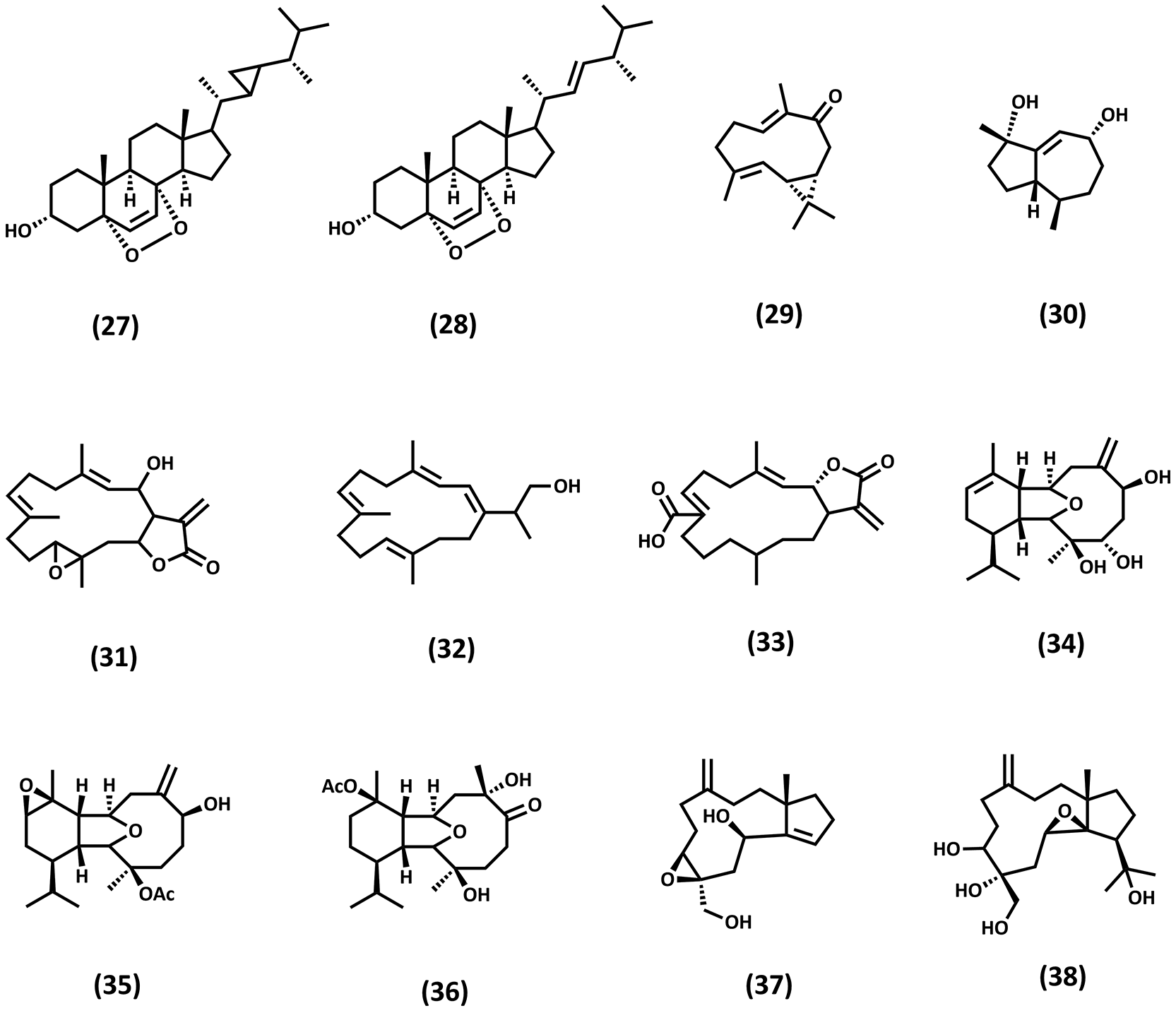
Another compound from the sesquiterpenoid class, as can be seen in Figure 10, Capgermacrene A (29), isolated from Capnella imbricata, and Trinor-guaiane sesquiterpene (30) isolated from Anthelia sp.35,36 Soft corals such as Capnella and Anthelia lack physical protection and rely on secondary metabolites for defense against predators, fouling organisms, and microbial infection. Based on the structure, Compound (29) has conjugated π-systems that can interact with cell-cycle regulatory proteins, disrupt mitochondrial membranes, and trigger apoptosis. While the trinor-guaiane compound shows weak activity against NBT-T2 cells, this might be due to its rigid structure. The trinor-guaiane compound retains anticancer activity because its hydrophobic core can associate with lipid membranes.
Corals of the genus Sarcophyton are particularly rich in cembranoid diterpenoids, including 2-hydroxy-crassocolide E (31), 16-hydroxycembra-1,3,7,11-tetraene (32), and (7Z)-lobohedleolide (33).37–39 The macrocyclic structure of cembranoid efficiently targets key cellular processes. Compound (31) contains a hydroxyl group at C-2 and lactone/ester moieties. These features enhance hydrogen bonding with biological targets and promote interaction with estrogen-related and apoptosis-regulating proteins. Then, compound (32) is characterized by multiple conjugated double bonds and a terminal hydroxyl group. This structure has biological consequences, which are increased membrane perturbation and enhanced interaction with Staphylococcus aureus bacterial membranes. Compound (33) contains a γ-lactone ring and a defined Z-configuration at C-7. The lactone acts as a reactive electrophilic center, capable of modifying nucleophilic amino acid residues (Cysteine, Lysine) and inhibiting proteins involved in cell proliferation.
Beyond cembranoids, corals also produce structurally complex eunicellin-type diterpenoids, particularly from Cladiella sp.40 Eunicellins are characterized by a polycyclic ether framework with multiple stereogenic centers, which provides a rigid three-dimensional architecture capable of precise interactions with biological macromolecules. Isolated compounds such as Cladieunicellins (34–36) demonstrate cytotoxic or anti-inflammatory properties, including inhibition of superoxide anion generation and suppression of tumor cell proliferation. Their bioactivity is often associated with oxygenated functional groups (hydroxyls, epoxides, acetates) that facilitate hydrogen bonding and modulation of inflammatory signaling pathways. The structural complexity of eunicellins also suggests high biosynthetic specialization, reflecting evolutionary adaptation for ecological defense.
Moreover, several studies have reported novel diterpenoids from Indonesian soft corals, including newly characterized scaffolds such as Sangiangol A and B (37) and (38) from Anthelia sp.41 These novel diterpenoids expand the chemical diversity of coral metabolites and frequently exhibit moderate cytotoxic activity against an NBT-T2 cell line. The discovery of new carbon skeletons indicates that coral biosynthetic pathways remain underexplored and represent a promising source of lead compounds for drug discovery. Structural novelty, combined with functional groups capable of redox modulation or protein interaction due to their unsaturated.42,43 This diverse habitat led researchers to study bioprospecting of marine tunicates, for example, in the Sangkarang Archipelago, which is part of the Coral Triangle. There were 55 species of tunicate in the Sangkarang Archipelago.44 In comparison, a total of 19 species of pelagic tunicates were observed in the China Sea.45 The Coral Triangle stands out as a significant hotspot for tunicate biodiversity, with a higher number of species. This diversity is crucial for understanding marine ecosystems and for conservation efforts in this biologically rich area.
The Tunicata class, especially Ascidiacea, has a sessile lifestyle. They act as filter feeders for plankton, facilitated by their barrelshaped bodies and two siphons. due to their barrel-shaped bodies and two.46 As a filter feeder, Tunicata helps to maintain water quality by filtering out plankton and other particles. Additionally, tunicates have symbiotic relationships with various microorganisms, including bacteria, which contribute to their chemical defense mechanisms.
The chemical defense mechanism of Tunicata release the unique compound that also has biological activity, their chemical structure is presented in Figure 11. For instance, Siladenoserinols A and B (39–40), isolated from the colonial ascidian Didemnum guttatum collected in Siladen, North Sulawesi, Indonesia, belong to the class of amino alcohol derivatives.47,48 More than half ascidian metabolites are amino acid-derived. These compounds exhibited inhibitory activity against the p53–Hdm2 protein–protein interaction, a critical pathway involved in cancer cell survival. The biological activity of siladenoserinols is likely related to the presence of polar amino and hydroxyl functional groups, which enable hydrogen bonding and electrostatic interactions with protein binding sites. Such interactions can disrupt protein–protein interfaces, including the p53-Hdm2 complex, therefore restoring p53 tumor suppressor function and inducing apoptosis in cancer cells.
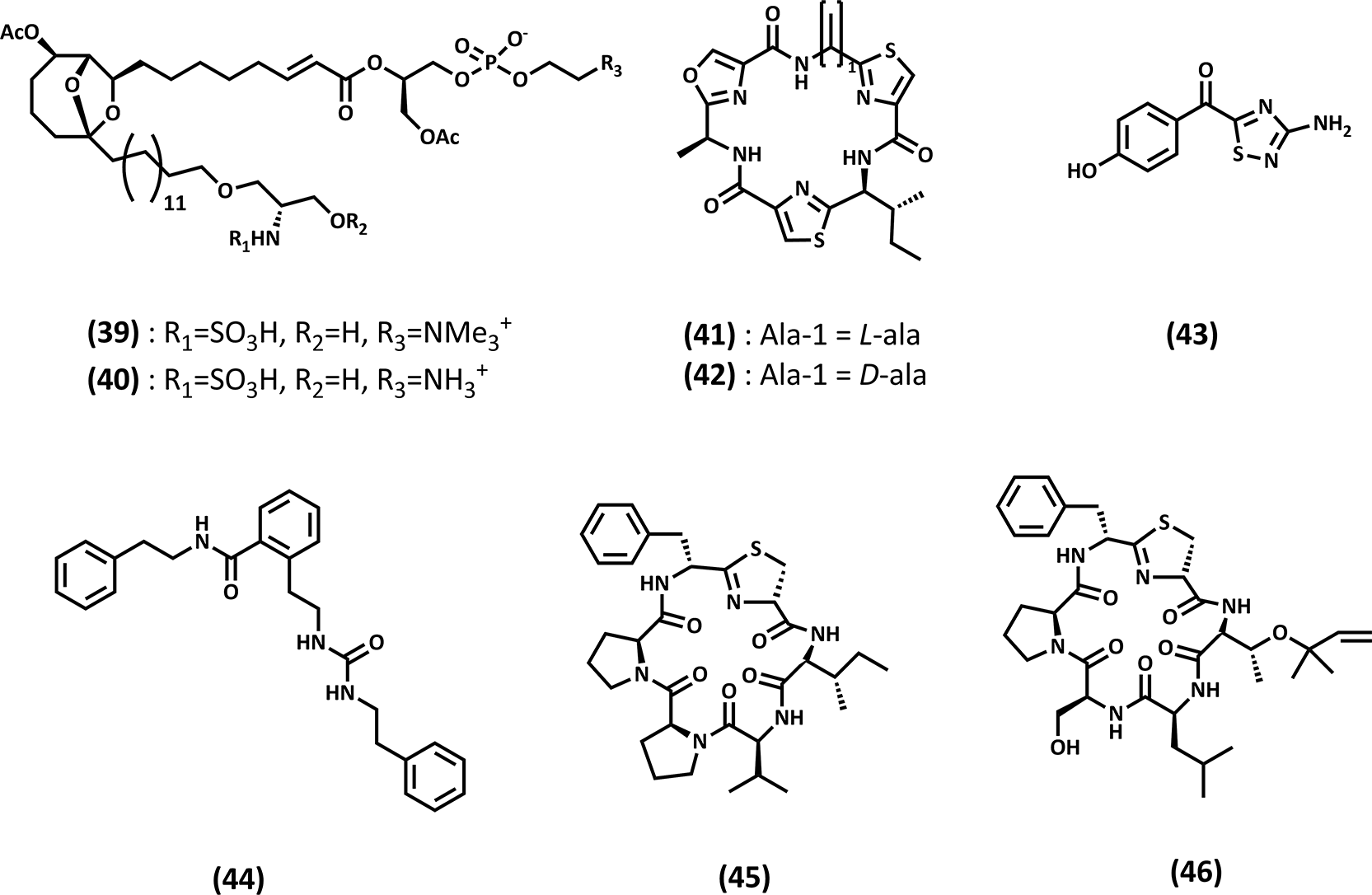
Bistratamides M and N (41–42) are cyclic peptide metabolites isolated from Lissoclinum bistratum collected in Raja Ampat, Papua, Indonesia. These compounds demonstrated moderate cytotoxic activity against multiple human tumor cell lines.49 The cytotoxicity of cyclic peptides is often caused by their conformational rigidity and high structural preorganization. This conformation enhances binding specificity toward intracellular targets such as enzymes, cytoskeletal proteins, or DNA-associated proteins. Moreover, the presence of heterocyclic and modified amino acid residues in bistratamides may facilitate membrane permeability and interaction with essential cellular pathways, ultimately leading to growth inhibition or apoptosis in cancer cells.
Another notable example is Polycarpathiamine A (43), an alkaloid isolated from the solitary ascidian Polycarpa aurata, which is found in Indonesian waters, exhibiting potent cytotoxic activity against L5178Y murine lymphoma cells.50 Thus, suggesting a key role for the nitrogen-containing heterocyclic structures in their interactions with nucleic acids and proteins. The cytotoxicity of polycarpathiamine A is likely due to its ability to interfere with DNA replication or key enzymatic processes involved in cell cycle regulation. Molleurea A (44), a urea-containing alkaloid isolated from the ascidian Didemnum molle, exhibited biological activity exclusively in a cytoprotective cell-based assay, with an IC50 value of 60 μM.51 The absence of pronounced cytotoxic or enzyme-inhibitory effects suggests that compound (44) does not directly interfere with essential cellular processes. Instead, its moderate cytoprotective activity is likely associated with the presence of the urea and amide moieties, which enable hydrogen-bonding interactions with cellular proteins involved in stress response or survival pathways. Additionally, the aromatic rings may contribute to membrane interactions or modulation of intracellular signaling rather than inducing cell death. Such mechanisms are consistent with the general bioactivity profile of marine alkaloids, which frequently act as chemical defense agents and display high potency against rapidly proliferating cells.
In addition to anticancer activity, tunicates also yield compounds with antiviral potential. Mollamide E (45) and F (46), a cyclic depsipeptide isolated from Didemnum molle collected in New Britain, Papua New Guinea, exhibited moderate inhibition of HIV integrase as well as cytoprotective effects in a cell-based assay.51 The biological activity of depsipeptides is closely linked to their mixed peptide–ester backbone, which confers both structural flexibility and enhanced lipophilicity. These features may facilitate binding to viral enzymes such as HIV integrase by mimicking natural substrate conformations or by occupying hydrophobic pockets within the active site. Additionally, the cyclic nature of compounds (45) and (46) likely contribute to their metabolic stability and sustained biological activity.
Mollusca is the second-largest phylum on Earth, with over 90,000 species, of which up to 55,000 are marine, approximately 30,000 are terrestrial, and up to 7,000 are freshwater. Molluscs can be found from the deep sea to the top of mountains, it is playing a fundamental role in a wide range of ecosystem functions and processes.52 There are diverse mollusca classes such as gastropods (snails and slugs), bivalves (clams and oysters), and cephalopods (squids and octopuses). Mollusks play important roles in ecosystems as ecosystem engineers, providing habitat, protection, and food for various other species. They are also economically significant, serving as a food source and making a contribution to aquaculture.53
One of the global centers of molluscan diversity is the Indo-Pacific Convergence Zone (IPCZ), which includes parts of the Coral Triangle. This region is a global marine biodiversity hotspot. Research in 2024 found that a total of 47.097 mollusk occurrence records were obtained, with the majority belonging to the class Gastropoda, which covered 19 orders, followed by Bivalvia, Cephalopoda, Polyplacophora, and Scaphopoda. These area contains 47.9% of mollusk species.54 The diverse species of Mollusca in the Coral Triangle might be due to the region’s diverse geographic factors.
Intertidal and coastal habitats are often subject to larger fluctuations in pH, temperature, salinity, and carbonate chemistry. Species inhabiting these environments are likely better adapted to these fluctuations, show greater tolerance, and will eventually be less affected by warming trends, reflecting their potential to cope with and survive environmental changes.53 Due to its habitat, mollusca release secondary metabolites to survive in fluctuating conditions.
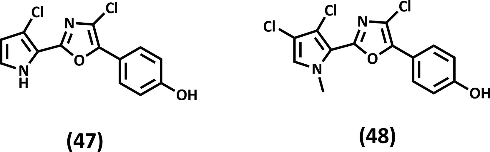
Notably, secondary metabolites were identified from the nudibranch Aldisa andersoni from the chemical class halogenated heteroaromatic alkaloids (Phorbazole alkaloids), as presented in Figure 12. Here, gastropod nudibranchs are among the most interesting, for example, the compounds of the sea slug Aldisa andersoni, displaying a wide biological activity, including several anticancer properties. The compounds are 9-chloro-phorbazole D (47) and N1-methyl-phorbazole A (48). They had cytotoxicity against MCF-7 mammary adenocarcinoma, the Hs683 oligodendroglioma, the A549 non-small-cell lung cancer, the SKMEL-28 melanoma, and the U373 glioblastoma, causing apoptosis at 18 to 34 μM.55
While these findings highlight the potential of molluscan-derived metabolites for drug discovery, several limitations in the available evidence should be acknowledged. The available evidence on bioactive compounds from molluscan species remains limited, and many reported metabolites are suggested to originate from associated organisms such as sponges or symbiotic microorganisms rather than the molluscs themselves.
Echinoderma is a phylum of marine invertebrates that consists of sea stars (Asteroidea), sea cucumbers (Holothuroidea), sea urchins (Echinoidea), brittle stars (Ophiuroidea), and crinoids (Crinoidea).56 Echinoderms can be found in many diverse benthic habitats, ranging from intertidal to deep-sea, with an estimated 7000 to 13000 species. These organisms are characterized by their pentamerous radial symmetry, a water vascular system, and a calcareous endoskeleton.57 They exhibit a variety of reproductive strategies, including both sexual and asexual reproduction.
Their unique character and various habitats make them valuable for bioprospecting. Echinoderms possess specialized connective tissues that can rapidly and reversibly alter their mechanical properties. This mutability is crucial for their survival and ecological success, influencing their nutrition, reproduction, and defense mechanisms. The regeneration process itself may be facilitated by the presence of MCTs (Mutable Collagenous Tissues).58 Additionally, Echinoderms are a rich source of bioactive compounds, including triterpene glycosides, steroids, peptides, and polysaccharides. These compounds exhibit a wide range of biological activities, such as antimicrobial, anticancer, anti-inflammatory, and antiviral effects.59
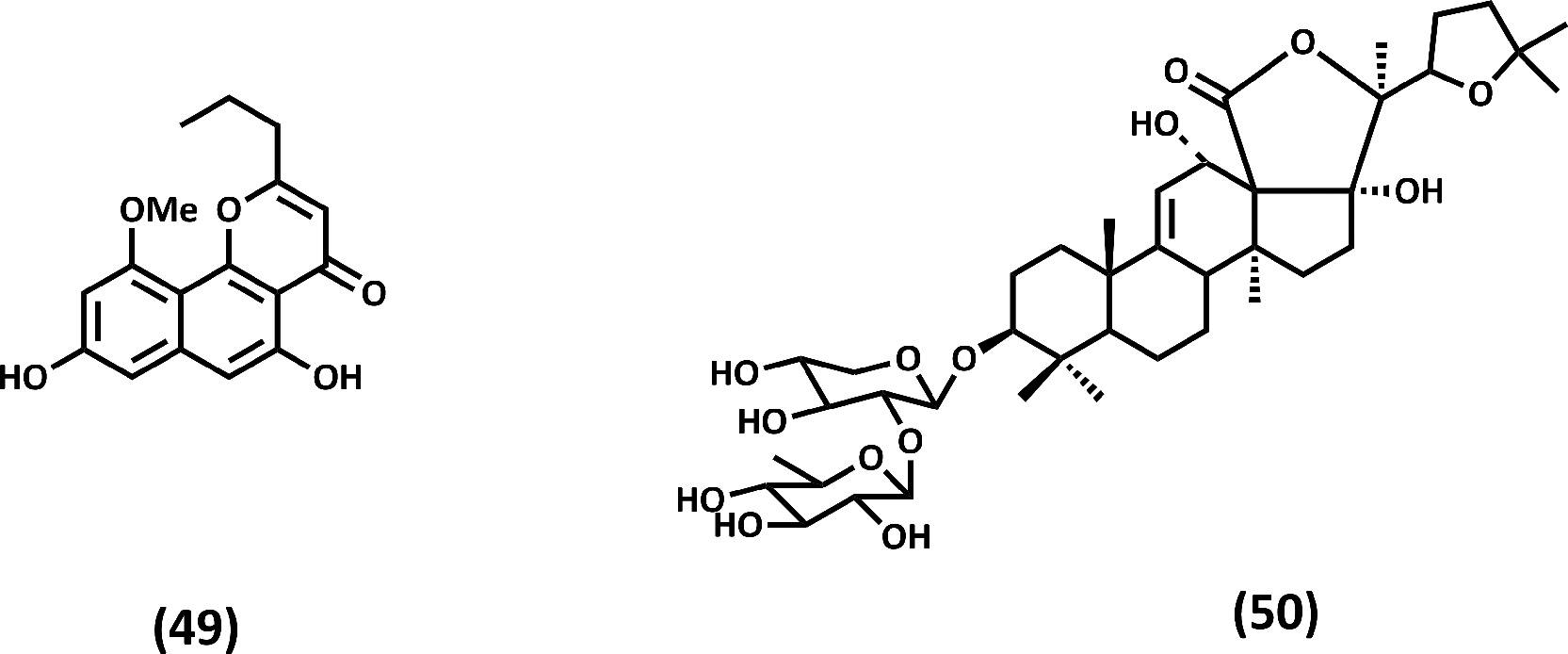
Several studies have investigated various compounds derived from echinoderms collected from the Coral Triangle area, as shown in Figure 13. These studies focus on their biological activities and potential therapeutic applications. Naphthopyrone Comaparvin (49) isolated from the marine echinoderm Comanthus sp. is identified as a new inhibitor of the NF-κB signaling pathway, acting by targeting both proteasome function and IκB phosphorylation.60 Saponin compound found in sea cucumbers also showed biological activity. 12-epi-desholothurin B (50) isolated from Holothuria atra body walls showed α-glucosidase inhibitory activity.61 Compound (50) has an aglycone triterpenoid moiety that acts as a hydrophobic side and a hydrophilic side (D-xylose and D-quinovose). This amphiphilic structure provides the ability to interact with the active site enzyme.
Despite the promising biological activities reported from echinoderm-derived compounds, several limitations in the current body of evidence should be considered. Research on bioactive compounds from echinoderms is still relatively limited and often concentrated on specific groups such as sea cucumbers. Many studies report biological activity based on crude extracts or partially purified fractions, which complicates the identification of the responsible compounds.
Algae are a diverse group of photosynthetic organisms found in various aquatic environments. They range from microscopic phytoplankton to large seaweeds and play crucial roles in marine ecosystems, including primary production, habitat formation, and nutrient cycling.62 The Coral Triangle, renowned for its exceptional marine biodiversity, encompasses a diverse array of algae. For instance, a study in the Spermonde Archipelago, Indonesia, identified 63 molecular operational taxonomic units (OTUs) of crustose coralline algae (CCA) from 11 genera, tripling the previously reported species richness.63 This number is compared to a study in New Zealand, which identified 122 species of coralline algae, indicating high regional diversity, but still less than the Coral.61 Natural products derived from marine environments demonstrate substantially higher efficiency in the drug discovery process compared to those obtained from other natural sources. The success rate of developing marine-derived compounds into therapeutic applications is reported to be nearly twice as high, reflecting the significant advantage of marine chemical diversity. To date, tens of thousands of marine natural products have been successfully identified, with thousands of new compounds added each year, underscoring the vast potential of marine resources for human health applications Given that many bioactive compounds represent advanced developments of previously biological systems, this highlights the importance of continued exploration of marine biodiversity. Characterizits (OTUs) of crustose coralline algae (CCA) from 11 genera, tripling the previously reported species richness.63 This number is compared to a study in New Zealand, which identified 122 species of coralline algae, indicating high regional diversity, but still less than the Coral. Isolated compound, (E)-17-(8-ethyl-4,5,9-trimethyldec-6-en-2-yl)-13-methyl-2,3,4,7,8,9,10,11,12,13,14,15,16,17-tetradecahydro-1H-cyclopenta [a]phenanthren-3-ol (51) can act as an antimicrobial agent against Staphylococcus aureus and an anticancer agent against MCF-7 with an IC50 value of 142.18 μg/mL. This compound has a steroid (cyclopenta [a]phenanthrene) core that is highly lipophilic. Gram-positive bacteria like S. aureus rely heavily on membrane integrity. Steroid-like molecules can insert into the phospholipid bilayer and disrupt the membrane. This compound is also structurally similar to endogenous steroids, estrogen, so that it can compete with estrogen, act as a partial antagonist, and disrupt estrogen-dependent signaling pathways ( Figure 14).
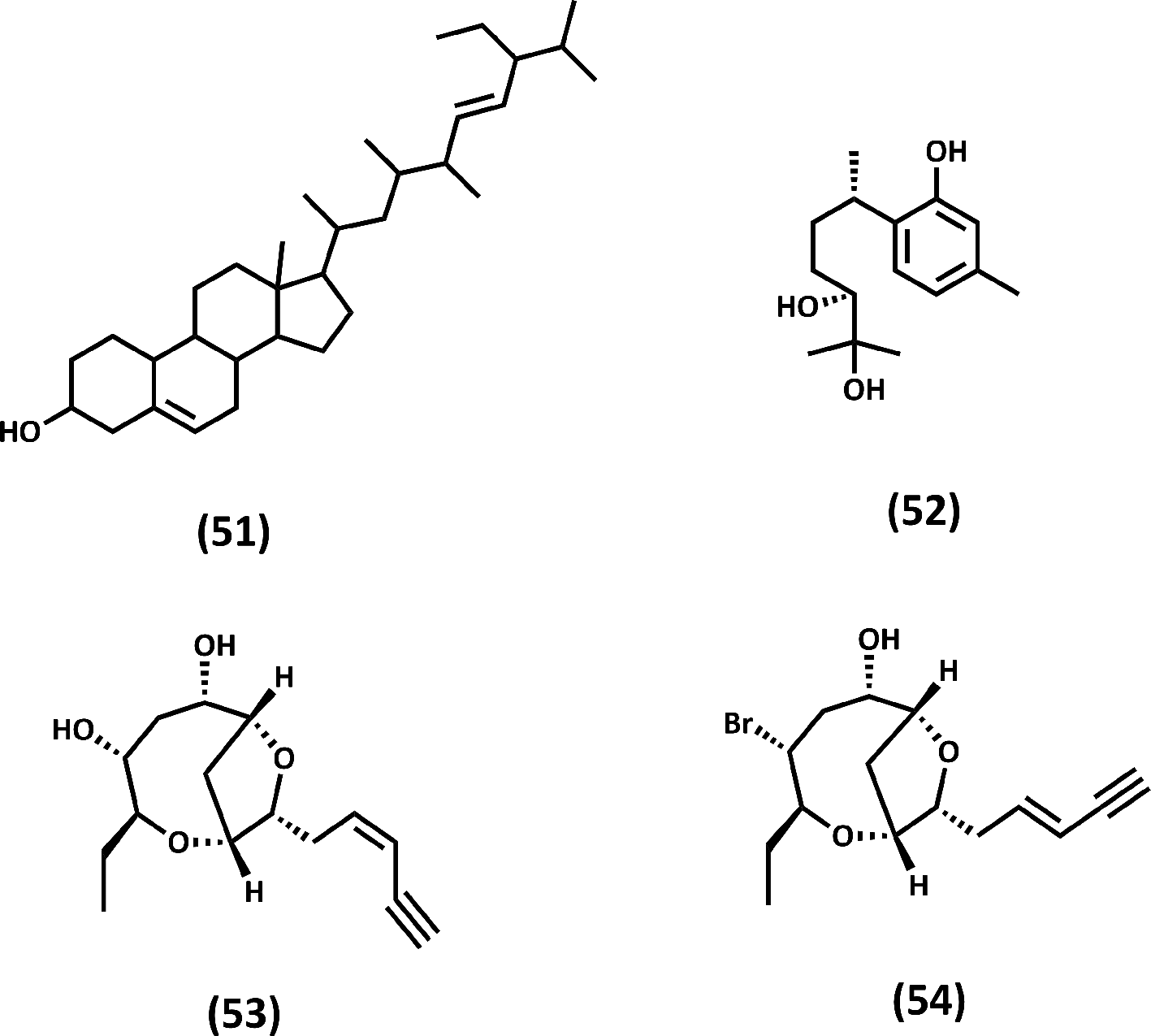
Another compound was (+)-10α-hydroxycurcudiol (52), isolated from the sesquiterpene alcohol group.64 This compound was collected from Papua New Guinea, from the algae Udotea orientalis. (+)-10α-hydroxycurcudiol can act as an antitrypanosome. The mode of action of (+)-10α-hydroxycurcudiol remains limited, but its similar compound, curcumin, has been shown to disrupt microtubules in Trypanosoma cruzi, thereby altering the parasite’s cytoskeleton structure.65 Due to its similarity, (+)-10α-hydroxycurcudiol may interact with tubulin or other structural proteins in the parasite.
A polyketide acetogenin compound was identified from Laurencia sp, a marine red algae. These are Laurefurenyne C (53) and F (54) collected from the Philippines. Compounds (53) and (54) have cytotoxicity against tumor cells (Leukemia L1210, Colon 38, Human colon H116, and Human lung H125).66 Laurefurenyne C and F possess a lipophilic carbon backbone and multiple oxygenated functionalities, such as alcohols and ethers. It can insert into the lipid bilayer, causing membrane dysfunction.
Marine-derived fungi are widely recognized as prolific sources of structurally diverse secondary metabolites with pronounced cytotoxic properties, rendering them highly attractive for anticancer drug discovery. In one study, five polyketide metabolites were isolated from a mixed coculture of two mangrove-derived endophytic fungi, Clonostachys rosea B5–2, and Nectria pseudotrichia B69–160. These compounds-furanocochliquinol (55), furanocochliquinone (56), nectrianolin D (57), cochlioquinone (58), and cochlioquinol (59) ( Figure 15)-belong to the cochlioquinone-related polyketide family, a scaffold frequently associated with fungal cytotoxins. All five metabolites exhibited potent cytotoxic activity against the human leukemia cell line HL-60, with IC50 values ranging from 0.47 to 1.61 μM. Among them, furanocochliquinol (55) showed the strongest activity (IC50 = 0.47 μM), indicating that this compound represents a promising lead structure within the series.67 The consistently low micromolar potency observed underscores the inherent cytotoxic potential of cochlioquinone-derived polyketides.
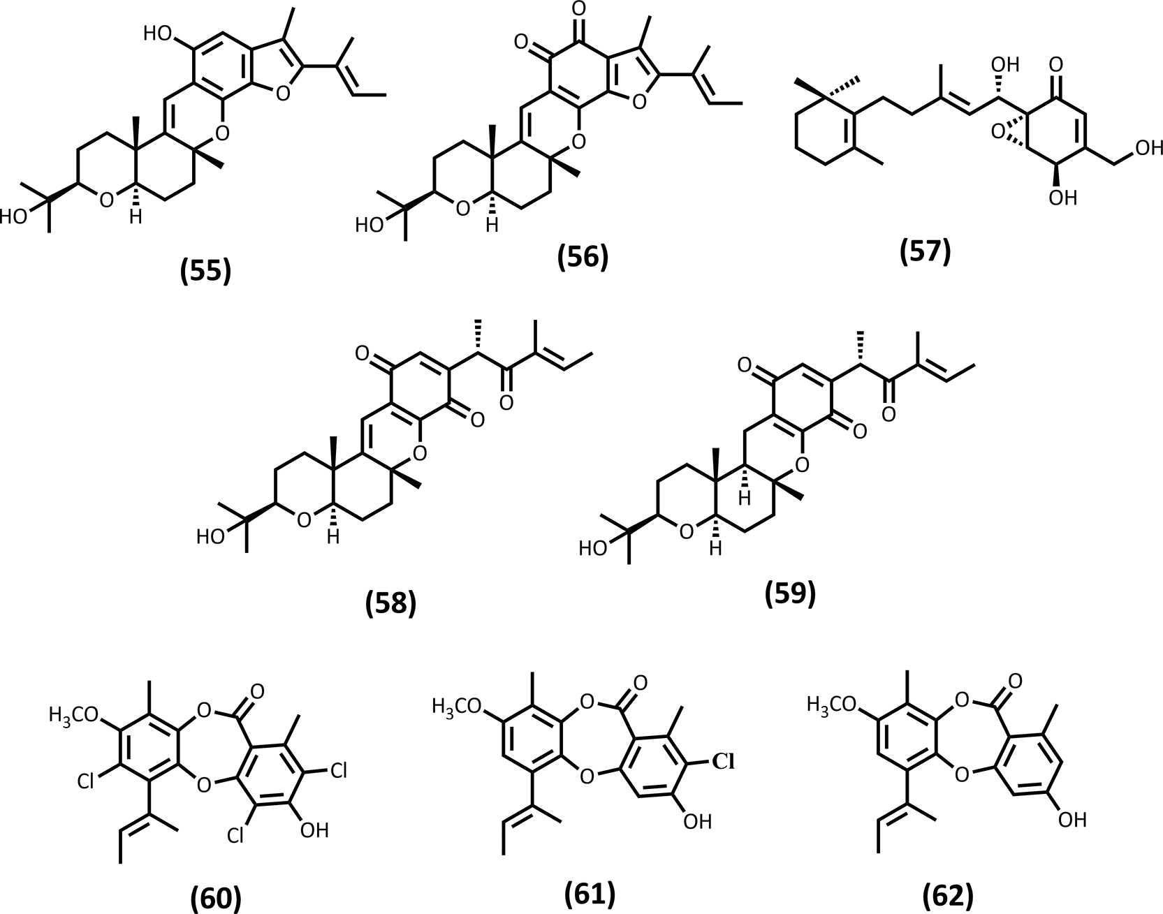
Structure-activity relationship (SAR) analysis revealed that specific structural features play a decisive role in modulating cytotoxic potency. Notably, the presence of a dehydrogenated double bond between C-12 and C-13 (Δ12(13)) was identified as a critical determinant of activity, as reflected in the cytotoxic profiles of cochlioquinone (58) and cochlioquinol (59). This observation is consistent with previous reports demonstrating that Δ12(13) unsaturation enhances cytotoxicity in cochlioquinone derivatives.68 From a mechanistic perspective, such unsaturation may influence molecular planarity and redox behavior, thereby facilitating interactions with intracellular targets involved in oxidative stress or apoptosis. Comparative analysis between furan-containing derivatives (55–56) and cochlioquinone (58) further suggests that incorporation of a fused furan moiety contributes positively to cytotoxic activity. The furan ring may enhance molecular rigidity or enable additional π–π and hydrogen-bonding interactions with cellular macromolecules. Moreover, the hydroquinone framework present in furanocochliquinol (55) appears to confer higher potency than the corresponding quinonoid structure in furanocochliquinone (56), indicating that redox-active hydroquinone motifs may promote increased intracellular reactivity, potentially through modulation of reactive oxygen species (ROS).
Nectrianolin D (57), a structural analogue of nectrianolin A, also exhibited notable cytotoxic activity. The enhanced potency of this compound has been associated with the presence of a trimethylcyclohexene moiety, which may strengthen hydrophobic interactions with cellular membranes or protein targets, as reported for related nectrianolin analogues.69 Collectively, these findings underscore how relatively subtle structural variations within polyketide frameworks can result in substantial differences in biological activity.
In another study, a marine-derived fungus, Aspergillus unguis, isolated from the sponge Haliclona fascigera collected in West Sumatra, Indonesia, was shown to produce three aromatic metabolites-nidulin (60), 2-chlorounguinol (61), and unguinol (62) ( Figure 15).70 These compounds exhibited moderate cytotoxic activity against the human colon cancer cell line WiDR, with IC50 values of 28.65, 39.13, and 27.57 μg/mL, respectively. Compared with cochlioquinone-derived polyketides, these halogenated aromatics displayed lower potency, potentially reflecting differences in molecular complexity, cellular uptake, or target specificity. Notably, nidulin (60), 2-chlorounguinol (61), and unguinol (62) also showed comparable toxicity toward normal Vero cells, indicating limited selectivity between cancerous and non-cancerous cell lines.70 This recurring issue highlights the importance of incorporating early-stage selectivity profiling, including selectivity index calculations and the use of normal cell controls.
Overall, these studies demonstrate that marine-derived fungi consistently yield cytotoxic secondary metabolites with diverse structural scaffolds, ranging from polyketides to halogenated aromatics. While many compounds exhibit moderate to strong cytotoxicity at low micromolar concentrations, insufficient selectivity toward cancer cells remains a major limitation. Nonetheless, such metabolites remain valuable as chemical probes and lead-like scaffolds for elucidating structure-activity relationships and guiding future biosynthetic, semisynthetic, or medicinal chemistry optimization.
In addition to their anticancer potential, marine-derived fungi associated with sponges and mangroves have emerged as important sources of antibacterial and antifungal secondary metabolites. Several studies summarized in this review demonstrate that marine fungi originating from the Coral Triangle region, one of the most biodiverse marine ecosystems globally, produce structurally diverse metabolites with notable antimicrobial activities.
In one study, twenty fungal strains were isolated from ten sponge specimens collected from Hoga Island, Wakatobi National Park, Indonesia72 Among these, Penicillium citrinum strain WK-P9, isolated from the sponge Suberea sp., was identified as a particularly promising producer of bioactive metabolites. Chemical investigation of this strain led to the isolation of one new citrinin-derived compound, penicitrinone G (63), together with three known metabolites, penicitrinone A (64), penicitrinone E (65), and penicitrinol J (66) ( Figure 16). Antibacterial evaluation against a panel of Gram-positive and Gram-negative bacteria, as well as selected fungal test organisms, revealed that only compounds 37 and 39 exhibited moderate antibacterial activity, primarily against Gram-positive bacteria. Penicitrinol J (66) showed inhibitory activity against Bacillus megaterium, Bacillus subtilis JH642, and Mycobacterium smegmatis, with minimum inhibitory concentration (MIC) values of 16, 16, and 32 μg/mL, respectively, whereas penicitrinone A (64) displayed weaker activity limited to M. smegmatis (MIC = 32 μg/mL).19 In contrast, penicitrinone E (65) and the new compound penicitrinone G (63) were inactive against all tested bacterial and fungal strains.
Structure-activity relationship (SAR) analysis suggested that subtle structural differences among citrinin-derived metabolites significantly influence antibacterial potency. Comparison between penicitrinone A (64) and penicitrinone E (65) indicated that the introduction of an additional carboxylic acid moiety in compound (65) may reduce antibacterial activity. Furthermore, comparison between compounds (65) and (66) suggested that the presence of a phenolic system in compound (66) confers higher antibacterial potency than the corresponding quinonoid system in compound (65). This observation is consistent with previous reports highlighting the importance of phenolic functionalities in supporting antibacterial activity.72 Notably, none of the isolated compounds exhibited antifungal activity under the tested conditions.
In another investigation, a marine-derived fungus, Aspergillus unguis, isolated from the sponge Haliclona fascigera collected from the coastal region of West Sumatra, Indonesia, was found to produce three halogenated aromatic metabolites, namely nidulin (60), 2-chlorounguinol (61), and unguinol (62) ( Figure 15).70 These compounds exhibited pronounced antibacterial activity against a broad panel of pathogenic bacteria, including methicillin-resistant Staphylococcus aureus (MRSA). Nidulin (60) displayed the strongest activity, with MIC values as low as 0.78 μg/mL against most tested Gram-positive bacteria, whereas compounds (61) and (62) showed comparable but slightly lower potency. The superior antibacterial activity of nidulin (60) has been attributed to the presence of multiple chlorine substituents, supporting its proposed role as a chlorine-releasing agent capable of disrupting bacterial protein function and redox homeostasis.
Further evidence of antimicrobial potential was reported from a mangrove-derived fungal strain, Penicillium chrysogenum ZZ1151, isolated from Indonesian mangrove sediment.73 Chemical investigation yielded one new prenylated phenolic compound, peniprenylphenol A (67), along with several known metabolites ( Figure 16). Antimicrobial screening against MRSA, Escherichia coli, and Candida albicans revealed that peniprenylphenol A (67) exhibited broad-spectrum activity, with MIC values of 6 μg/mL against MRSA and 13 μg/mL against E. coli and C. albicans. Several known metabolites also showed antibacterial or antifungal activity, although generally with lower potency. These findings suggest that prenylated phenolic scaffolds contribute positively to antimicrobial activity, potentially by enhancing membrane affinity and cellular uptake.
In contrast to antibacterial activity, antifungal activity among marine fungal metabolites appears to be less prevalent. For example, an endophytic fungal strain, Daldinia eschscholzii, isolated from the red alga Gracilaria sp., produced two lactone derivatives, helicascolide C (68) and helicascolide A (69) ( Figure 16), of which only compound (68) exhibited fungistatic activity against the phytopathogenic fungus Cladosporium cucumerinum at relatively high dose74 produced two lactone derivatives, helicascolide C (68) and helicascolide A (69) ( Figure 16), of which only compound (68) exhibited fungistatic activity against the phytopathogenic fungus Cladosporium cucumerinum at a relatively high dose.74 Neither compound showed antibacterial activity against representative Gram-positive or Gram-negative bacteria, underscoring the generally narrow antifungal spectrum observed for many marine fungal metabolites.
Overall, antibacterial secondary metabolites derived from marine-associated fungi display a pronounced preference toward Gram-positive bacteria, whereas activity against Gram-negative bacteria and fungi remains comparatively limited. This trend is consistent with the structural characteristics of many fungal metabolites, which often lack features required to penetrate the outer membrane barrier of Gram-negative bacteria. Importantly, several compounds exhibit MIC values in the low microgram per milliliter range, indicating genuine antibacterial potential rather than nonspecific cytotoxicity. Comparative structural analysis across studies further suggests that halogenation, phenolic functionalities, and prenylation play critical roles in enhancing antibacterial potency, particularly against resistant pathogens such as MRSA. Collectively, these findings support the continued exploration of marine-derived fungi as a promising source of novel antibacterial scaffolds.
Marine-derived fungi have also been recognized as a promising source of bioactive metabolites with inhibitory activity against protein tyrosine phosphatase 1B (PTP1B), an enzyme closely associated with insulin resistance and type 2 diabetes. In one study, a fungal strain, Penicillium verruculosum TPU1311, isolated from the ascidian Polycarpa aurata collected in Manado, North Sulawesi, Indonesia, was found to produce two new merosesquiterpenes, verruculide A (70) and verruculide B (74), along with three known related compounds, chrodrimanins A (71), B (72), and H (73) ( Figure 17).75 All five compounds were evaluated for their inhibitory activity against PTP1B. Verruculide A (70), chrodrimanin A (71), and chrodrimanin H (73) exhibited notable inhibitory activity, with IC50 values of 8.4, 8.5, and 14.9 μM, respectively. In contrast, verruculide B (74) showed only moderate inhibition, achieving approximately 40% inhibition at a concentration of 23.1 μM, while chrodrimanin B (72) did not exhibit measurable inhibitory activity at a concentration of 20.7 μM.75 These results indicate that relatively small structural variations within the merosesquiterpene framework can lead to substantial differences in PTP1B inhibitory potency.
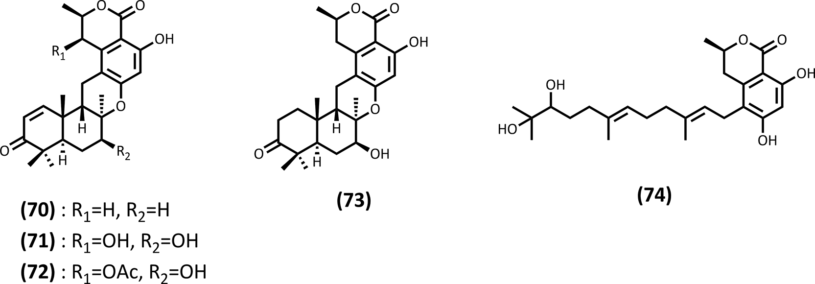
Structure-activity relationship (SAR) analysis suggested that the presence of two hydroxyl groups at the C-7 and C-7′ positions does not significantly enhance PTP1B inhibitory activity, as evidenced by the comparable potencies of compounds bearing or lacking these functionalities. In contrast, acetylation of the 4′-hydroxyl group resulted in a pronounced reduction in inhibitory activity, indicating that a free hydroxyl group at this position plays a critical role in mediating interactions with the PTP1B active site. This observation suggests that hydrogen-bonding interactions involving the 4′-hydroxyl group may be essential for effective enzyme inhibition.
Overall, these findings highlight marine-derived merosesquiterpenes as a structurally distinct class of PTP1B inhibitors and underscore the importance of specific hydroxyl substitution patterns in modulating inhibitory potency. Such compounds represent valuable lead-like scaffolds for the development of selective PTP1B inhibitors and provide insights into key structural features required for enzyme targeting.
In contrast to marine-derived fungi, which predominantly produce polyketides and aromatic metabolites, marine bacteria-particularly cyanobacteria and actinomycetes-are well known for generating structurally complex peptides, lipopeptides, and alkaloids. This fundamental biosynthetic distinction is reflected in differences in molecular architecture, bioactivity profiles, mechanisms of action, and translational potential, as discussed in the following section.
Marine bacteria, especially cyanobacteria and actinomycetes, have yielded a wide array of cytotoxic secondary metabolites with diverse structural features and biological activities. In one study, three new lipopeptides, tasiamides C-E (75–77), were isolated from a marine cyanobacterium (Symploca sp.) collected in Kimbe Bay, Papua New Guinea ( Figure 18).76 Cytotoxic evaluation against the human colon cancer cell line HCT-116 revealed that tasiamides C and D exhibited IC50 values greater than 25 μM and approximately 25 μM, respectively, and were therefore considered inactive under the tested conditions.
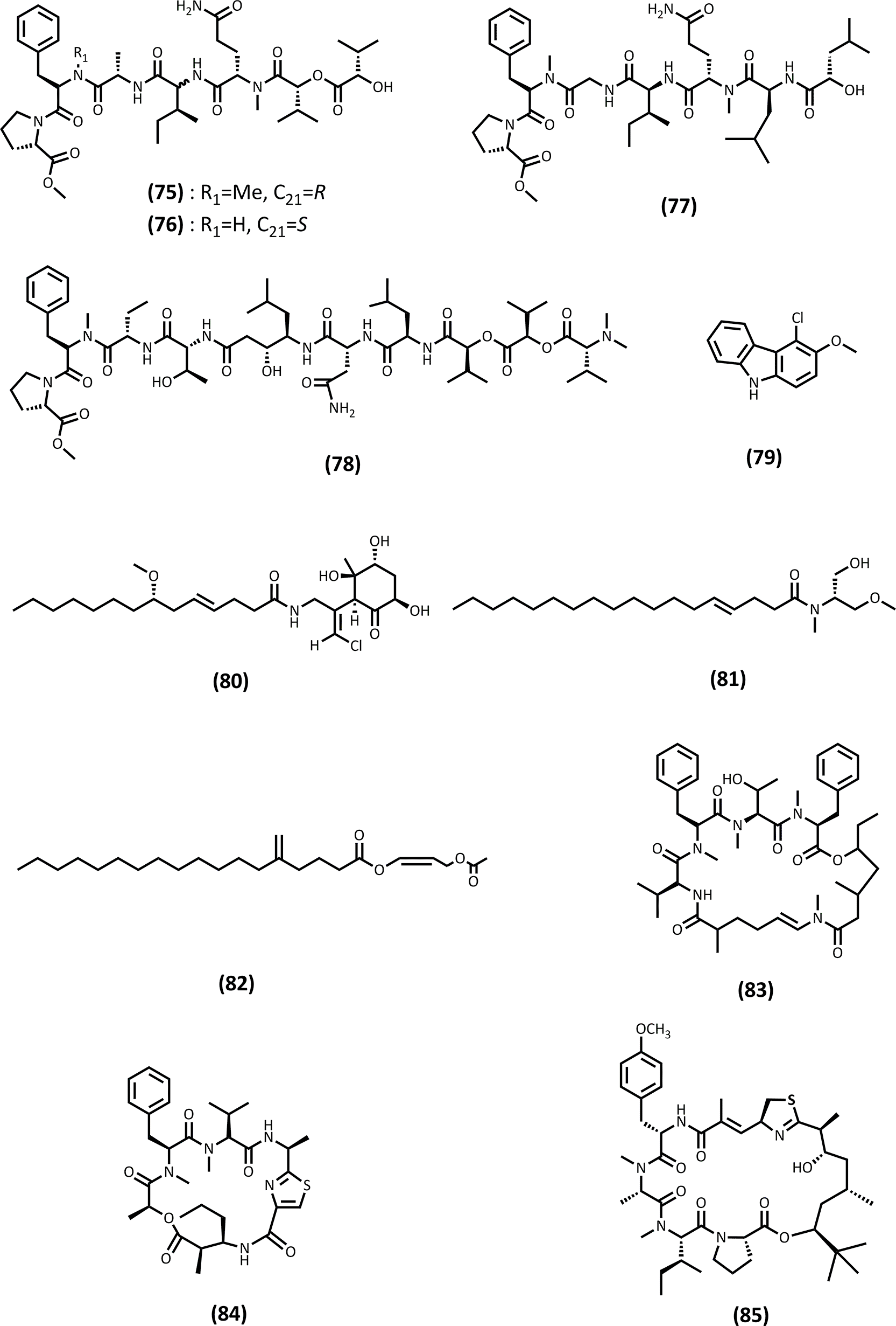
Notably, comparison with structurally related cyanobacterial metabolites, such as grassystatin B (78) and symplocin A ( Figure 18), which exhibit potent biological activity as cathepsin E inhibitors with IC50 values of 886 pM and 300 pM, respectively, highlights the critical role of the statine residue. The absence of this residue in tasiamides C and D appears to account for their markedly reduced activity. These findings underscore how minor modifications in amino acid composition can profoundly influence biological activity and target specificity. Accordingly, although tasiamides C and D do not display pronounced anticancer activity, they remain valuable for elucidating biosynthetic flexibility and structural requirements associated with potent cytotoxic effects.
In another investigation, secondary metabolites produced by a novel actinomycete strain (DSD3025T) isolated from marine sediments of Tubbataha Reefs Natural Park, Sulu Sea, Philippines, were examined.77 This study led to the isolation of six rare halogenated carbazole alkaloids, with chlorocarbazomycin A (79) identified as the major constituent ( Figure 18). At a concentration of 125 μg/mL, compound (79) inhibited the growth of human colorectal cancer cells (HCT-116) by 55.4% and human ovarian cancer cells (A2780) by 50.2%, indicating moderate cytotoxic potential.
Further examples of cytotoxic metabolites from marine cyanobacteria were reported from a strain tentatively identified as cf. Lyngbya sordida, collected in Papua New Guinea.78 Chemical investigation yielded one new malyngamide, malyngamide 2 (80), together with several known compounds ( Figure 18). Compound (80) exhibited moderate cytotoxic activity against the human lung carcinoma cell line H-460, with an IC50 value of 27.3 μM (95% confidence interval: 22.4–32.9 μM). In addition, the known compound wewakazole showed stronger activity, with an IC50 value of 10.1 μM (95% confidence interval: 8.4–12.1 μM), while a mixture of majusculamides A and B displayed the highest cytotoxicity and was likely responsible for the activity observed in the crude extract.
Additional lipid-derived cytotoxic metabolites were isolated from marine cyanobacteria collected in different geographical regions.79 Two new compounds, serinolamide A (81) and propanediester (82) ( Figure 18), exhibited in vitro cytotoxicity against the human lung cancer cell line H-460. Serinolamide A (82) induced complete loss of cell viability at a concentration of 50 μg/mL and reduced cell survival to approximately 60% at 5 μg/mL, indicating concentration-dependent cytotoxic effects.
Finally, a tropical marine cyanobacterium, Moorea bouillonii, collected from New Britain, Papua New Guinea, was reported to produce a new cytotoxic cyclic depsipeptide, bouillonamide (83), together with the previously known compounds ulongamide A (84) and apratoxin A (85) ( Figure 18).80 Compounds (83) and (84) were evaluated for in vitro cytotoxicity against Neuro-2a cells, yielding IC50 values of 6.0 μM and 16.0 μM, respectively, demonstrating moderate to strong cytotoxic activity within this compound class.
Collectively, cytotoxic metabolites produced by marine bacteria exhibit a broad spectrum of biological activities, ranging from weakly active or inactive compounds to potent enzyme inhibitors and cytotoxic agents. While several bacterial metabolites display only moderate anticancer activity, their structural complexity and biosynthetic uniqueness render them highly informative for understanding structure-activity relationships and enzymatic target specificity. In particular, statine-containing lipopeptides exemplify how subtle amino acid substitutions can dramatically alter biological outcomes. These observations reinforce the concept that moderate cytotoxicity does not diminish the scientific value of a metabolite but instead provides critical insight into biosynthetic plasticity and functional group relevance in marine bacterial natural products.
In addition to their well-recognized cytotoxic and anticancer properties, marine symbiotic bacteria, particularly cyanobacteria and actinobacteria, represent an important reservoir of antibacterial secondary metabolites. Recent studies from the Indo-Pacific region, including Southeast Asia, have highlighted the antibacterial potential of marine-derived bacterial symbionts, although the extent of chemical characterization and biological validation varies considerably among reports.
In one study, a novel actinomycete species, Streptomyces tubbatahanensis DSD3025T, isolated from marine sediments of the Tubbataha Reefs Natural Park, Philippines, was shown to produce six rare halogenated carbazole alkaloids, with chlorocarbazomycin A (79) identified as the major metabolite ( Figure 18).77 This compound exhibited selective antibacterial activity against Gram-positive pathogens, including Staphylococcus aureus and Streptococcus pyogenes, although growth inhibition was observed at relatively elevated concentrations. While the antibacterial potency remains moderate from a pharmacological standpoint, this study is notable for revealing chemically uncommon halogenated carbazole scaffolds from a previously unexplored marine Streptomyces species, underscoring the biosynthetic novelty associated with tropical marine sediments.
Despite these advances, extract-based antibacterial screening continues to dominate investigations of marine symbiotic bacteria. In one report, bacterial symbionts associated with nudibranchs and their prey were evaluated, with Streptomyces werraensis and Ruegeria sp. identified as the most active isolates, particularly against Gram-negative pathogens such as Pseudomonas aeruginosa and Acinetobacter baumannii.81 However, pronounced differences in antibacterial activity between preliminary and scaled-up fermentations highlighted the strong influence of cultivation conditions on secondary metabolite production. Moreover, although several known metabolites were detected in bioactive extracts, the absence of compound-level antibacterial evaluation precluded definitive attribution of the observed bioactivity to specific chemical entities.
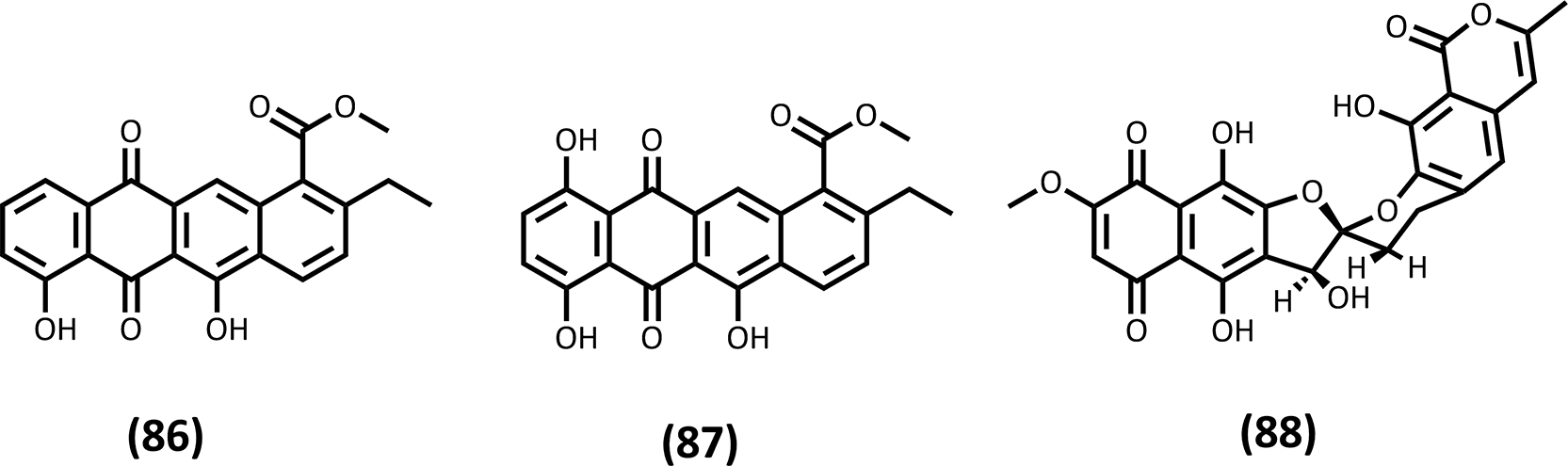
More rigorous compound-level validation was achieved in a study involving marine sediment-derived Streptomyces griseorubens, from which two anthracycline shunt metabolites, bisanhydroaklavinone (86) and 1-hydroxybisanhydroaklavinone (87), were isolated ( Figure 19).82 Bisanhydroaklavinone displayed potent antibacterial activity against multidrug-resistant Staphylococcus aureus, with low MIC values, and mechanistic investigations demonstrated disruption of bacterial membrane integrity. Notably, the accumulation of these shunt metabolites occurred without genetic manipulation, highlighting the intrinsic biosynthetic flexibility of marine Streptomyces to generate antibacterial scaffolds distinct from classical antitumor anthracyclines.
Additional studies from Indonesian waters further support the antibacterial potential of marine symbiotic Streptomyces. In one investigation, symbionts associated with sponges and corals produced extracts active against both Gram-positive and Gram-negative bacteria, including P. aeruginosa.83 Metabolomic analysis revealed actinomycin D and its analogues as dominant constituents, suggesting that the observed antibacterial activity was largely attributable to known antibiotics, thereby limiting chemical novelty. Although molecular networking indicated the presence of unannotated metabolites, the lack of isolation and bioactivity testing hindered evaluation of their true contribution.
Bioautography-guided approaches have been proposed as an effective strategy to directly correlate chromatographic profiles with antibacterial activity. In one such study, a symbiotic bacterium associated with Polycarpa aurata yielded adenosine and bis(2-ethylhexyl) benzene-1,2-dicarboxylate as compounds associated with inhibitory zones on TLC plates.84 Nevertheless, quantitative antibacterial evaluation and biosynthetic verification remain essential, particularly given the frequent occurrence of phthalates as laboratory contaminants and their ambiguous biological relevance.
A compelling example of a marine-derived antibacterial lead was reported for 7,8-dideoxygriseorhodin C (88), which was isolated from a marine-associated microorganism and exhibited sub-micromolar activity against Gram-positive bacteria, including methicillin-resistant Staphylococcus aureus (MRSA), with minimal cytotoxicity toward mammalian cells ( Figure 19).85 Furthermore, strong synergistic effects with oxacillin were observed, effectively restoring β-lactam efficacy against resistant strains. These findings position 7,8-dideoxygriseorhodin C (88) not only as a promising antibacterial lead but also as a potential antibiotic adjuvant.
Overall, marine symbiotic bacteria-particularly Streptomyces species-constitute a rich source of antibacterial metabolites with diverse structural scaffolds. However, translating these discoveries toward clinically relevant leads requires a systematic shift from extract-level screening to compound isolation, quantitative activity profiling, and mechanistic validation. Such approaches are essential for establishing robust structure-activity relationships and fully realizing the antibacterial potential of marine bacterial natural products.
Marine microorganisms, particularly symbiotic bacteria, have emerged as a prolific source of antimalarial natural products, offering structurally novel chemotypes and alternative mechanisms of action to address the persistent challenge posed by drug-resistant Plasmodium falciparum. Recent studies demonstrate that integration of advanced screening strategies with structure-activity relationship (SAR) analysis can substantially improve the efficiency of antimalarial lead discovery from complex marine-derived natural product extracts (NPEs).
In one study, a pharmacology-driven quantitative high-throughput screening (qHTS) platform was applied to prioritize antimalarial activity from a large library of minimally enriched marine microbial extracts.87 Unlike conventional single-point assays, qHTS enabled concentration-response profiling of NPEs, providing apparent EC50 values, efficacy estimates, and curve-class assignments that allowed discrimination between true antimalarial activity and nonspecific cytotoxic effects. Application of this approach to thousands of marine microbial extracts identified two phylogenetically related Streptomyces species as prominent sources of antimalarial activity. Subsequent chemical investigation yielded a series of modified tetrapeptides belonging to the actinoramide/padanamide family. Among these metabolites, actinoramide A exhibited potent and reproducible activity against multiple P. falciparum strains, with EC50 values around 200 nM, while showing negligible cytotoxicity toward human HEK293 and HepG2 cells. A pronounced diastereomer-dependent effect was observed, as 25-epi-actinoramide A (89) ( Figure 20) displayed approximately 20–50-fold lower potency, underscoring the critical role of stereochemistry in antimalarial activity. Although the presence of a statine-like moiety suggested potential inhibition of aspartic proteases such as plasmepsins, biochemical assays revealed no inhibition of plasmepsin activity, indicating that the molecular target of actinoramides in P. falciparum remains unresolved. Beyond the identification of potent compounds, this study highlights the utility of qHTS in minimizing false positives and optimizing extract prioritization under realistic resource constraints, thereby providing a broadly applicable framework for antimalarial natural product discovery.
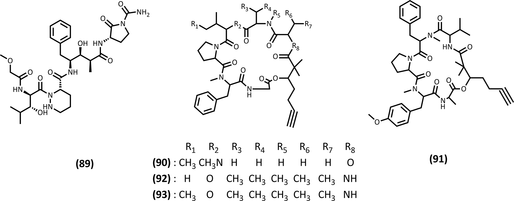
Complementary insights into marine-derived antimalarial agents were obtained from a study involving the isolation of four cyclic depsipeptides, dudawalamides A-D (90–93) ( Figure 20), from a marine cyanobacterium (Moorea producens) collected in Papua New Guinea.87 These compounds belong to the kulolide superfamily of NRPS-PKS-derived lipopeptides characterized by the presence of the distinctive Dhoya (2,2-dimethyl-3-hydroxy-7-octynoic acid) unit. The dudawalamides exhibited selective antiparasitic activity with minimal cytotoxicity toward mammalian cells, reinforcing the therapeutic relevance of this structural class. Within this series, dudawalamides A (90) and D (93) displayed the most pronounced antimalarial activity against P. falciparum, with IC50 values of 3.6 and 3.5 μM, respectively, while exhibiting divergent activity profiles against other protozoan parasites such as Leishmania donovani and Trypanosoma cruzi. Detailed SAR analysis revealed that subtle variations in residue composition and stereochemistry, particularly at specific amino acid positions, resulted in multi-fold differences in antiparasitic potency. Notably, inversion of stereochemistry or modification of a single methyl group was sufficient to markedly alter biological activity, emphasizing the finely tuned structure-activity landscape of Dhoya-containing depsipeptides.
Collectively, these studies demonstrate that marine microorganisms yield chemically diverse antimalarial natural products, ranging from highly potent nanomolar tetrapeptides to micromolar cyclic depsipeptides with broader antiparasitic profiles. Importantly, they illustrate how the integration of advanced screening methodologies with rigorous SAR analysis can substantially enhance the identification and prioritization of marine-derived antimalarial leads, while simultaneously revealing key structural features governing potency and selectivity.
Marine cyanobacteria have emerged as a valuable source of potent molluscicidal natural products with potential applications in the control of schistosomiasis, a parasitic disease that remains a major global health burden. The complex life cycle of Schistosoma species, which requires freshwater snails of the genus Biomphalaria as intermediate hosts, renders molluscicidal intervention a critical complementary strategy to chemotherapy-based disease control. Although niclosamide remains the most widely used molluscicide, its high cost, limited water solubility, and ecotoxicological concerns have stimulated the search for alternative agents with improved selectivity and potency.
A notable advance in this area was the discovery of cyanolide A (94), a highly potent molluscicidal metabolite isolated from the marine cyanobacterium Lyngbya bouillonii collected in Papua New Guinea ( Figure 21).88 Cyanolide A exhibited exceptional activity against the snail vector Biomphalaria glabrata, with an LC50 value of 1.2 μM, markedly surpassing the potency of the reference molluscicide niclosamide (LC100 = 4.6 μM). This level of activity positions cyanolide A (94) among the most potent molluscicidal agents reported to date from marine microorganisms. Structural elucidation based on extensive NMR spectroscopic analysis revealed that cyanolide A is a symmetric dimeric macrolide glycoside, representing a new member of the glycosylated macrolide family produced by cyanobacteria. From a biosynthetic perspective, cyanolide A (94) is proposed to arise from a polyketide-derived monomer assembled from acetate units and subsequently modified by S-adenosylmethionine-dependent methylation events. Notably, the presence of a gem-dimethyl group and an unusual methyl extension pattern suggests noncanonical methylation steps, consistent with previous observations that cyanobacteria rarely utilize propionate starter units in polyketide biosynthesis.
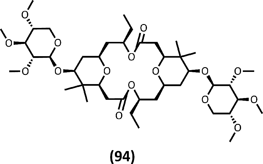
The pronounced molluscicidal activity of cyanolide A (94) is hypothesized to reflect an ecological role in deterring herbivory by mollusks, a function supported by its high toxicity toward B. glabrata. Importantly, biological evaluation indicated that cyanolide A displays only moderate toxicity toward brine shrimp (LC50 = 10.8 μM) and negligible cytotoxicity against mammalian cell lines, including human lung adenocarcinoma (H-460) and murine neuroblastoma (Neuro-2a), at concentrations up to 35 μM.88 This favorable selectivity profile underscores its potential utility as a lead compound for environmentally targeted molluscicidal applications.
Collectively, the discovery of cyanolide A highlights the significant potential of marine cyanobacteria as sources of highly potent and structurally novel molluscicidal agents. Beyond its immediate application prospects, cyanolide A also provides valuable insights into the structural features and biosynthetic strategies that underlie molluscicidal activity. These findings reinforce the importance of continued exploration of marine microbial natural products as alternative tools for vector control in schistosomiasis-endemic regions.
Marine microorganisms, particularly marine cyanobacteria and symbiotic actinobacteria, have emerged as a rich source of neuromodulatory secondary metabolites targeting ion channels and neuronal signaling pathways. Among these, compounds that modulate voltage-gated sodium channels (VGSCs) or alter intracellular calcium dynamics have attracted increasing interest due to their therapeutic relevance in pain management, epilepsy, neurodegenerative disorders, and central nervous system (CNS) dysfunctions.
One notable class of marine-derived neuromodulators is represented by halogenated lipoamides isolated from tropical marine cyanobacteria. From two geographically distinct cyanobacterial collections originating from Curaçao and Papua New Guinea, a series of vinyl chloride-containing metabolites, including janthielamide A (95), kimbeamides A-C (96–98), and the polyketide pyranone kimbelactone A (99), were isolated through bioassay-guided fractionation ( Figure 22).89 Janthielamide A (95) exhibited moderate inhibition of VGSC activity in murine Neuro-2a cells, with an IC50 value of 11.5 μM, and effectively antagonized veratridine-induced sodium influx in murine cerebrocortical neurons (IC50 = 5.2 μM). Kimbeamide A (96) also showed VGSC inhibitory activity at micromolar concentrations, although further biological evaluation was limited by the intrinsic chemical instability of these polyunsaturated lipoamides. Collectively, these findings highlight halogenated lipoamides as structurally unique cyanobacterial neuromodulators with sodium channel-targeting potential.
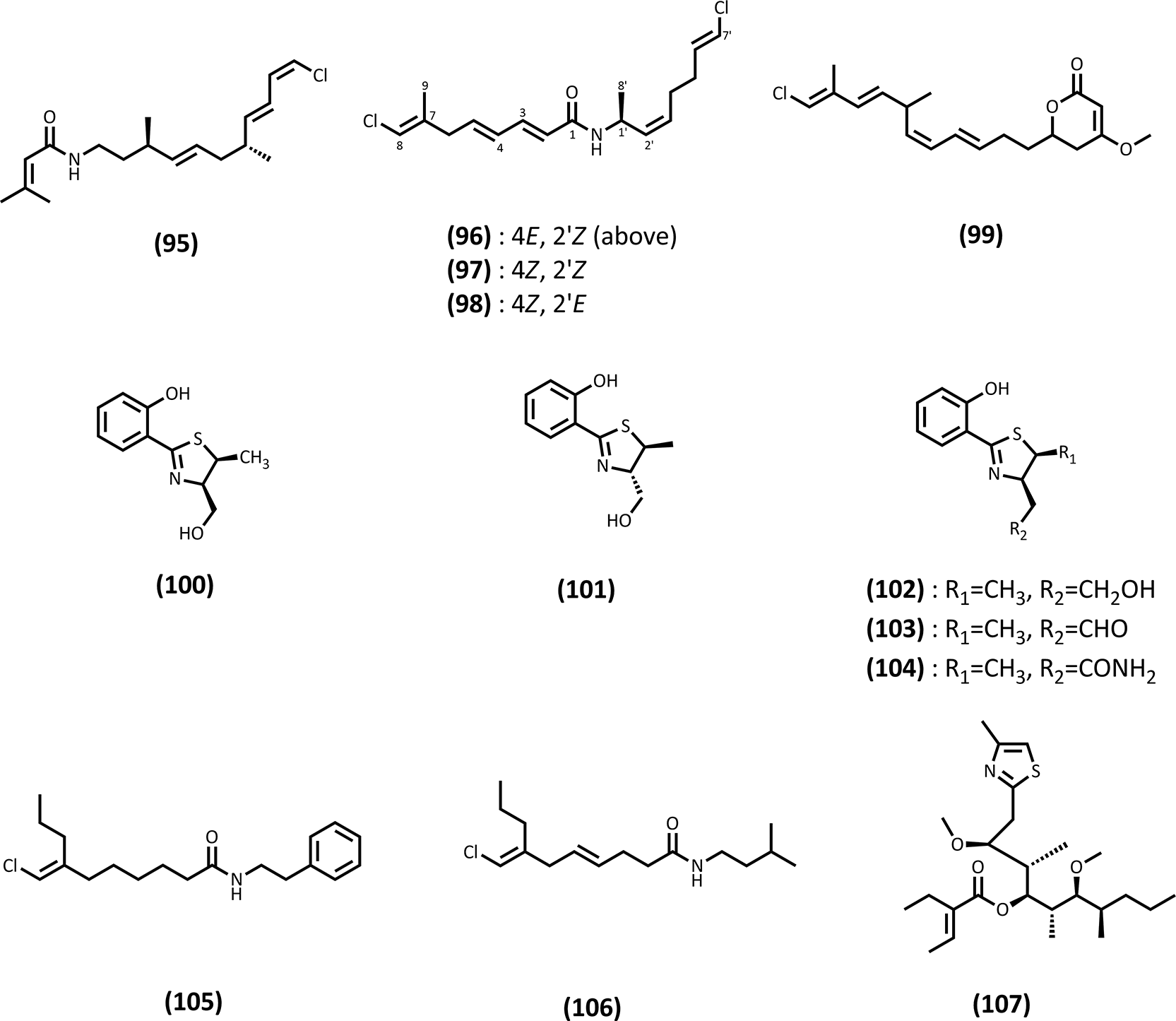
Complementary insights into neuronal modulation were provided by studies employing phenotypic screening using primary dorsal root ganglion (DRG) neurons. A large-scale screening of bacterial symbionts associated with the cone snail Conus pulicarius led to the identification of a Streptomyces strain (CP32) producing a series of benzyl thiazole and thiazoline derivatives, termed pulikatins A-E (100–104) ( Figure 22).90 These compounds induced bidirectional modulation of Ca2+ flux in rat DRG neurons, with certain analogues enhancing and others suppressing calcium influx. Remarkably, minor structural variations, such as the presence or absence of a single methyl group, resulted in opposing phenotypic effects. Subsequent receptor-binding assays revealed selective affinity toward the human serotonin receptor 5-HT2B, suggesting that pulikatins may act through GPCR-mediated pathways rather than direct ion channel blockade. This study underscores the value of DRG-based phenotypic assays in uncovering subtle yet biologically meaningful neuromodulatory activities.
Additional neuromodulatory activity has been reported for vinyl chloride-containing fatty acid amides isolated from filamentous marine cyanobacteria. Credneramides A (105) and B (106) ( Figure 22), along with their fatty acid precursor credneric acid, were isolated from a cyanobacterial assemblage collected in Papua New Guinea.91 Although these compounds showed weak VGSC inhibition, they potently suppressed spontaneous calcium oscillations in primary murine cortical neurons at low micromolar concentrations. Structure-activity analysis suggested that the presence of phenethylamine or isopentylamine head groups plays a critical role in enhancing calcium-modulatory activity, potentially through interactions with neurotransmitter-related receptors or signaling cascades.
Among the most potent marine-derived neuromodulators identified to date are the hoiamides, a family of structurally complex lipopeptides produced by tropical marine cyanobacteria. Hoiamide B (107) ( Figure 22), a cyclic depsipeptide, and its linear analogue, hoiamide C were isolated from cyanobacterial collections from Papua New Guinea.92 Hoiamide B (107) strongly stimulated sodium influx in murine neocortical neurons (EC50 = 3.9 μM) and potently inhibited spontaneous calcium oscillations at nanomolar concentrations (EC50 = 79.8 nM). These activities were comparable to those of the previously reported hoiamide A, a partial agonist of VGSC neurotoxin site 2. In contrast, the linear analogue hoiamide C lacked significant neuromodulatory activity, emphasizing the importance of macrocylization and hydrogen-bond donor functionality in mediating biological effects. Collectively, these findings indicate that hoiamides may exert their neuromodulatory actions through multiple molecular targets, including VGSC activation and disruption of neuronal network-dependent calcium signaling.
Overall, marine microorganisms provide a diverse repertoire of neuromodulatory natural products with distinct mechanisms of action, ranging from direct modulation of VGSCs to indirect regulation of calcium signaling and GPCR pathways. While many of these compounds display only moderate potency, their pronounced structural novelty and sensitivity to subtle chemical modifications make them valuable chemical probes for elucidating neuronal signaling mechanisms. These studies further demonstrate that phenotypic assays, such as VGSC and DRG-based screening platforms, are powerful tools for identifying functionally relevant neuromodulators from complex marine natural product sources.
Despite the exceptional potential of the Coral Triangle as a source of bioactive marine natural products, bioprospecting efforts in this region face several scientific, environmental, and technical challenges. One of the most pressing challenges arises from the increasing vulnerability of coral reef ecosystems to climate change-driven stressors, including ocean warming, coral bleaching, and ocean acidification. Long-term ecological assessments have demonstrated that repeated bleaching events can drastically alter reef community structure, reduce coral cover, and disrupt the complex biological interactions that underpin the production of secondary metabolites.93 These environmental disturbances may indirectly affect the availability and chemical diversity of marine natural products by altering host-microbe associations and metabolic pathways.
Another major challenge lies in the limited accessibility and sustainability of marine macroorganisms traditionally used as sources of natural products. Many reef-associated organisms exhibit slow growth rates, restricted geographic distributions, or ecological sensitivity, making large-scale collection impractical and environmentally detrimental. Historical and geological analyses of Indo-Pacific reef systems further highlight that reef development is strongly influenced by long-term environmental stability, hydrodynamics, and nutrient regimes, emphasizing that coral reef ecosystems are inherently vulnerable to rapid anthropogenic change.94 These constraints underscore the need to minimize direct harvesting of macroorganisms in bioprospecting initiatives.
From a microbiological perspective, a significant challenge remains the limited cultivability of marine symbiotic microorganisms. Although sponge- and coral-associated microbial consortia are now recognized as the true producers of many complex bioactive metabolites, the majority of these microorganisms remain uncultured under standard laboratory conditions. Metagenomic investigations of Indonesian marine sponges have revealed a high diversity of biosynthetic gene clusters, including those encoding polyketide synthases, within uncultured bacterial taxa such as Candidatus Entotheonella.95 While these findings provide compelling evidence for microbial biosynthetic potential, translating genomic information into actual compound production remains a major bottleneck.
Nevertheless, these challenges are accompanied by significant opportunities that are reshaping the future of sustainable marine bioprospecting. Advances in culture-independent approaches, such as metagenome mining and genome-guided natural product discovery, now allow researchers to identify promising biosynthetic pathways without relying on large-scale organism collection. Such approaches are particularly valuable in biodiverse yet environmentally sensitive regions like the Coral Triangle, where conservation considerations are paramount.
An improved understanding of coral-microbiome interactions also presents new opportunities for sustainable exploitation of marine resources. Recent studies have demonstrated that microbial community composition plays a critical role in coral health, resilience, and disease resistance, and that environmental stressors can rapidly reshape these communities.94 From a bioprospecting standpoint, these dynamic microbial assemblages represent both a challenge and an opportunity, as stress-induced shifts in microbial metabolism may activate otherwise silent biosynthetic pathways, potentially leading to the production of novel secondary metabolites.
Furthermore, insights into microbial dynamics within reef ecosystems have highlighted the role of “unseen players,” such as bacteria, archaea, and viruses, in shaping benthic competition and chemical interactions on coral reefs.93 These microbial processes respond rapidly to environmental perturbations and may serve as early indicators of changes in secondary metabolite production. Incorporating microbial ecology into bioprospecting strategies could therefore enhance the efficiency and predictability of natural product discovery.
Looking forward, the integration of sustainable bioprospecting with conservation-oriented frameworks represents a critical future direction. The ability to culture or heterologously express biosynthetic gene clusters from marine microorganisms offers a pathway toward scalable and environmentally responsible production of bioactive compounds. In parallel, emerging strategies such as microbiome manipulation and transplantation, originally developed to enhance coral resilience, may also provide novel platforms for accessing beneficial microbial functions and metabolites without further stressing natural reef systems.94
In conclusion, while environmental degradation, limited cultivability of microorganisms, and ecosystem fragility present substantial challenges to marine bioprospecting in the Coral Triangle, recent methodological and conceptual advances offer promising solutions. By integrating natural product chemistry with microbial genomics, ecological insight, and sustainability principles, future bioprospecting efforts can balance drug discovery objectives with the long-term preservation of one of the world’s most valuable marine biodiversity hotspots.
The Coral Triangle represents one of the most exceptional yet underexplored marine biodiversity hotspots for natural product-based drug discovery. This review demonstrates that marine organisms and their associated microorganisms from this region constitute a prolific source of structurally diverse secondary metabolites exhibiting a broad spectrum of pharmacological activities, including anticancer, antibacterial, antimalarial, antidiabetic, neuromodulatory, and antimolluscicidal effects. Across multiple taxonomic groups-sponges, corals, tunicates, mollusks, echinoderms, algae, and symbiotic or free-living microorganisms-the Coral Triangle consistently yields chemically novel scaffolds that are rarely encountered in terrestrial systems. A key insight emerging from this synthesis is that chemical novelty and biological relevance are closely intertwined with the ecological and evolutionary pressures characteristic of Coral Triangle ecosystems. Sessile or slow-moving organisms, such as sponges, corals, and tunicates, rely heavily on chemical defense mechanisms, leading to the evolution of metabolites with high potency and target specificity. Likewise, marine microorganisms-particularly fungi, cyanobacteria, and actinobacteria-have proven to be major contributors to bioactive compound discovery, often producing metabolites with finely tuned structure-activity relationships that respond sensitively to subtle chemical modifications. These features render marine-derived compounds not only promising drug leads but also valuable chemical probes for elucidating fundamental biological pathways.
Importantly, this review highlights a recurring pattern in Coral Triangle bioprospecting studies: while numerous compounds display moderate to strong in vitro activity, challenges related to selectivity, mechanism-of-action elucidation, and translational development remain pervasive. In antibacterial research, for example, many metabolites exhibit preferential activity against Gram-positive pathogens, including multidrug-resistant strains, yet limited penetration of Gram-negative bacteria remains a bottleneck. Similarly, anticancer and neuromodulatory compounds frequently show potent bioactivity but require deeper mechanistic validation and selectivity profiling to advance beyond early-stage discovery. These observations emphasize that moderate bioactivity does not diminish scientific value, as such compounds provide essential insights into biosynthetic plasticity, SAR trends, and functional group relevance. From a methodological standpoint, the integration of advanced screening platforms-such as quantitative high-throughput screening (qHTS), phenotypic neuronal assays, and enzyme-targeted bioassays-has markedly improved the efficiency of identifying meaningful bioactivities from complex marine extracts. When coupled with structure-guided analysis, metabolomics, and microbial genomics, these approaches enable a more rational prioritization of leads and reduce the risk of rediscovering known compounds. In this context, marine microorganisms emerge as particularly attractive targets for sustainable bioprospecting, offering scalable alternatives to the direct harvesting of ecologically sensitive macroorganisms.
Nevertheless, bioprospecting efforts in the Coral Triangle cannot be decoupled from conservation considerations. Climate change-driven stressors, habitat degradation, and the limited cultivability of symbiotic microorganisms pose significant challenges to both biodiversity preservation and natural product discovery. Addressing these challenges requires a paradigm shift toward sustainability-driven bioprospecting, incorporating culture-independent strategies, genome mining, heterologous expression, and microbiome-informed approaches. Such strategies not only minimize ecological impact but also unlock access to cryptic biosynthetic potential harbored by uncultured microbial taxa.
In conclusion, the Coral Triangle stands at the intersection of extraordinary marine biodiversity, chemical innovation, and urgent global health needs. By integrating natural product chemistry, microbial ecology, genomics, and conservation science, future bioprospecting initiatives in this region can generate high-impact drug leads while safeguarding ecosystem integrity. Sustained interdisciplinary collaboration and responsible resource management will be essential to fully realize the Coral Triangle’s potential as a global engine for marine-derived drug discovery.
| Views | Downloads | |
|---|---|---|
| F1000Research | - | - |
|
PubMed Central
Data from PMC are received and updated monthly.
|
- | - |
Provide sufficient details of any financial or non-financial competing interests to enable users to assess whether your comments might lead a reasonable person to question your impartiality. Consider the following examples, but note that this is not an exhaustive list:
Sign up for content alerts and receive a weekly or monthly email with all newly published articles
Already registered? Sign in
The email address should be the one you originally registered with F1000.
You registered with F1000 via Google, so we cannot reset your password.
To sign in, please click here.
If you still need help with your Google account password, please click here.
You registered with F1000 via Facebook, so we cannot reset your password.
To sign in, please click here.
If you still need help with your Facebook account password, please click here.
If your email address is registered with us, we will email you instructions to reset your password.
If you think you should have received this email but it has not arrived, please check your spam filters and/or contact for further assistance.
Comments on this article Comments (0)