Keywords
Dysgerminoma, Pseudohermaphroditism, Gonadal dysgenesis, Ovarian germ cell tumor.
Dysgerminoma, Pseudohermaphroditism, Gonadal dysgenesis, Ovarian germ cell tumor.
Ovarian dysgerminoma (OD) is a malignant neoplasm derived from the primordial germ cells of the ovary. Dysgerminomas, unlike other types of ovarian tumors, do not have precursor lesions. The cause of OD is not completely understood. The World Health Organization (WHO) defines them as caners made up of basic germ cells that lack a distinct pattern of development.1,2 Dysgerminoma the ovarian equivalent of testicular seminoma, is the most prevalent ovarian malignant germ cell tumor (OMGCT), accounting for 1–2% of primary ovarian neoplasms and 32.8–37.5 percent of all OMGCTs.3
This tumor is frequently linked to increased levels of beta human chorionic gonadotrophin (beta-HCG) and lactate dehydrogenase (LDH). Cancer Antigen 125 (CA-125) and alpha-fetoprotein (AFP) elevations are less frequent.4 There are no particular laboratory tumor markers associated with histotype, in contrast to a number of ovarian neoplasms. Most of the time, women who are asymptomatic are inadvertently identified with dysgerminoma.5 Although they can arise at any age, germinal cell tumors are most common in children and adolescents, with a peak frequency occurring between the ages of 10 and 30. 90 percent of patients are under 30 years old, according to epidemiological data.6 Gonadal dysgenesis (pure and mixed form) can be linked to ovarian dysgerminoma.7
Congenital possession of both male and female sex organs by one individual is referred to as hermaphroditism. There are two types; pseudohermaphroditism and true hermaphroditism. Both ovary and testes must be present for the former to be diagnosed, either alone or in combination to create unilateral or bilateral ovotestes. In contrast, in pseudohermaphroditism, only the secondary sex organs are both male and female, and the gonads are either ovarian or testicular.8 However, a consensus statement was released in 2006 suggesting that the Disorder of Sex Development (DSD) categorization be used to replace a number of no longer used names, including sex reversal, intersex, and pseudohermaphrodite.9 DSDs are congenital conditions marked by abnormal chromosomal, gonadal, or anatomical sex development.10 A kind of gonadal dysgenesis known as female pseudohermaphroditism (46 XX) is typified by a female karyotype and masculine or ambiguous genitalia.11 In this instance, a female fetus exposed to an excessively androgenic environment congenitally masculinizes her external genitalia.12
In addition to having dysgenetic gonads, patients with DSD are more likely to develop gonadal malignancies and may exhibit variable degrees of ambiguous genitalia. For proper therapy, including tumor surveillance and prompt intervention, early detection of these disorders is essential. Here we describe a rare instance of ovarian dysgerminoma in a male patient who is 22 years of age. This case emphasizes how crucial is to take gonadal cancer into account when treating patients who exhibit abnormal genital development.
A male patient in his 20s presented to the Oncology center of Aliabad Teaching Hospital, Kabul University of Medical Sciences, with complaints of a noticeable, asymmetrical bulging in the lower abdomen on the left side. The mass had been gradually enlarging over the past four months, accompanied by significant anorexia and weight loss. His medical history was otherwise unremarkable, with no history of previous illness or family history of relevant conditions. However, an unusual event occurred approximately eight months prior, during which the patient experienced vaginal bleeding, similar to menstruation, occurring once or twice. Upon physical examination, patient had an abdominopelvic solid mass, with irregular borders with signs of ascites. Abdominal palpation revealed a firm, mobile, non-tender mass. While on genital examination the genitalia were noted to be intermediate in appearance between a typical clitoris and penis, with small phallus and the urethral opening located below it. No testes were palpable in the scrotum, which was empty.
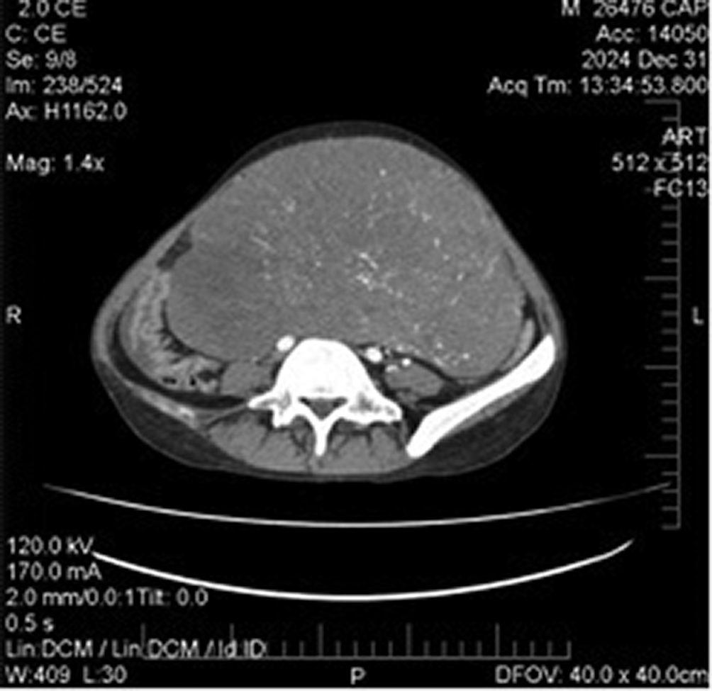
On laboratory assessment, complete blood cells count, urinalysis, renal and liver function tests, and thyroid function tests were normal. Ultrasound revealed a large, solid mass occupying most of the abdominal cavity. Doppler imaging showed low resistance blood flow within the mass. CT imaging of the chest, abdomen, and pelvis with intravenous contrast showed a substantial solid mass measuring approximately 20 cm x 19 cm x 12 cm. The mass was heterogeneous with multiple calcifications and was located in the mid and lower abdomen, likely arising from the adnexal region. There were no signs of vascular invasion, but the mass was in close proximity of vital structures, including uterus, urinary bladder, and iliac vessels. Mild free fluid in the abdomen cavity and enlarged lymph nodes in the left para-aortic region were noted ( Figure 1). Considering the patient’s clinical presentation and imaging findings, the main differential diagnosis included; gonadal tumors particularly germ cell tumor such as dysgerminoma, ovarian neoplasm, and mixed gonadal dysgenesis. Given the presence of a large abdominal mass, and the patient’s ambiguous genitalia, a gonadal germ cell tumor was highly suspected.
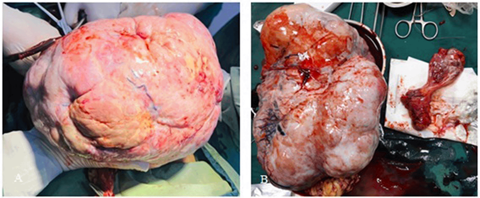
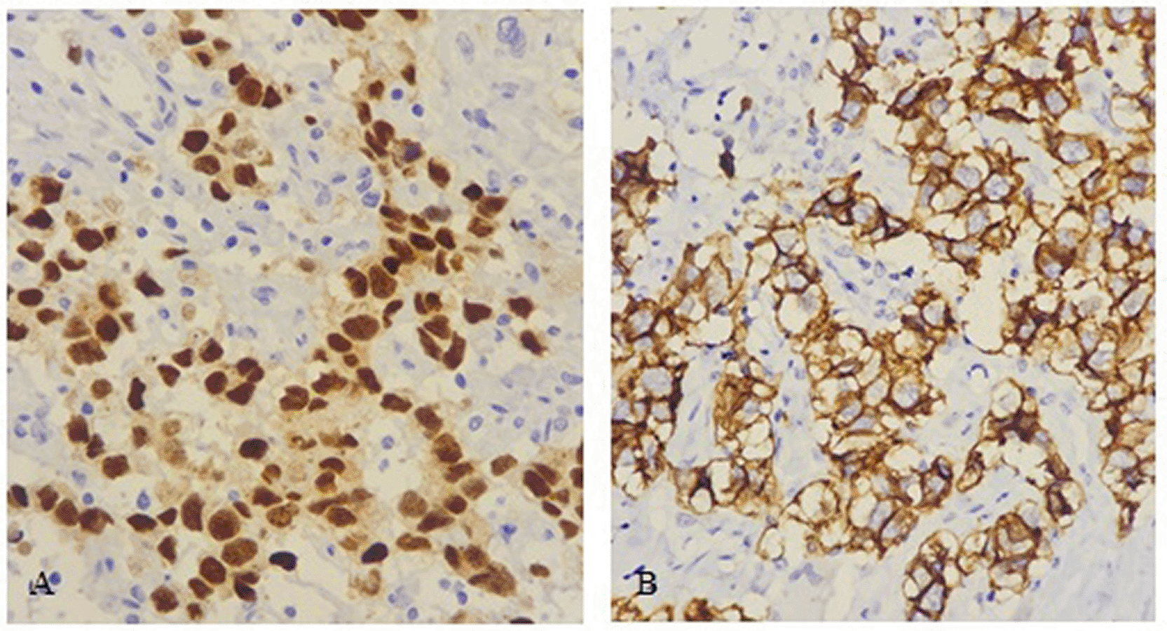
The patient underwent bilateral salpingo-oophorectomy, with total transabdominal hysterectomy and partial omentectomy. The decision for hysterectomy was influenced by the patient’s strong identification as male and his personal desire not to retain female reproductive structures. Surgical findings revealed a large mass in the left ovary, measuring approximately 20 cm x 18 cm x 11 cm, with a solid and cystic cut surface ( Figure 2). The mass was sent for histopathological examination, which confirmed poorly differentiated malignant neoplasm, the microscopic examination of the left ovarian mass revealed an infiltrative neoplasm with extensive necrosis, arranged in nests and cords ( Figure 3). Immunohistochemical staining for OCT3/4 was diffuse and positive in the neoplastic cells, supporting the diagnosis of a germ cell tumor, specifically dysgerminoma. CD117 staining was also positive, further confirming the diagnosis ( Figure 4).
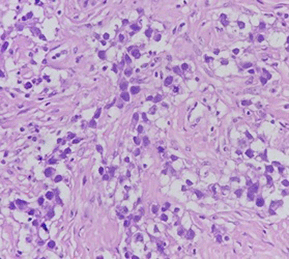
The neoplastic cells exhibit distinct cell borders, clear cytoplasm, and centrally located nuclei. Characteristic of dysgerminoma.
The patient’s post-operative recovery was monitored, and the patient underwent 1 cycle of the BEP chemotherapy (Bleomycin, Etoposide, and Cisplatin), which is a standard treatment for dysgerminoma, and the treatment is still in progress. At the time of this report, the patient had returned to daily activities and has responded very well to the treatment, but further surveillance for recurrence of the tumor was scheduled.
Ovarian dysgerminomas (OD) are uncommon, constituting around 2% of all malignant ovarian neoplasms.13 Large, transparent epithelial cells developing in diffuse sheets of nests separated by a lymphocyte-rich stroma make up this extremely radiosensitive malignant tumor. This disorder, which is a tumor of the embryo’s neuter germ cells, can develop in both ovarian and testicular tissue. In the latter, it is more commonly referred to as seminoma and embryonal carcinoma.14 Recent data indicates that among patients with dysgenetic gonads, OD is the most prevalent malignant tumor of the gonads. The WHO Classification of Tumors 2020 states that OD typically develops as a part of gonadal dysgenesis.1 Reports of a study done by Howitt et al. shows that about 5–10% of patients with chromosomal abnormalities or aberrant genital development will develop dysgerminoma.15
Although the exact cause and pathophysiology of hermaphroditism are still unknown, sex chromosomal abnormalities, aberrant gonadal formation, and associated endocrine problems during embryonic development may be a contributing factor.16 SRY-genes (Sex-determining region Y protein) triggers the development of a testis from the embryonic gonad by activating a series of genes that determine male sexuality.13 If the patient’s gonad is the testis, fetal testicular Sertoli cells release Müllerian inhibitory substance, which causes Müllerian duct regression. At the same time, embryonic testicular Leydig cells make testosterone from cholesterol, resulting in activated dihydrotestosterone, which is then used to differentiate male external genitalia. If the gonad is the ovary, it does not produce the Müllerian inhibitory substance, and the Müllerian ducts would otherwise become the uterus, fallopian tubes, and cervix.17
The pathophysiology of dysgerminoma in hermaphrodites involves complex interactions between genetic factors and gonadal development. Individuals with a 46, XY karyotype may develop dysgerminoma due to the presence of Y-chromosome material, which is linked to gonadal dysgenesis.18 Tumors in hermaphrodites can develop from either testicular or ovarian tissue, and since the gonads are frequently destroyed, the origin cannot be identified. The gonads are particularly vulnerable to gonadal malignancies, which are usually dysgerminomas and/or gonadoblastomas.19,20 Twenty-seven cases of hermaphrodite dysgerminoma were documented by Meyer R. Apart from hermaphrodite dysgerminoma, it can also occur in people with less obvious sexual abnormalities, such as female hypoplasia of genital organs and male cryptorchidism.14
The diagnosis of dysgerminoma particularly in those with disorder of sex development present unique challenges. Patients may present with abdominal pain, distention, or masses, often leading to imaging studies. For instance, a case of a 24-year-old male patient with abdominal pain revealed a malignant gonadal mass through CT imaging, ultimately diagnosed as dysgerminoma.21 Dysgerminoma can mimic other conditions, such as abdominal tuberculosis, complicating diagnosis.22 Histopathological examination is essential, as dysgerminomas exhibit distinct microscopic features, including lymphocytic infiltrate and various growth patterns.23 Human chorionic gonadotropin (hCG), which is released by syncytiotrophoblastic giant cells, is modestly increased in 5% of dysgerminoma patients.4,24,25 Elevated blood β-hCG levels are typically the cause of estrogenic hormonal manifestations, including menstrual abnormalities, vaginal bleeding, sexual pseudoprecocity, and pregnancy signs. Furthermore, serum LDH might be a tumor marker and is frequently increased. Additionally, these markers can be used to detect recurrences as well as to track the effectiveness of therapy.15,26 Although β-hCG and LDH assessments are crucial for diagnosis, and could provide valuable insights, they were not performed for the patient in this case due to the patient’s financial constraints, as he could only afford imaging studies.
A combination of chemotherapy and surgery is usually used to treat dysgerminoma, and the prognosis is generally good, particularly in instances that are detected early. The patient’s circumstances, including pregnancy, and the disease’s stage can influence the management approaches. In the majority of cases, surgical resection is necessary, and staging laparotomy may be necessary to determine the disease’s extent.27 Fertility-sparing surgery is frequently seen as safe and successful in younger patients.28 Adjuvant chemotherapy after surgery, usually with a BEP regimen, is advised, particularly for advanced stages.29 Combining chemotherapy and salvage surgery in recurrent instances may enhance long-term survival.30 In the present case, the patient and his parents provided written informed consent to proceed with the surgical intervention, including hysterectomy. The patient had always identified as male, and lived as a boy. Given the social and cultural context of the country, living as a girl would have posed significant psychological and societal challenges for him. Therefore, a bilateral salpingo-oophorectomy, partial omentectomy along with total hysterectomy was performed, removing the uterus and cervix to align with the patient’s gender identity and alleviate associated distress.
It is widely acknowledged that localized dysgerminomas do not metastasize or reoccur. As a result, dysgerminomas are among the disorders that have a good prognosis and may be cured.31 The stage of the tumor determines the prognosis. The overall five-year survival rate is satisfactory, surpassing 75% (even 90% in stage I); in patients with illness that extends beyond the ovaries, it drops to about 63%.32 The complete ovarian tissue, the entire genital tract, the lumbar muscles, the iliac vessels, the pubic symphysis, the rectum, and the spine are all destroyed by the local invasion of advanced dysgerminoma.25
This case highlights a rare presentation of ovarian dysgerminoma in a phenotypic male patient, emphasizing the diagnostic challenges in disorder of sexual development (DSDs) and gonadal tumors. Limited access to genetic and hormonal testing complicated the evaluation, but clinical findings, imaging, and histopathology guided appropriate management. The decision for the treatment was influenced by both oncologic necessity and the patient’s gender identity. This case underscores the importance of early recognition, multidisciplinary care, and patient centered decision-making in management of such complex conditions.
This study was approved by the Research Ethics Committee of Kabul University of Medical Sciences, Kabul, Afghanistan (Approval No: 10; Agenda No: 14; Date: March 8, 2026). The study was conducted in accordance with the principles of the Declaration of Helsinki.
Written informed consent was obtained from the patient for participation in this study and for publication of this case report and accompanying images.
I first noticed the swelling in my abdomen a few months ago, but I didn’t think it was serious until it started getting bigger. The previous bleeding episode was confusing, but I didn’t seek medical help at that time. When I was diagnosed with a tumor, I was concerned, so when the doctors explained the treatment option, I decided to proceed with the surgery to remove the tumor and female reproductive organs. I am grateful for the medical care I received and look forward to moving on with my life.
| Views | Downloads | |
|---|---|---|
| F1000Research | - | - |
|
PubMed Central
Data from PMC are received and updated monthly.
|
- | - |
Provide sufficient details of any financial or non-financial competing interests to enable users to assess whether your comments might lead a reasonable person to question your impartiality. Consider the following examples, but note that this is not an exhaustive list:
Sign up for content alerts and receive a weekly or monthly email with all newly published articles
Already registered? Sign in
The email address should be the one you originally registered with F1000.
You registered with F1000 via Google, so we cannot reset your password.
To sign in, please click here.
If you still need help with your Google account password, please click here.
You registered with F1000 via Facebook, so we cannot reset your password.
To sign in, please click here.
If you still need help with your Facebook account password, please click here.
If your email address is registered with us, we will email you instructions to reset your password.
If you think you should have received this email but it has not arrived, please check your spam filters and/or contact for further assistance.
Comments on this article Comments (0)