Keywords
Cardiac hydatid cyst, Echinococcosis, Cardiac surgery.
This article is included in the Fallujah Multidisciplinary Science and Innovation gateway.
Cardiac hydatid cyst, Echinococcosis, Cardiac surgery.
I made minor changes to the discussion part only according to the valuable comments of the reviewers. no major changes were done to other parts of the article.
See the authors' detailed response to the review by Ali Bilal Ulas
See the authors' detailed response to the review by Laila Jedidi
Hydatid cysts are parasitic infections mostly caused by Echinococcus granulosus and are mostly observed in the liver and lungs. Echinococcosis is a human infection caused by the larval stage of any species of the E. granulosus complex, E. multilocularis, or E. vogeli. E. granulosus complex parasites that cause unilocular cystic lesions are common in areas where livestock are kept in close proximity to dogs.1
Echinococcus granulosus has a 2-host life cycle. Dogs and other canines are definitive hosts, whereas sheep and other herbivorous animals are intermediate hosts. Humans serve as an incidental intermediate host (dead end), infected by the ingestion of food contaminated with feces from dogs harboring E. granulosus eggs.2
Eggs develop into a larva in the duodenum. The larva pierces the intestinal wall, enters the portal circulation, and is transported to the liver, lungs, or seldom, to other organs. The host immune response attempts to eliminate the parasites after tissue invasion, where an inflammatory reaction occurs around the sites where the parasites are lodged. Although the immune response destroy many parasites, some survive, escape elimination, and continue to develop into hydatid cysts, surrounded by fibrous connective tissue, and become fluid-filled bladder-like cysts, usually in the liver (60-70%, in the right lobe) or lung (20-30%). Hydatid cysts can also occur in other organs such as the spleen and kidney (35%), brain and heart (11.5%), and bones in rare cases.2
The hexacanth embryo (six-hooked, first-stage larva) reaches the heart through the coronary arteries and gives rise to a primary, often solitary, hydatid cyst within the myocardial layers. Secondary cardiac involvement may follow rupture of a primary cyst into the pericardial cavity. In such cases, the resulting cysts are initially superficial and subepicardial but may later extend into the myocardium, where they often become multiple.3
After cardiac hydatid cysts develop, their progressive enlargement may produce characteristic structural and functional effects. Patients from endemic regions who present with an abnormal cardiac shadow on chest radiography (CXR) should, therefore, raise suspicion of cardiac hydatid disease. As the cyst enlarges, it compresses the surrounding myocardium and may displace the coronary vessels, cause rhythm disturbances, and interfere mechanically with atrioventricular valve and ventricular function. Echocardiography remains the preferred imaging modality for the diagnosis of cardiac hydatidosis.
Computed tomography may help confirm the diagnosis and exclude hydatid involvement of the liver, lungs, and brain when uncertainty remains. Case management depends on cyst size, location, symptoms, and the general condition of the patient. Surgery remains the definitive treatment for cardiac echinococcosis. Operative mortality in cystic echinococcosis has generally ranged from 0.5% to 4%, whereas long-term recurrence has ranged from 2% to 25%, with a greater risk after repeat intervention. Older series of cardiac hydatid disease reported operative mortality rates of 4.8% to 10%, although more recent small series have shown better outcomes. Albendazole and mebendazole have demonstrated efficacy against cystic echinococcosis, but albendazole remains the preferred agent due to its superior systemic absorption and greater penetration into hydatid cysts. Medical therapy alone is reserved for inoperable cysts, poor clinical condition, or multiple-organ involvement, and albendazole is also used before and after surgery to reduce recurrence.4,5
The study aims to characterize the different presentations of cardiac hydatid cysts in an endemic setting and to emphasize that early diagnosis and timely treatment may prevent major complications, particularly infection and rupture.
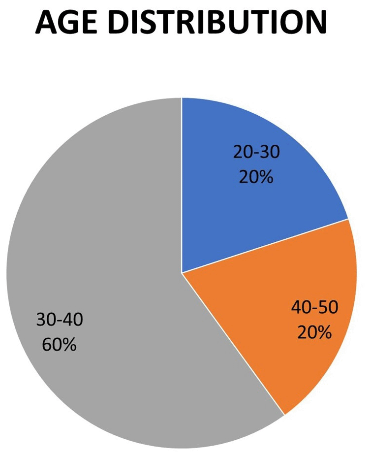
The present retrospective case-series study included five consecutive patients with cardiac hydatid cysts who underwent surgical treatment at the Iraqi Center for Heart Diseases, Medical City, Baghdad, between January 2018 and July 2024. The series included one case in 2018, one case in 2020, two cases in 2023, and one case in June 2024. Inclusion criteria comprised all patients diagnosed with cardiac hydatid cysts who underwent surgical management at our center during the study period, whereas patients with incomplete records or non-surgical management were excluded. Patient age ranged from 20 to 50 years, and the female-to-male ratio was 4:1, as shown in Figure 1. All patients were referred by a cardiologist to a cardiac surgeon, and all underwent open-heart surgery through median sternotomy with cardiopulmonary bypass and cardioplegic arrest. After exposure of the cyst, the surrounding operative field was isolated with pads soaked in povidone iodine The cyst content was aspirated carefully before excision in order to reduce the risk of spillage. The residual cavity was managed according to cyst location and myocardial involvement. The myocardial defect was repaired according to the size and site of the defect; patch repair was used in cases with a large defect or friable ventricular wall. Synthetic patch was used (PTFE patch).
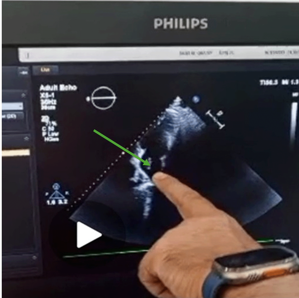
Clinical presentation varied across the series. Palpitation was the most frequent presenting complaint and occurred in three patients. One patient presented with ischemic chest pain associated with elevated cardiac enzyme levels and electrocardiographic changes. One patient presented with weakness and headache secondary to a brain hydatid cyst. Diagnosis was established in all patients by echocardiographic examination and chest computed tomography. Serological testing for echinococcosis was performed in some patients when available. Figure 2 shows an echocardiographic image of a left atrial hydatid cyst located posterior to the heart with daughter cysts. In the left atrial case, the diagnosis favored hydatid cyst rather than myxoma because imaging showed a cystic lesion with daughter cysts and without the typical pedunculated attachment expected for atrial myxoma. Four patients had isolated cardiac hydatid disease, whereas one patient had combined cardiac, splenic, and brain hydatid cysts. In the patient with multi-organ hydatid disease, cardiac surgery was prioritized because of the immediate risk associated with the intracardiac lesion. The extracardiac lesions were managed in a staged manner with postoperative follow-up and adjunct medical therapy.
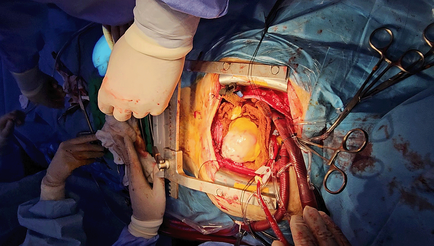
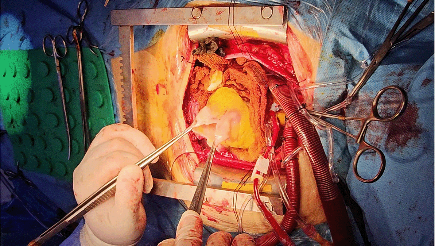
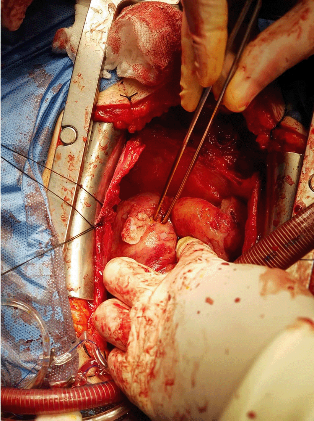
Cardiac involvement affected the left ventricle in four patients. One patient had a left atrial hydatid cyst with daughter cyst dissemination to the brain. Three patients had ruptured hydatid cysts. Two of those patients had intrapericardial rupture associated with pericardial adhesions, whereas one patient had rupture of a left atrial cyst into the cardiac cavity. Two patients had intact cysts. Figure 3 shows an intact left ventricular hydatid cyst during aspiration of the cyst content, Figure 4 shows the left ventricular cavity after cyst excision, and Figure 5 shows intrapericardial rupture of a cardiac hydatid cyst. Three patients had a single cardiac hydatid cyst, whereas two patients had multiple cardiac hydatid cysts.
Operative course was reviewed in all cases, where two operations were complicated by adhesions and extension of the aortic incision, which the operative notes attributed to friable tissue related to the inflammatory process. Three operations proceeded without major intraoperative difficulty. Early postoperative review identified one patient who developed significant bleeding and required re-exploration on cardiopulmonary bypass with re-suturing of the left ventricular free wall. Elevated blood pressure during recovery contributed to delayed extubation until the first postoperative day, massive blood transfusion, and prolonged intensive care unit stay.
Albendazole was administered in all patients at a high dose of 400 mg twice daily. Follow-up of the first two cases extended to five years and showed no recurrence with good cardiac function. Follow-up of the last three cases was shorter, but no recurrence was detected and cardiac function remained good. Two female patients later completed uncomplicated pregnancy and labor two years after surgery. A summary of patient demographics, clinical presentation, cyst characteristics, operative management, follow-up, and outcomes is presented in Table 1.
Isolated cardiac hydatid cysts are rare events,6 as constant myocardial contractility and high intraventricular pressure create a mechanical environment that limits the growth and attachment of hydatid cysts. Cardiac hydatid cysts posing a high risk of spontaneous rupture and subsequent anaphylaxis; therefore, they should be diagnosed early and treated seriously. There are two types of rupture, intrapericardial rupture and intracardiac rupture, and both complications were present in our cases.
The female: male ratio was 4:1, unlike Oraha et al. (2018),7 which was conducted in Kurdistan, north of Iraq, and included four cases, three male and one female.
In this study, we noticed that the incidence in the last two years was 60%. Eighty percent of patients were from urban areas rather than rural areas, which might be due to contaminated food from restaurants. The presence of loose dogs in the cities also represents a risk factor for transmission of eggs of E. granulosus to cattle and, accidentally, to human beings. Regarding the presentation of patients, 60% presented with palpitation, which agrees with the findings of Oraha et al. (2018).7 Twenty percent of patients presented with CNS manifestations due to rupture of the left atrial hydatid cyst with a daughter cyst delivered to the brain. Another 20% of patients presented with features of IHD due to rupture of a large LV H.C to the pericardium, which stimulated the inflammatory process and caused pericarditis with myocarditis and elevated cardiac enzymes.
Four patients had LV H.C because the left ventricle has large muscle mass and receives blood from coronary circulation more than other chambers of the heart. That finding agrees with Oraha et al. (2018).7,8 Regarding diagnostic investigations of choice, echocardiography and chest CT scan agree with Oraha et al. (2018),7 as the diagnosis of cardiac hydatid cyst is easy with a typical cystic appearance on echocardiography. However, it is difficult to distinguish it from myxoma in rare cases.9,10 Surgery is the treatment modality of choice; all patients underwent median sternotomy open heart surgery. The patient who had multiple cardiac hydatid cysts after rupture of the main cyst and spillage of daughter cysts into the pericardial space developed extension of the aortic cannulation site during surgery, but the complication was controlled successfully.
During the post-operative period, the patient who developed significant bleeding required re-exploration on cardiopulmonary bypass due to elevated blood pressure, while there was a friable LV wall due to the inflammatory process that caused separation of the LV wall patch. The friability in that case was most likely related to inflammatory involvement of the myocardial tissue by the ruptured hydatid cyst. In similarly fragile cases, reinforcement with a biological patch may offer additional protection against separation at the repair site, although this possibility requires further evaluation. However, the mortality rate was zero, with good surgical outcomes. In our study we use synthetic patch as biological patch was not available in our center at that time. However, the mortality rate was zero, with good surgical outcomes.
The study has several limitations as the sample size was small because cardiac hydatid disease is rare, the study was retrospective, and follow-up duration was not uniform across all patients. Serological testing and advanced imaging were not available in a standardized manner for every case. These limitations reduce the generalizability of the findings, although the series still provides useful clinical and operative observations from an endemic setting.
• Improve public hygiene and sanitation practices.
• Increase public awareness of hydatid disease and its modes of transmission through television programs, social media, and other health education platforms.
• Slaughterhouses should ensure proper disposal of the viscera and bowel of infected sheep in order to limit further transmission.
• Functional or non-organic diagnoses should be considered only after exclusion of organic causes, especially in patients with repeated presentations to the emergency department in endemic areas.
Cardiac hydatid cyst is an uncommon but important diagnosis in endemic settings and requires early recognition because delayed treatment may lead to serious complications. The present case-series showed favorable surgical outcomes, with no mortality, no recurrence during follow-up period, and good postoperative cardiac function in all patients. Echocardiography and computed tomography played an important role in diagnosis, whereas timely surgical intervention remained central to management. The limited sample size and non-uniform follow-up restrict generalization, but the findings still support early diagnosis and surgical treatment as key components of care in operable cases.
The study was based on a retrospective review of hospital-admitted patients who had already received standard clinical care. According to institutional practice, separate ethics committee approval was not required for retrospective analysis of anonymized clinical data. Despite that, ethical approval was obtained from the University of Fallujah.
Written informed consent for treatment had been obtained at hospital admission, and written informed consent for publication of clinical details and images was obtained from the patients or their legal guardians.
| Views | Downloads | |
|---|---|---|
| F1000Research | - | - |
|
PubMed Central
Data from PMC are received and updated monthly.
|
- | - |
Provide sufficient details of any financial or non-financial competing interests to enable users to assess whether your comments might lead a reasonable person to question your impartiality. Consider the following examples, but note that this is not an exhaustive list:
Sign up for content alerts and receive a weekly or monthly email with all newly published articles
Already registered? Sign in
The email address should be the one you originally registered with F1000.
You registered with F1000 via Google, so we cannot reset your password.
To sign in, please click here.
If you still need help with your Google account password, please click here.
You registered with F1000 via Facebook, so we cannot reset your password.
To sign in, please click here.
If you still need help with your Facebook account password, please click here.
If your email address is registered with us, we will email you instructions to reset your password.
If you think you should have received this email but it has not arrived, please check your spam filters and/or contact for further assistance.
Comments on this article Comments (0)