Keywords
Q14108, SCARB2, SCARB2, antibody characterization, antibody validation, western blot, immunoprecipitation, immunofluorescence
This article is included in the YCharOS (Antibody Characterization through Open Science) gateway.
Q14108, SCARB2, SCARB2, antibody characterization, antibody validation, western blot, immunoprecipitation, immunofluorescence
SCARB2 serves as a receptor for the lysosomal enzyme β-glucocerebrosidase (GCase), mediating its trafficking from the endoplasmic reticulum to lysosomes.1 Dysfunction of this pathway contributes to the accumulation of glycolipids and lysosomal stress, mechanisms strongly implicated in neurodegenerative disorders such as Parkinson’s disease (PD) and action myoclonus–renal failure (AMRF) syndrome.2,3
Here, we characterized twelve antibodies against SCARB2, for use in western blot, immunoprecipitation, and immunofluorescence using standardized protocols. Antibody performance was assessed by comparing signal in wild-type (WT) and knockout (KO) cell lines. The resulting data can guide the selection of antibodies best suited to specific research needs, enabling robust biochemical and cellular assessment of the target.
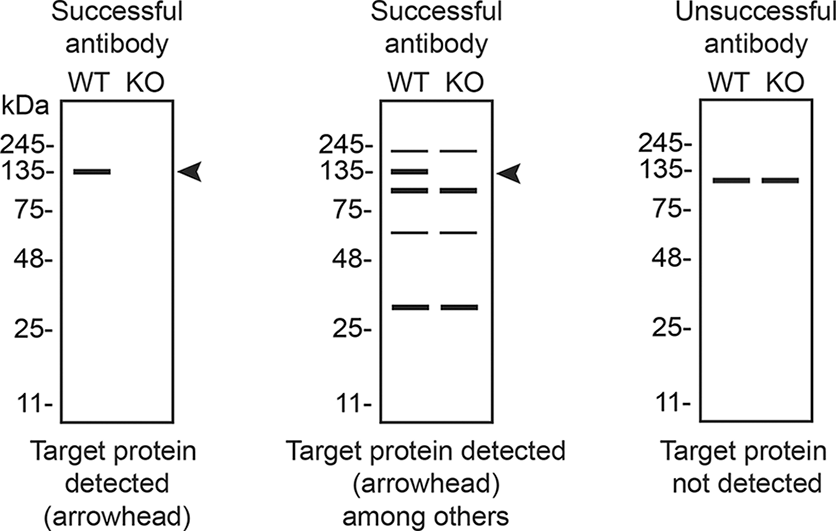
This work is part of a broader collaborative initiative involving academics, funders, and antibody manufacturers, aimed at improving biomedical research reproducibility through the systematic characterization of commercial antibodies against human proteins and open data sharing. Antibodies are provided in-kind by participating manufacturers. The approach involves identifying cell lines with sufficient target expression, generating or sourcing corresponding KO cell lines, and evaluating commercially available antibodies, with a focus on renewable (monoclonal and recombinant) reagents.4 We do not provide explicit antibody recommendations; rather, the data are presented to enable independent interpretation. Guidance on data interpretation is available via the YCharOS gateway5 and in Table 5 of this data note.
Antibody performance was assessed by comparing readouts from WT and KO cells.4 The first step is to identify a cell line expressing sufficient levels of the target protein to generate a measurable signal using antibodies. To this end, we examined the DepMap transcriptomics database (Cancer Dependency Map Portal, RRID:SCR_017655) to identify cell lines with expression levels greater than 2.5 log2 (transcripts per million “TPM” + 1), a threshold we have found to be suitable.6 The MCF7 cell line expresses the SCARB2 transcript at 7.2 log2(TPM + 1), and a corresponding SCARB2 KO MCF7 cell line was obtained from Abcam ( Table 1). Moreover, as seen on DepMap, the MCF7 does not carry mutations in the SCARB2 gene that could affect antibody-epitope binding.
| Institution | Catalog number | RRID (Cellosaurus) | Cell line | Genotype |
|---|---|---|---|---|
| Abcam | ab271144 | CVCL_0031 | MCF7 | WT |
| Abcam | ab274952 | CVCL_B9BE | MCF7 | SCARB2 KO |
Primary antibodies are listed in Table 2, with usage conditions summarized in Table 3 and secondary antibodies in Table 4. Antibody usage is reported as dilution ratios to align with manufacturer recommendations and account for proprietary formulations.
| Company | Catalog number | Lot number | RRID (Antibody Registry) | Clonality | Clone ID | Host | Concentration (μg/μL) | Vendors recommended applications |
|---|---|---|---|---|---|---|---|---|
| Abcam | ab176317** | 1068975–2 | AB_2620169 | recombinant mono | EPR12080 | rabbit | 0.10 | Wb, IP |
| Abcam | ab196651** | GR312647–3 | AB_3101952 | recombinant mono | EPR12081 | rabbit | 0.84 | Wb |
| Abcam | ab314217** | 1065721-4 | AB_3101784 | recombinant mono | EPR26243–125 | rabbit | 0.53 | Wb, IP, IF |
| ABclonal | A9185** | 4000001467 | AB_2863681 | recombinant mono | ARC1467 | rabbit | 0.24 | Wb |
| Bio-Techne (R&D Systems) | MAB1888* | JUZ024071 | AB_2182970 | monoclonal | 220411 | rat | 1.00 | Wb |
| Bio-Techne (Novus Biologicals) | NBP3–22186** | 240465 | AB_3097844 | recombinant mono | SR2211 | rabbit | NA | Wb, IP |
| Cell Signaling Technology | 27960** | 1 | AB_3083079 | recombinant mono | E2Z5F | rabbit | 0.33 | Wb, IP, IF |
| Proteintech | 27102–1-AP | 00050639 | AB_2880756 | polyclonal | - | rabbit | 0.50 | Wb |
| Thermo Fisher Scientific | 702770** | 2477085 | AB_2723321 | recombinant mono | 12H5L1 | rabbit | 0.50 | Wb |
| Thermo Fisher Scientific | 703037** | 2942968 | AB_2734813 | recombinant mono | 22H6L14 | rabbit | 0.50 | Wb, IF |
| Thermo Fisher Scientific | 711805** | 2384977 | AB_2723322 | recombinant poly | 12HCLC | rabbit | 0.50 | Wb |
| Thermo Fisher Scientific | 712072** | 2854061 | AB_2724602 | recombinant poly | - | rabbit | 0.50 | Wb, IF |
| Catalog number | Concentration (μg/μL) | Vendor-recommended Wb dilution | Wb dilution used | IP volume (μL, for 2 μg input) | Vendor-recommended IF dilution | IF dilution tested (1) | IF dilution tested (2) | IF dilution shown |
|---|---|---|---|---|---|---|---|---|
| ab176317** | 0.10 | 1/1000–1/5000 | 1/1000 | 20.2 | - | 1/10 | 1/100 | 1/100 |
| ab196651** | 0.84 | 1/20000 | 1/20000 | 2.4 | 1/100 | 1/100 | 1/800 | 1/800 |
| ab314217** | 0.53 | 1/1000 | 1/1000 | 3.8 | 1/50 | 1/50 | 1/500 | 1/50 |
| A9185** | 0.24 | 1/500–1/1000 | 1/500 | 8.3 | - | 1/200 | 1/500 | 1/200 |
| MAB1888* | 1.00 | 1/1000 | 1/1000 | 2.0 | - | - | - | - |
| NBP3–22186** | - | 1/500–1/2000 | 1/500 | 5.0 | - | 1/200 | 1/500 | 1/200 |
| 27960** | 0.33 | 1/1000 | 1/200 | 6.1 | 1/50–1/100 | 1/100 | 1/300 | 1/300 |
| 27102–1-AP | 0.50 | 1/1000–1/8000 | 1/1000 | 4.0 | - | - | - | - |
| 702770** | 0.50 | 1/500 | 1/500 | 4.0 | - | 1/250 | 1/500 | 1/500 |
| 703037** | 0.50 | 1/1000 | 1/1000 | 4.0 | 1/100 | 1/100 | 1/500 | 1/500 |
| 711805** | 0.50 | 1/200 | 1/200 | 4.0 | - | 1/250 | 1/500 | 1/500 |
| 712072** | 0.50 | 1/1000 | 1/1000 | 4.0 | 1/100 | 1/100 | 1/500 | 1/500 |
| Company | Secondary antibody | Catalog number | RRID (Antibody Registry) | Clonality | Concentration (μg/μL) | Working concentration (μg/mL) |
|---|---|---|---|---|---|---|
| Proteintech | HRP-Goat Anti-Rabbit Antibody (H + L) | RGAR001 | AB_3073505 | recombinant polyclonal | 1.0 | 0.05 |
| Invitrogen | HRP-Goat Anti-Rat Antibody (H + L) | 31470 | AB_228356 | polyclonal | 0.8 | 0.4 |
| Cell Signaling Technology | Protein A, HRP conjugate | 12291 | NA | polyclonal | 0.125 | 0.5 |
| Proteintech | CoraLite Plus 555-Goat Anti-Rabbit Antibody (H + L) | RGAR003 | AB_3073507 | recombinant polyclonal | 0.5 | 0.5 |
| Invitrogen | Alexa Fluor™ 555-Goat anti-Rat (H + L) | A-21434 | AB_141733 | polyclonal | 2 | 0.5 |
All twelve antibodies were first tested in western blot using WT and SCARB2 KO protein lysates ( Figure 1). This step enables concurrent validation of the KO cell line (absence of detectable SCARB2 signal) and identification of antibodies that detect SCARB2 with a band that is absent in the KO lane. Dilutions were selected based on supplier recommendations and adjusted when necessary ( Table 3).
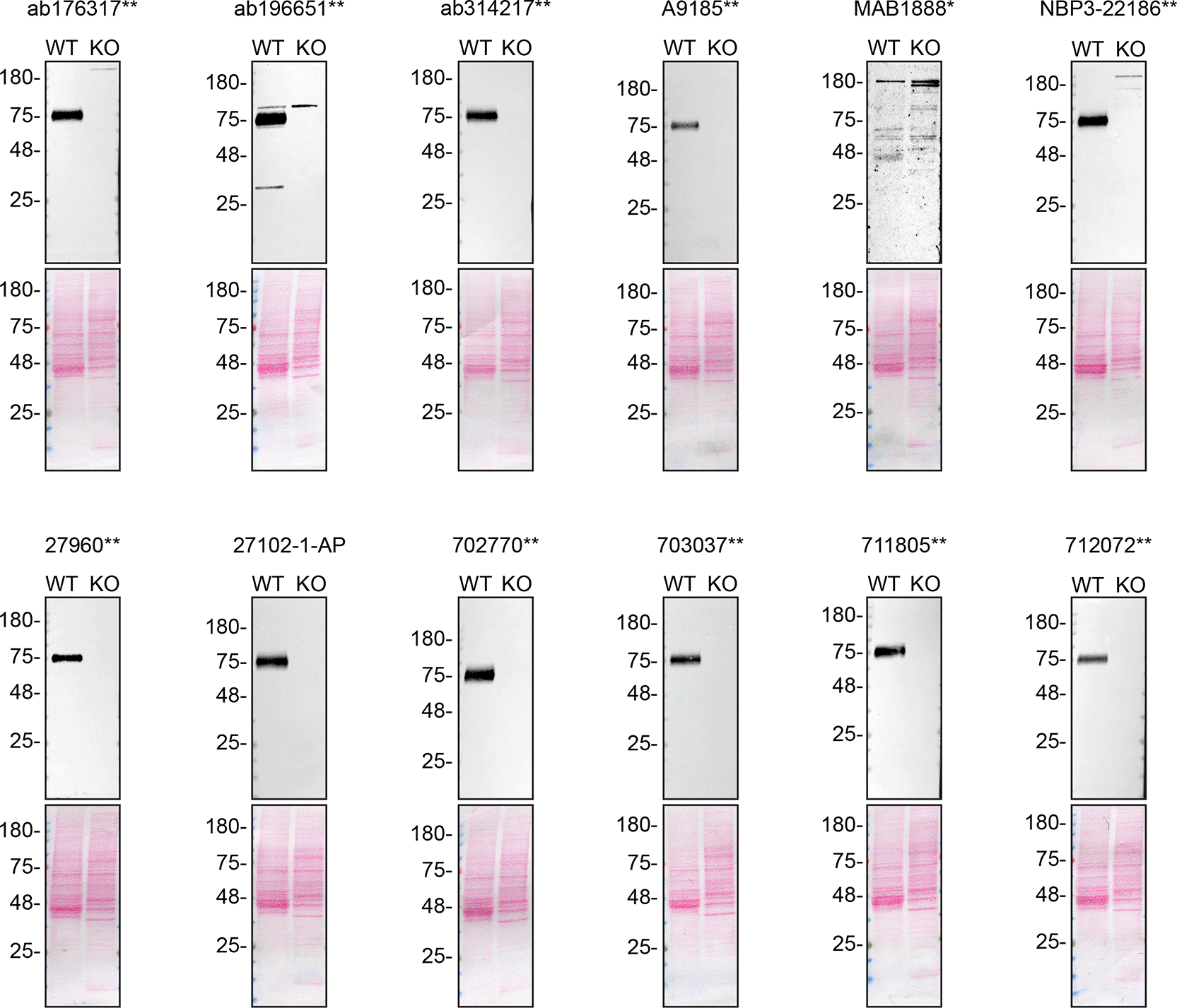
Lysates from MCF7 WT and SCARB2 KO were prepared and analyzed in western blot using the indicated SCARB2 antibodies. Ponceau-stained membranes are shown to confirm equal loading and efficient protein transfer from gel to membrane. Predicted molecular weight: 54 kDa. **Recombinant antibody; *Monoclonal antibody.
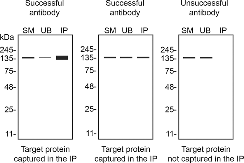
The ability of each antibody to capture SCARB2 from MCF7 lysates was assessed by immunoprecipitation followed by western blot analysis ( Figure 2). Each immunoprecipitation was performed using 2 μg of antibody, or 5 μL when the concentration was not available ( Table 3). Starting material (SM), unbound (UB), and immunoprecipitated (IP) fractions were analyzed by SDS-PAGE and probed using antibody 702770 identified in the western blot screen. This assay evaluates target capture but does not assess selectivity (i.e., binding to SCARB2 versus off-target proteins).
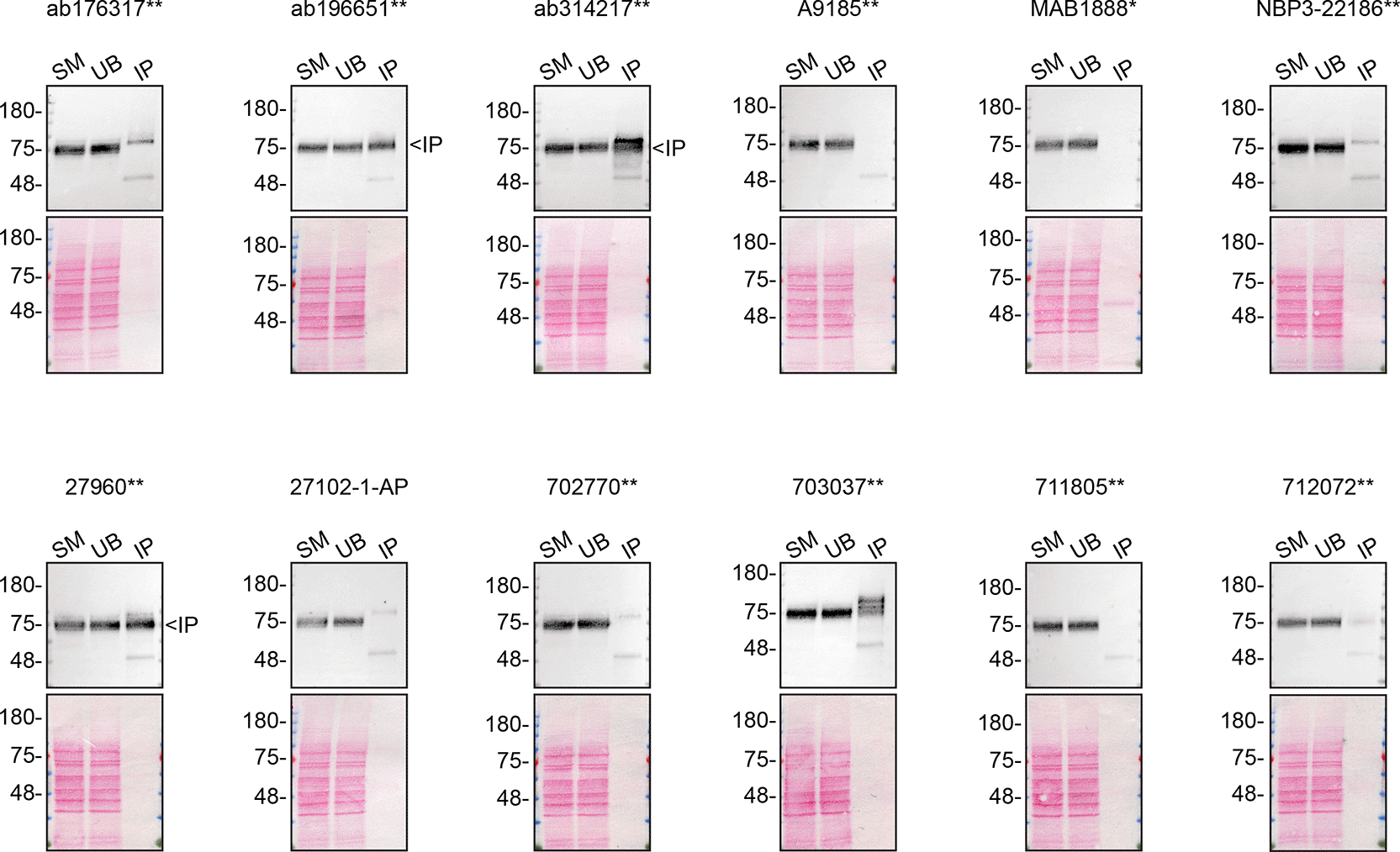
MCF7 lysates were prepared, and immunoprecipitation was performed using the indicated SCARB2 antibodies. Immunoprecipitates were analyzed by western blot using SCARB2 antibody 702770** (1/500). Ponceau-stained membranes are shown. SM = 6% starting material; UB = 6% unbound fraction; IP = immunoprecipitate. ** = recombinant antibody; * = monoclonal antibody.
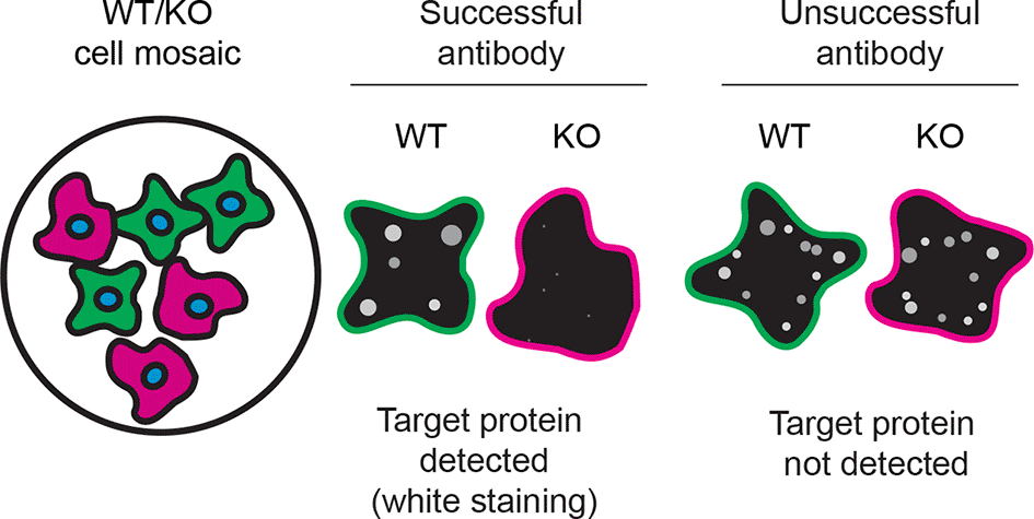
For immunofluorescence, ten recombinant antibodies were screened at two dilutions using a mosaic strategy in which WT and KO cells, pre-labelled with distinct dyes, were imaged within the same field of view to minimize experimental bias ( Figure 3). Antibodies were tested at supplier-recommended dilutions when available; otherwise, they were tested at 1 and 2 μg/mL ( Table 3). Because detergent choice can strongly influence staining of lysosomal proteins,7 both saponin and Triton X-100 were evaluated.
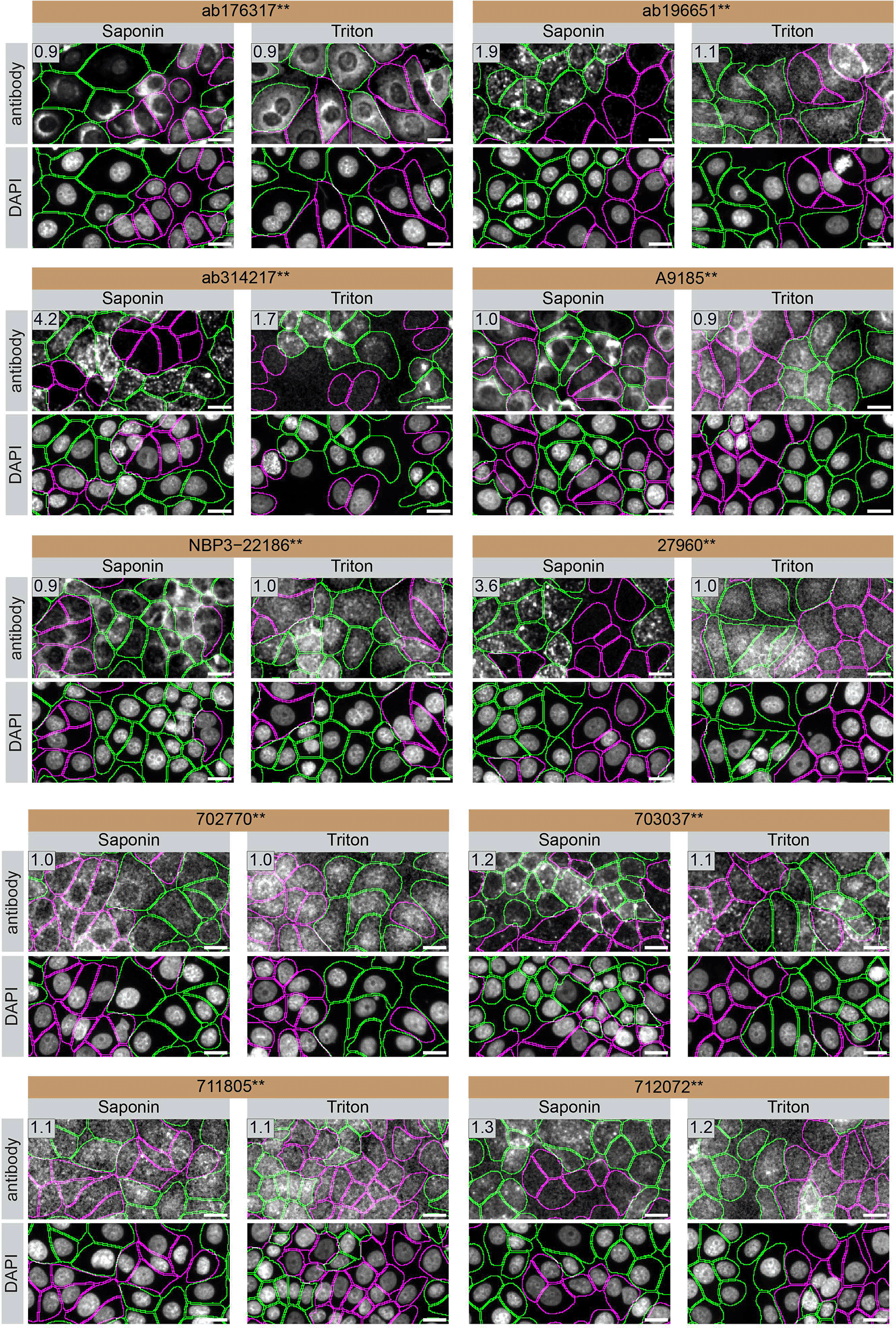
MCF7 WT and SCARB2 KO cells were pre-labelled with green and far-red fluorescent dyes, respectively, mixed at a 1:1 ratio, and plated in 96-well plates. Cells were permeabilized with saponin or Triton X-100 and stained with the indicated SCARB2 antibodies. Representative images of the antibody and DAPI channels (grayscale) are shown. WT and KO cells are outlined with green and magenta dashed lines, respectively. WT/KO signal intensity ratios are indicated in the top-left corner of each antibody channel image. Scale bar = 10 μm. **Recombinant antibody; *Monoclonal antibody.
Signal was quantified across at least 500 WT and KO cells,4 and representative images are shown in Figure 3. For each antibody, the WT/KO signal intensity ratio is reported: values near 1 indicate comparable signal in WT and KO cells, values >1 indicate enrichment in WT cells, and values <1 indicate higher signal in KO cells. The value shown corresponds to the condition yielding the highest ratio. These ratios facilitate comparison under the conditions tested but should be interpreted within the context of the specific assay and cell type.
In conclusion, we screened twelve SCARB2 commercial antibodies in western blot, immunoprecipitation, and ten antibodies by immunofluorescence by comparing signal in WT and SCARB2 KO MCF7 cell lines. To assist with data interpretation, Table 5 outlines common antibody performance scenarios across applications. High-quality renewable antibodies detecting SCARB2 were identified for each application. Researchers studying SCARB2 in other species are encouraged to consider these results and verify predicted species reactivity with the manufacturer before proceeding.
| Western blot | Immunoprecipitation | Immunofluorescence |
|---|---|---|
| 
| 
|
Inherent limitations are associated with the antibody characterization platform used in this study. First, the YCharOS project focuses on renewable (recombinant and monoclonal) antibodies and does not test all available antibodies against any given targets. While YCharOS partners provide access to the majority of renewable antibodies, some widely used polyclonal antibodies may not be included.
Second, the YCharOS approach is protein-agnostic and aims to provide objective data on antibody performance without predefined expectations regarding molecular weight or localization. As such, only a brief overview of the protein target is provided, and expert interpretation of banding patterns and subcellular localization is encouraged.
Third, experiments are not performed in biological replicates due to the parallel testing of multiple antibodies recognizing distinct epitopes. The identification of at least one specific antibody supports target expression in WT cells and its absence in KO cells, providing a reference for evaluating other antibodies. Experiments are performed using standardized conditions and master mixes to minimize variability. In immunofluorescence, testing at multiple antibody concentrations provides an additional assessment of specificity. Experiments may be repeated when no signal is detected. Furthermore, these data are generated independently of manufacturers’ validation processes, effectively constituting an external replication.
Finally, conclusions are limited to the experimental conditions and cell line used. The reliance on a single cell type represents a constraint, as protein expression levels can influence antibody performance. The use of cancer cell lines also introduces potential confounders, including mutations that may affect antibody-epitope binding. These limitations are inherent to cell line-based validation approaches.
The standardized protocols used for this antibody characterization study were established and approved by a collaborative group of academics, industry researchers, and antibody manufacturers.4 Brief descriptions of the experimental procedures used in this study are described below.
The cell lines used in this study are listed in Table 1. To facilitate proper citation and unambiguous identification, all cell lines and antibodies are referenced with their corresponding Research Resource Identifiers (RRIDs).8 All cell lines used in this study were regularly tested for mycoplasma contamination and were confirmed to be mycoplasma-free.
Primary antibodies are listed in Table 2 with RRIDs.9 Working dilutions for western blot and immunofluorescence, and volumes corresponding to 2 μg input for immunoprecipitation, are provided in Table 3. Secondary antibodies and their working concentrations are listed in Table 4. Antibody usage is reported as dilution ratios.
MCF7 WT and SCARB2 KO cells were collected in RIPA buffer (25 mM Tris-HCl pH 7.6, 150 mM NaCl, 1% NP-40, 1% sodium deoxycholate, 0.1% SDS) (Thermo Fisher Scientific, cat. number 89901) supplemented with 1x protease inhibitor cocktail mix (MilliporeSigma, cat. number P8340). Lysates were sonicated briefly and incubated 30 min on ice. Lysates were spun at ~110,000 × g for 15 min at 4°C and 30 μg of protein aliquots of the supernatants were analyzed by SDS-PAGE and western blot. BLUelf prestained protein ladder (GeneDireX, cat. number PM008-0500) was used.
Western blots were performed with precast midi 4-20% Tris-Glycine polyacrylamide gels (Thermo Fisher Scientific, cat. number WXP42012BOX) ran with Tris/Glycine/SDS buffer (Bio-Rad, cat. number 1610772), loaded in Laemmli loading sample buffer (Thermo Fisher Scientific, cat. number AAJ61337AD) and transferred on nitrocellulose membranes. Proteins on the blots were visualized with Ponceau S staining (Thermo Fisher Scientific, cat. number BP103-10) which is scanned to show together with individual western blot. Blots were blocked with 5% milk for 1 hr, and antibodies were incubated O/N at 4°C with 5% milk in TBS with 0.1% Tween 20 (TBST) (Cell Signalling Technology, cat. number 9997). Following three washes with TBST, the peroxidase conjugated secondary antibody was incubated at a dilution of ~0.2 μg/ml in TBST with 5% milk for 1 hr at room temperature followed by three washes with TBST. Membranes were incubated with Pierce ECL (Thermo Fisher Scientific, cat. number 32106) prior to detection with the iBright™ CL1500 Imaging System (Thermo Fisher Scientific, cat. number A44240).
Antibody-bead conjugates were prepared by adding 2 μg to 500 μl of Pierce IP Lysis Buffer from Thermo Fisher Scientific (cat. number 87788) in a microcentrifuge tube, together with 30 μl of Dynabeads protein A- (for rabbit antibodies) or protein G- (for rat antibodies) (Thermo Fisher Scientific, cat. number 10002D and 10004D, respectively). Tubes were rocked for ~1 h at 4°C followed by two washes to remove unbound antibodies.
MCF7 WT were collected in Pierce IP buffer (25 mM Tris-HCl pH 7.4, 150 mM NaCl, 1 mM EDTA, 1% NP-40 and 5% glycerol) supplemented with protease inhibitor. Lysates were rocked 30 min at 4°C and spun at 110,000 × g for 15 min at 4°C. 0.5 ml aliquots at 1 mg/ml of lysate were incubated with an antibody-bead conjugate for ~1 h at 4 °C. The unbound fractions were collected, and beads were subsequently washed three times with 1.0 ml of IP buffer and processed for SDS-PAGE and western blot on precast midi 4–20% Tris-Glycine polyacrylamide gels.
MCF7 WT and SCARB2 KO cells were labelled with a green and a far-red fluorescence dye, respectively (Thermo Fisher Scientific, cat. number C2925 and C34565). The nuclei were labelled with DAPI (Thermo Fisher Scientific, cat. Number D3571) fluorescent stain. WT and KO cells were plated on 96-well plate with optically clear flat-bottom (Perkin Elmer, cat. number 6055300) as a mosaic and incubated for 24 hrs in a cell culture incubator at 37°C, 5% CO2. Culture medium was removed, and cells were fixed in 4% paraformaldehyde (PFA) (VWR, cat. number 100503-917) in phosphate buffered saline (PBS) (Wisent, cat. number 311-010-CL) for 10 min at room temperature. Cells were permeabilized in PBS1x with 0.05% Saponin (MilliporeSigma, cat. number 47036) or 0.1% Triton X-100 (Thermo Fisher Scientific, cat. number BP151-500) for 10 min at room temperature. Cells were blocked in IF buffer (PBS1x with 5% BSA and 0.005% Saponin or 0.01% Triton X-100) with 5% goat serum (Gibco, cat. number 16210-064) for 1 h at room temperature. Cells were incubated with the corresponding IF buffer containing the primary SCARB2 antibodies overnight at 4°C. Cells were then washed 3 × 10 min with IF buffer and incubated with the corresponding Fluor 555-conjugated secondary antibody in IF buffer for 1 hr at room temperature. Cells were washed 3 × 10 min with PBS1x then incubated with DAPI and washed once with PBS1x.
Images were acquired on an ImageXpress micro confocal high-content microscopy system (Molecular Devices), using a 20x NA 0.94 air objective and scientific CMOS cameras, equipped with 395, 475, 555 and 635 nm solid state LED lights (lumencor Aura III light engine) and bandpass filters to excite DAPI, Cellmask Green, Alexa-555 and Cellmask Red, respectively. Images had pixel sizes of 0.68 × 0.68 microns, and a z-interval of 4 microns. A minimum of 500 WT and KO cells were imaged per antibody. For analysis and visualization, shading correction (shade only) was carried out for all images. Then, maximum intensity projections were generated using 3 z-slices. Segmentation was carried out separately on maximum intensity projections of Cellmask channels using CellPose 1.0, and masks were used to generate outlines and for intensity quantification.10 We have developed a collection of scripts in Python and in ImageJ/FIJII made openly available on GitHub (https://github.com/ABIF-McGill/YCharOS_IF_characterization ).4 Figures were assembled with Adobe Illustrator.
| Views | Downloads | |
|---|---|---|
| F1000Research | - | - |
|
PubMed Central
Data from PMC are received and updated monthly.
|
- | - |
Provide sufficient details of any financial or non-financial competing interests to enable users to assess whether your comments might lead a reasonable person to question your impartiality. Consider the following examples, but note that this is not an exhaustive list:
Sign up for content alerts and receive a weekly or monthly email with all newly published articles
Already registered? Sign in
The email address should be the one you originally registered with F1000.
You registered with F1000 via Google, so we cannot reset your password.
To sign in, please click here.
If you still need help with your Google account password, please click here.
You registered with F1000 via Facebook, so we cannot reset your password.
To sign in, please click here.
If you still need help with your Facebook account password, please click here.
If your email address is registered with us, we will email you instructions to reset your password.
If you think you should have received this email but it has not arrived, please check your spam filters and/or contact for further assistance.
Comments on this article Comments (0)