Keywords
Preeclampsia, foetal biometry, Doppler velocimetry
Preeclampsia, foetal biometry, Doppler velocimetry
Preeclampsia is a hypertensive disorder of pregnancy, affecting 3–5% of pregnancies globally.1 This disorder is responsible for 2% to 8% of pregnancy-related complications, more than 50,000 maternal deaths, and over 500,000 foetal deaths worldwide.2 Wagner defined the diagnostic criteria for preeclampsia as a systolic blood pressure of 140 mmHg or higher or a diastolic blood pressure of 90 mmHg or higher on two separate occasions, at least 4 hours apart, with traces of protein in urine.3 As an alternative, 110 mmHg or higher for diastolic blood pressure or 160 mmHg for systolic blood pressure with proteinuria after 20 weeks of gestation. These are consistent with the Mayo and Cleveland Clinics’ definitions of the disorder. The American College of Obstetricians and Gynaecologists and the European Society of Cardiology define preeclampsia as a pregnancy-specific disorder characterised by new-onset hypertension with end-organ dysfunction, occurring after the 20th week of gestation in a previously normotensive woman.4,5 The pathophysiology of preeclampsia is multifaceted and not yet fully understood, though placental insufficiency, endothelial dysfunction, and oxidative stress are considered central mechanisms.6 Although the implications of preeclampsia can be serious for the mother, it also has profound effects on foetal development, often leading to growth restriction, preterm delivery, and increased perinatal mortality.
Doppler velocimetry and foetal biometry are crucial ultrasonographic tools for assessing foetal health in preeclamptic pregnancies. These techniques provide insight into foetal growth parameters and placental blood flow, providing a window into the effects of maternal vascular dysfunction on the developing foetus.7 Previous studies have established that abnormal Doppler indices, such as elevated uterine artery pulsatile index (UtA PI) and middle cerebral artery resistance index (MCA RI), are indicative of impaired placental perfusion and foetal hypoxia.8 However, the extent to which these indices correlate with specific growth parameters remains an area of active investigation.
In South Africa, where socio-economic disparities influence maternal and neonatal health outcomes, understanding the impact of preeclampsia on foetal growth is critical for improving clinical care and reducing adverse outcomes. This study focuses on mother-perinate pairs who received antenatal and postnatal care at Nelson Mandela Academic Hospital in Mthatha, Eastern Cape Province. This study aims to elucidate the relationship between maternal vascular health and foetal development in preeclampsia-related preeclamptic pregnancies by evaluating foetal biometry and Doppler indices.
Ethical clearance to conduct this study was granted by the Walter Sisulu University Health Sciences Research Ethics Committee (WSU HREC 019/2025). This study was authorised by the Eastern Cape Department of Health and the Nelson Mandela Academic Hospital’s Gynaecology and Obstetrics Department. Informed written consent and assent were obtained from the mothers during recruitment. This study adhered to the principles of the Declaration of Helsinki to protect participants.
Preeclamptic and normotensive pregnant women aged between 18–35 years with singleton pregnancies were included in the study.
Pregnant women with chronic hypertension, type 2 diabetes, gestational diabetes, renal and CVDs, or any critical health condition were excluded from the study. Furthermore, data were not collected from eligible pregnant women who revoked their consent to participate in the study.
Ethical clearance to conduct this study was obtained from the Walter Sisulu University Health Sciences Research Ethics Committee (WSU HREC 019/2025). This study is part of the study on the Assessment of the Cardiovascular Risk Profile of Infants Exposed to Pre-eclampsia in-utero : A Prospective Case-Control Study in South African Children of African Ancestry, registered on 2021-10-25, with the NIH ClinicalTrials.gov (Protocol https://ClinicalTrials.gov Identifier: NCT05091827; https://clinicaltrials.gov/ct2/show/NCT05091827).9
A standard interviewer-administered structured questionnaire10 collected information on maternal health and cardiovascular risk factors. This information included sociodemographics (age, ethnicity, and location), socio-economic status (marital status, profession, income/revenue, and living standard), maternal obstetric history, family history, cardiometabolic health history, and cardiovascular risk factors (physical exercise, smoking, alcohol consumption, and feeding habits.
Foetal biometry and Doppler indices were assessed using transabdominal and Doppler ultrasound per the SCoR/BMUS Guidelines for Professional Ultrasound Practice.11 The foetal biometry parameters assessed included the deepest vertical pocket (DVP), head circumference (HC), abdominal circumference (AC), femoral length (FL), biparietal diameter (BPD), estimated foetal weight (EFW); the uterine arteries (both the resistive and pulsatile indices), umbilical pulsatile index and resistive index as well as the middle cerebral artery pulsatile index (MCA PI) and its resistive index.
The IBM Statistical Package for Social Sciences (SPSS) Version 23.0 (IBM Corp) was used for data analysis. The Smirmov-Kolmogorov test was used to check for normality, and the data were corrected for outliers where necessary. Descriptive statistics were employed for data presentation as mean ± standard deviation (SD) and proportions (percentages) for categorical variables. An independent sample t-test was used to compare mean differences between groups (perinates of pre-eclamptic and normotensive pregnant women). Pearson correlations were used to assess the relationship between foetal biometry and Doppler indices. Multiple linear regression analyses were performed to predict foetal outcomes. Differences were considered statistically significant at p ≤ 0.05.
A total of 60 pregnant women (30 preeclamptic and 30 normotensive) and their perinates were recruited into the study. Women with preeclampsia were older than their normotensive counterparts, with a mean age of 28.96 ± 1.92 vs. 26.19 ± 0.98 years, and most of them were multiparous. 64% of the preeclamptic group had been diagnosed with preeclampsia before, compared to 17% in the normotensive group. Preeclamptic women reported 24% and 65% tobacco and alcohol use compared to 7% and 13%, respectively, in the normotensive group (see Table 1).
Perinates from preeclamptic pregnancies tended to be smaller compared to those from normotensive pregnancies. They had smaller head circumference (255 ± 74.41 mm vs 293.85 ± 13.18 mm, p = 0.05), abdominal circumference (236.84 ± 69.44 mm vs 288.29 ± 23.83 mm, p = 0.06) and estimated foetal weight (1609 ± 326.91 g vs 1987.53 ± 403.43 g, p = 0.05) (see Table 2).
The right uterine artery pulsatile index (UtA PI) was significantly higher in preeclamptic pregnancies compared to normotensive pregnancies (1.52 ± 0.70 vs 1.06 ± 0.37, p = 0.05). The middle cerebral artery pulsatile index (MCA PI) was significantly higher in preeclamptic pregnancies compared to normotensive pregnancies (2.17 ± 0.76 vs 1.99 ± 0.39, p = 0.04). The MCA resistance index (MCA RI) was also significantly higher in preeclamptic pregnancies (0.88 ± 0.12) compared to normotensive pregnancies (0.86 ± 0.07), with a p-value of 0.01 ( Table 3).
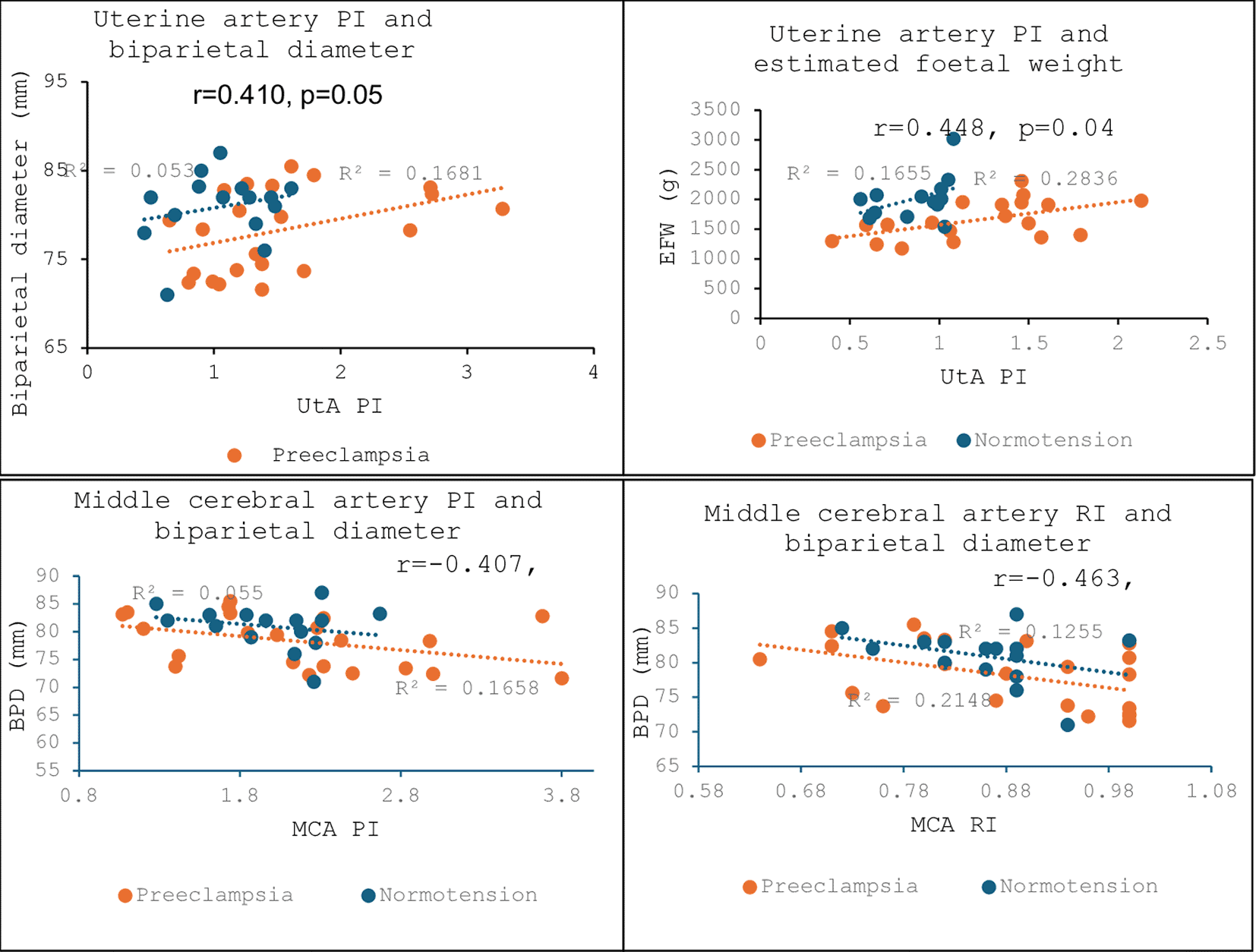
Pearson’s correlations were performed to determine the relationships between Doppler indices and foetal biometry. The uterine pulsatile index showed a positive association with biparietal diameter (r = 0.410, p = 0.05) and the estimated foetal weight in preeclamptic pregnancies (r = 0.448, p = 0.04). The R2 for these associations implied that approximately 16.81% and 28.36% variability in biparietal diameters and estimated foetal weights in this group may be attributed to the fluctuations in uterine artery pulsatile indices. However, these relationships were not statistically significant in the normotensive group. The middle cerebral artery pulsatile index and its associated resistive index exhibited negative correlations with biparietal diameter in the preeclampsia-affected group (r = −0.407, p = 0.06 and r = −0.463, p = 0.03) (Figure 1).
Multiple regression models were designed to assess the relationship between foetal head circumference and middle cerebral artery pulsatile and resistive indices (MCA PI & MCA RI) as key predictors. The overall model had an R2 of 0.337 and p = 0.04, the adjusted R2 = 0.204 and p = 0.04. The middle cerebral artery pulsatile index (MCA PI) emerged as a strong positive predictor for foetal head circumference (β = 0.568, p = 0.02). This implies that for every unit increase in the MCA pulsatile index, foetal head circumference increases by an average of 0.568 cm. Conversely, the MCA resistive index significantly predicted foetal head circumference (β = −0.716, p = 0.004), suggesting that each unit increase in the resistive index results in a 0.716 cm decrease in foetal head circumference. The model showed that MCA PI and MCA RI contributed significantly to foetal cranial development ( Table 4).
| Adjusted R2 = 0.204 | |||||
|---|---|---|---|---|---|
| Standardized β | Standard error (SE) | 95% CI | t-value | P-value | |
| MCA PI | 0.568 | 22.401 | [8.477; 99.976] | 2.421 | 0.02 |
| MCA RI | −0.716 | 137.765 | [−714.946; −152.239] | −3.147 | 0.004 |
The study highlights significant differences in maternal characteristics, foetal biometry, and Doppler indices between preeclamptic and normotensive pregnancies. Women with preeclampsia had a higher prevalence of a history of preeclampsia, smoking, and alcohol consumption. Perinates in preeclamptic pregnancies were smaller and had higher uterine artery resistive indices.
The current study aligns with previous reports suggesting that a history of preeclampsia is an important contributor to the development of preeclampsia.7 In fact, a retrospective study of the Swedish Birth Register from 1987–2004, in which a total of 1,430,464 deliveries were included, showed that preeclampsia occurred more commonly in women who had had preeclampsia before. The study showed a 14.7% reoccurrence rate of preeclampsia in a second pregnancy and 31.9% in a third pregnancy.12
In terms of foetal biometry, foetuses from preeclamptic pregnancies exhibited significantly smaller head circumferences and lower estimated foetal weights compared to foetuses in normotensive pregnancies. These findings were consistent with studies indicating that maternal preeclampsia impedes placental function, ultimately restricting foetal growth.13 This study also noted non-significant differences in abdominal circumferences, femur length, and biparietal diameter, suggesting variability in the impact of preeclampsia on foetal growth parameters. Similar studies have reported that while some foetal measurements are consistently affected by preeclampsia, others may vary depending on the severity and onset of the condition.8
Doppler velocimetry revealed higher uterine artery pulsatile index (UtA PI) and middle cerebral artery pulsatile index (MCA PI) in preeclamptic pregnancies, both being statistically significant. These findings are indicative of altered blood flow dynamics in preeclampsia, which can compromise oxygen and nutrient delivery to the foetus. The elevated MCA resistance index (MCA RI) further supports the notion of increased vascular resistance in preeclampsia.14 Increased placental vascular resistance can lead to foetal hypoxemia and potentially trigger compensatory mechanisms, where blood flow is shunted to vital organs such as the foetal brain at the expense of peripheral growth.15 Contrasting studies have suggested that while Doppler parameters are helpful in assessing placental and foetal health, their predictive value for adverse outcomes may vary across populations.16,17
The uterine artery pulsatile index (UtA PI) demonstrated a strong positive association with estimated foetal weight. This may be due to placental adaptation, where the placenta compensates for increased vascular resistance by enhancing nutrient transport efficiency, allowing the developing foetus to maintain or even increase its weight irrespective of compromised uteroplacental blood flow.18,19 Another mechanism that may explain this is the shunting resulting from the redistribution of blood, where the foetus prioritises blood flow to essential organs like the brain, ensuring adequate growth irrespective of placental insufficiency.20,21 The current findings are supported by Wu et al, who showed that the right UTA PI of 1.27 (95%CI: 0.84–1.63) was associated with the early onset of preeclampsia.22 Participants in our study were diagnosed with preeclampsia before 26 weeks, even though data were only collected at 33 weeks of gestation. Furthermore, some studies have shown that higher UTA PI is associated with small for gestational age foetuses, indicating its important contribution to uteroplacental blood flow.23 Other contrasting findings observed no significant association between UtA PI and foetal weight in mild cases of preeclampsia, suggesting variability based on disease severity.24,25
The relationship between UtA PI and foetal biparietal diameter (BPD) was also notable, with a positive correlation, though not statistically significant. Similar findings have been reported in studies emphasising the role of uteroplacental blood flow in determining foetal cranial growth. However, the lack of statistical significance in this study may reflect sample size limitations or variability in the impact of preeclampsia on cranial growth parameters.
Interestingly, the middle cerebral artery pulsatile index (MCA PI) correlated negatively with biparietal diameter, indicating a potential compensatory mechanism where increased cerebral blood flow prioritises brain development in response to hypoxic conditions. Elevated MCA PI reflects increased vascular resistance within foetal cerebral circulation. Over time, this can impede brain growth, sometimes reflected by reduced biparietal diameter.26 This aligns with the concept of “brain-sparing” observed in foetuses affected by preeclampsia, as reported in recent literature.27 However, the marginal significance of this finding underscores the need for further investigation to confirm its clinical relevance.
The middle cerebral artery resistive index (MCA RI) showed a strong negative correlation with BPD. Similar reports revealed that elevated vascular resistance in the foetal cerebral circulation is a compensatory response to placental insufficiency.28 However, contrasting studies have reported variability in MCA RI values, particularly in cases of late-onset preeclampsia, where cerebral haemodynamics may be less affected.29,30
The inferential output revealed significant associations between foetal biometry and middle cerebral artery (MCA) indices, shedding light on the hemodynamic adaptations in pregnancies affected by hypertensive disorders such as preeclampsia. The negative association between the MCA resistive index (MCA RI) and biparietal diameter (BPD) suggests that increased vascular resistance in the foetal cerebral circulation may impair cranial growth. This finding aligns with the concept of “brain-sparing,” where blood flow is redistributed to prioritise vital organs under hypoxic conditions.31 However, contrasting findings suggest that the degree of impact on BPD may vary depending on the severity and onset of preeclampsia, with late-onset cases enduring less severe effects.32,33
This study demonstrates that maternal preeclampsia in a resource-limited South African setting is linked to significant reductions in foetal biometric measurements and marked alterations in placental and cerebral blood-flow indices. Doppler velocimetry’s value as an early warning tool is highlighted by showing how elevated uterine and cerebral Doppler pulsatility and resistive indices predict impaired head growth and lower estimated foetal weight. Incorporating these ultrasound metrics into routine care could enable targeted interventions to improve perinatal outcomes among high-risk pregnancies.
Department of Human Biology, Walter Sisulu University, Nelson Mandela Drive, Mthatha, South Africa
Chuma Mabuto, Constance Rufaro Sewani-Rusike, Ebenezer Ackah, Ngoakoana Edna Matjuda
Department of Biological and Environmental Sciences, Walter Sisulu University, Mthatha, South Africa
Benedicta Nkeh-Chungag, Nomagugu Ndlovu, Ongeziwe Zamxaka, Yamkela Mfenyana, Roland Tiagha Akah, Jude Eteneneng Enoh
Department of Obstetrics and Gynaecology, Nelson Mandela Academic Hospital, Mthatha, South Africa
Charles Businge, Nontsikelelo Gubu-Ntaba, Nandipha Sotobe-Mbana, Malusi Madikane
Department of Obstetrics and Gynaecology, Mthatha Regional Hospital, Mthatha, South Africa
Mziwohlanga Mdondolo
Department of Obstetrics and Gynaecology, Frere Hospital, Mthatha, South Africa
Mfundo Feketshane, NP Selanto-Charman, Tandi Noah
Physiology Section, Otto Loewi Research Centre for Vascular Biology, Immunology and Inflammation, Medical University of Graz, Graz
Nandu Goswami
Corresponding authors
Correspondence to Chuma Mabuto ([email protected]).
| Views | Downloads | |
|---|---|---|
| F1000Research | - | - |
|
PubMed Central
Data from PMC are received and updated monthly.
|
- | - |
Provide sufficient details of any financial or non-financial competing interests to enable users to assess whether your comments might lead a reasonable person to question your impartiality. Consider the following examples, but note that this is not an exhaustive list:
Sign up for content alerts and receive a weekly or monthly email with all newly published articles
Already registered? Sign in
The email address should be the one you originally registered with F1000.
You registered with F1000 via Google, so we cannot reset your password.
To sign in, please click here.
If you still need help with your Google account password, please click here.
You registered with F1000 via Facebook, so we cannot reset your password.
To sign in, please click here.
If you still need help with your Facebook account password, please click here.
If your email address is registered with us, we will email you instructions to reset your password.
If you think you should have received this email but it has not arrived, please check your spam filters and/or contact for further assistance.
Comments on this article Comments (0)