Keywords
Bacterial infections; Staphylococcus aureus; Antibiotic-Resistant; Biofilm-Forming; Streptococcus agalactiae; Cranberry extract; gene expression; Vaginal Infections
This article is included in the Cell & Molecular Biology gateway.
This article is included in the Fallujah Multidisciplinary Science and Innovation gateway.
Bacterial infections; Staphylococcus aureus; Antibiotic-Resistant; Biofilm-Forming; Streptococcus agalactiae; Cranberry extract; gene expression; Vaginal Infections
The most frequent cause of vaginitis in women of childbearing age is bacterial vaginosis (BV). Its prevalence is estimated to be between 17%-19% in family planning clinics and 4% to 10% in student health clinics, with the prevalence rising to 24% to 40% in sexually transmitted disease (STD) clinics, depending on the population under study. 16–29% of pregnant women have been found to have BV, and 30% of women who visit infertility clinics have the disease. Although it can happen without sexual activity, it primarily affects young, sexually active girls (Hillier and Holmes, 1999). Numerous facets of fetal development, physiological function, immunology at mucosal surfaces, disease susceptibility, and nutritional absorption are influenced by commensal microbiota associated with the human body (Forney et al., 2012). Therefore, commensals in the lower female reproductive tract play a crucial role in both preventing infections and preserving vaginal health (Stojanovic et al., 2012). Women who have human vaginal infections are far more likely to give birth before their due dates (Mulu et al., 2015). Pelvic inflammatory disorders (PID), which can result in tubal infertility, ectopic pregnancy, reproductive dysfunction, and unfavorable pregnancy outcomes (such as low birth weight and preterm delivery), can arise if they are left untreated (Kaambo and Africa, 2017). The development of cervical dysplasia, an elevated risk of postpartum infections, HIV, and the acquisition and spread of herpes simplex virus-2 (HSV-2) can all be attributed to vaginal infections (Ravel et al., 2011). Aerobic vaginitis (AV) and bacterial vaginosis (BV) infections in prenatal health services among asymptomatic pregnant women, however, are associated with a high rate of unexpected pregnancy outcomes and premature birth in Africa and around the world (Krauss et al., 2017). Due to increased estrogen levels following puberty, sexually mature women have a lower reproductive tract that favors lactobacilli colonization. Estrogen results in thicker stratified epithelia and increased glycogen concentrations with Lactobacillus species contributing to a lower pH of ≤4.5 in the vagina (Danielsson et al., 2011), due to lactic acid and H2O2 generation. Staphylococcus aureus, and Streptococcus agalactiae (group B Streptococcus) are frequent commensal microorganisms found in the lower reproductive tract, alongside Lactobacillus species. Overgrowth of these might lead to discharge alterations, and it can cause vaginitis (Sangeetha et al., 2015). Streptococcus agalactiae (Group B Streptococcus) is a commensal microbiota found in the human intestine and genitourinary systems. It is also a major pathogen in AV and other diseases (Sørensen et al., 2010). GBS can cause complications in the elderly, pregnant women, and those with pre-existing illnesses (Brigtsen et al., 2015). Neonatal sepsis, meningitis, and pneumonia are linked to maternal colonization with GBS in pregnant women at delivery, It is believed that the microbiota found in the vagina differs greatly from that seen in other parts of the human body (Fredricks, 2011). GBS is currently thought to be the primary cause of early-onset sepsis globally (Cools et al., 2016). Furthermore, preterm birth, extremely low birth weight delivery, and puerperal sepsis—all of which contribute significantly to morbidity and death in sub-Saharan Africa—are linked to GBS and E. faecalis (Kim et al., 2014). The majority of pregnant women experience asymptomatic bacteriuria, despite the fact that GBS can also result in lower urinary tract infections, acute pyelonephritis, and silent bacteriuria (Edwards and Gonik, 2013). Women with untreated GBS bacteriuria are more likely to experience chorioamnionitis, early-onset newborn illness, and intrauterine fetal loss (Anderson et al., 2007). Although Staphylococcus. aureus infects women’s lower female genital tracts, newborns are exposed to this organism directly in the vaginal canal and perineum. The mouth, skin, and umbilical stump may get colonized by the transmitted S. aureus (Chan et al., 2013). Neonatal illnesses such cellulitis, umbilical stump infection (Omphalitis), arthritis, and Staphylococcal Scalded Skin Syndrome (SSSS) may arise from this. Sepsis, meningitis, and newborn bacteremia are some of the most serious side effects of these S.aureus infections. Vascular injury, extracellular fluid loss, hypotension, multiple organ failure, and death are the causes of sepsis. Infant sepsis alone accounts for 225,000 fatalities annually, making it the third most prevalent cause of infant mortality worldwide in sub Saharan Africa (Tumuhamye et al., 2020). The vaccinium macrocarpon (Cranberry) has been found to have many beneficial effects on human health, ranging from prevention of cancers and cardiovascular diseases to the ability to modify bacterial behaviour, interfering with virulence factors and leading to prevention of common bacterial infections.
A disposable cotton swab was used to collect 100 specimens from pregnant and non-pregnant women who had bacterial vaginosis. In October 2023, the samples were collected at a private clinic. As soon as samples were taken from the patient for diagnosis, they were cultured, and the patient’s information was documented.
Every sample was grown on MacConkey agar, chocolate agar, and human blood agar. After that, it was incubated for 24 hours at 37 °C in an aerobic environment. To determine the type of bacteria, it was then subjected to a battery of tests. First, their size, shape, color, pigments, and hemolytic activity were evaluated. They were then subjected to gram staining and microscopic analysis. They next went through biochemical testing like Voges-Proskauer, indole, and methyl red. Lastly, VITEK2 was used to confirm each isolate’s diagnosis.
In accordance with CLSI 2023, the antibiotic susceptibility test was performed using the disc diffusion method (Campbell, 2013). The effectiveness of antibiotics, which were produced by (Bioanalyse Company in Turkey) (Cefixime, Ciprofloxacin, Amikacin, Chloramphenicol, Levofloxacin, Gentamycin, Azithromycin, Tobramycin) against Staphylococcus. aureus, Streptococcus agalactiae.
Using the Resazurin Microtitre-plate Assay (REMA) with a few changes, the minimum inhibitory concentration (MIC) of the solutions Levofloxacin, Cefixime, Cranberry and mix (Levofloxacin + Cranberry) and (Cefixime + Cranberry) was found. Each well of the microtiter plate received 100 μl of Brain Heart Broth (BHI) under aseptic conditions. Next, the first row of the 96-well plates was filled with 100 μl of the test sample, which included (gentamicin, clove extract, and GCE). 100 μl of the test material was pipetted at descending concentrations (1/2,1/4,1/8,1/16,1/32,1/64, and 1/128) from one well to the next in order to perform serial dilutions. A 10 μl bacterial suspension containing 1.5 × 10⁸ CFU/ml was added to each well. For 18 to 24 hours, the plates were incubated at 35 ± 2 °C with loose wraps of Parafilm. Each well received 10 μl of resazurin solution (Alamar blue) after incubation. To see if the colour changed, the plate was incubated again for four hours. Resazurin’s hue was observed to determine the outcome visually; transitions from purple to pink, red, or colourless were considered positive (Sarker et al., 2007).
The micro-titer plate method was used to determine bacteria’s ability to create biofilm. Microtiter wells with 180 μl of Tryptone soya broth were injected with a 20 μl bacterial suspension from an overnight culture (similar to McFarland standard No. 0.5). A control set of microtiter wells had 200 μl of sterile Tryptone soya broth. All microtiter plates were then incubated at 37 °C for the entire day. After that, the wells were washed three times with phosphate-buffered saline (pH 7.2) and dried at room temperature. Each well was treated with 200 μl of methanol for 15 minutes. Next, the plates were dried at room temperature. Each well received 200 μl of 0.1% crystal violet solution for 15 minutes. Following that, the crystal violet solution was withdrawn and washed three times with phosphate-buffered saline (pH 7.2) to remove any remaining dye. Wells were dried at room temperature for 15 minutes. A microplate reader (Biotek ELX 800, UK) measured absorbance at 600 nm after adding 200 μl of 33% glacial acetic acid to each well. The cutoff value was calculated as the control’s mean OD600 plus three standard deviations. Any isolate with an OD above the cut-off value was designated a biofilm producer (Al-Shaabani et al., 2020).
The cranberry fruit was obtained from the local market. Air-dried at room temperature after being washed several times with distilled water (DW), then it was converted into powder form by using a sterile electric grinder. Then the active components of cranberry fruit were extracted by using an organic solvent (methanol). Where a quantity of 25 g of air-dried powder was extracted with 200 ml of methanol, by using the Soxhlet apparatus for 5 h at 60 °C. Thereafter, the methanol was evaporated by using the rotary evaporator at 40 °C. The final extract was kept at 4 °C before using (Akroum et al., 2009).
Written informed consent was obtained from all participants prior to data collection. As shown in Form 1 and Form 2, which are examples and part of these approvals.
RNA extraction
The gene expression was tested at ½MIC for three treatments for each isolate (Levofloxacin, Cefixime, Cranberry and mix (Levofloxacin + Cranberry) and (Cefixime + Cranberry), as well as a control without treatment (only bacteria growing in the broth). After growing the bacteria at ½MIC of each treatment solution, the RNA was extracted using the Trizol technique (Thermo Fisher Scientific, 2016).
Convert RNA to cDNA
The RNA is extracted and converted into cDNA using the Luna Universal qPCR&RT-qPCR kit’s instructions (10 μl of master mix, 1 μl of random primer, 1 μl of RT enzyme, 2 μl of nucleas-free water, and finally 6–8 μl of RNA) in a PCR machine (verity, Applied Biosystems). The PCR conditions were 25 °C for 5 minutes, 42 °C for 15 minutes, 85 °C for 15 seconds, and 4 °C hold (Jaafar & Shareef, 2023).
Two different genes were selected to each bacteria according to the bacteria the rgg2 gene was amplified used the forward primer (5′- GCGCCTTAGAATGAATCGACAC-3′) and reverse primer (5′- GCAGATGAGCACCTATCCAAGT-3′) and tetM gene with forward primer (5′-GCTTCCGTTGGGAAGTGGAA‐3′) and reverse primer (5′- CCTTGTTCGCAACCATAGCG-3′) for bacteria S. agalactiae. and for bacteria S. aureus the fnpB gene was amplified used forward primer (5′- TGGTGCCACTGCTTTAACCT-3′) and revers primer (5′- ACCAGTACCACCTGCCAAAG-3′) and also norA gene amplified used forward primer (5′- CCAGGTAAATTAGCCGATTGC-3′) and reverse primer (5′- AAATCGCCTGCGTTCTAGAG‐3′). Thereafter, a two-step gene expression was initiated by putting the cDNA samples into the real-time PCR instrument and making the additions according to the kit manufacturer’s instructions. (Luna Universal qPCR/RT-qPCR) The RT-PCR was performed using 10 μl of master mix, 3 μl of nuclease-free water, 1 μl of forward and reverse gene primers, and 5 μl of cDNA material. The thermal cycling conditions were as follows: 95 °C for 60 seconds (initial denaturation 1 cycle); 95 °C for 15 seconds (denaturation 40–45 cycles); and 60 °C for 30 seconds (extension 40–45 cycles) (Al-Taee et al., 2018).
After samples were collected from 100 patients cultivated on three culture mediums diagnosed microscopically and used the ViteK-2 system and biochemical tests. The following Table 1 displays the results achieved.
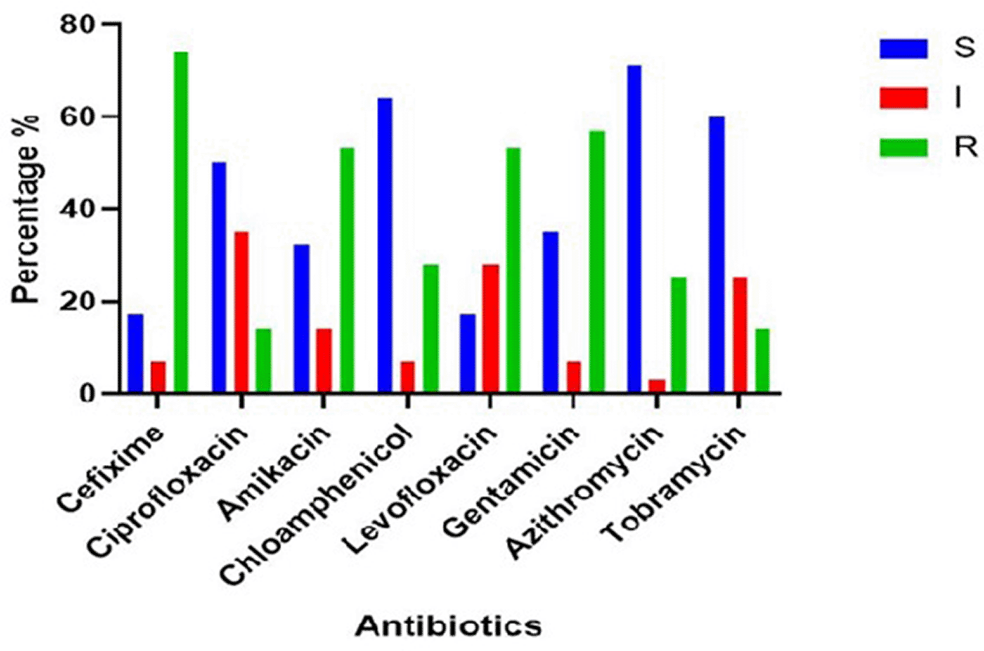
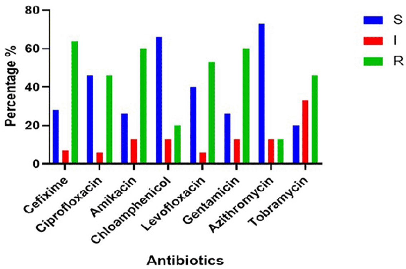
The disk diffusion method was used to determine antibiotic susceptibility. The susceptibility of bacteria varied based on the antibiotic used and species. The result shows the. Figure 1: the Staphylococcus. aureus isolates were sensitive to most antibiotics and resistant to Amikacin 53.5%, Levofloxacin 53.5%, Gentamycin 57,1% and Cefixime 74.8%. In Figure 2, the Streptococcus agalactiae is also sensitive to most antibiotics, but resistant to Cefixime 64.2%, Levofloxacin 53%, Gentamycin 60%, Tobramycin 47% Amikacin 60%.
In Figure 3 the Streptococcus agalactiae (86.66%)(13/15) of isolates were biofilm producers out of 13 biofilm producing isolates 3 (23.07%), 9(69.23%), and 1(7.69%) were weak, moderate, strong, respectively. Figure 4 demonstrated that (89%) (25/28) of Staphylococcus. aureus isolates were considered biofilm producers out of 25 biofilm-producing isolates; 10(40%), 12(48%), and 3(12%) were weak, moderate, and strong, respectively.
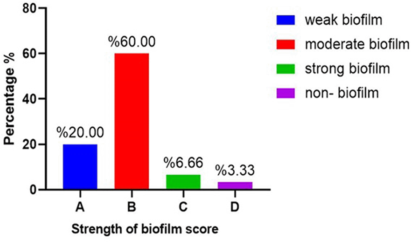
A: weak biofilm; B: moderate biofilm, C: strong biofilm, D: Non- biofilm.
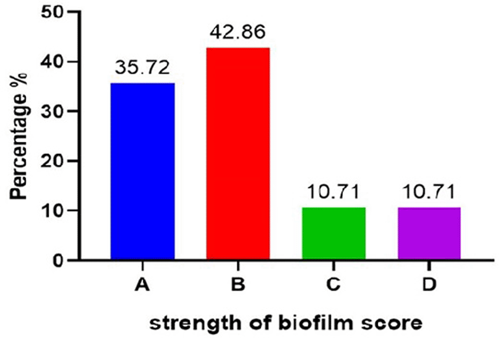
A: weak, B: moderate, C: strong, D: non.
The images were designed using the PowerRender software and were designed at a resolution of 600 dpi.
After the MIC was detect the gene expression results for rgg2 gene and tetM gene for S. agalactiae bacteria and norA gene and fnpB gene for S. aureus bacteria show ½MIC of Cefixime, Levofloxacin increase gene expression but ½MIC of cranberry, mix the (Cranberry + Cefixime), (Cranberry+ Levofloxacin) reduce gene expression (Table 2), (Table 3) and (Table 4), (Table 5).
The rise of antibiotic-resistant bacteria continues to challenge the pharmaceutical sector (Abed et al., 2024). Antibiotics are considered the first choice of treatment the infections; however, their effectiveness is often reduced due to multidrug-resistant strains of Staphylococcus spp and other bacteria (Mahal et al., 2023). With the increasing spread of antibiotic resistance among pathogenic bacteria, it has become essential to search for effective alternative treatments. The use of natural compounds, such as medicinal herbs, is considered one of the most promising options in this field. Numerous studies have been conducted to investigate the effects of medicinal herbs on various types of bacteria, including Staphylococcus aureus, Escherichia coli, Pseudomonas aeruginosa, and others. The findings of these studies have shown that certain medicinal compounds possess the ability to affect the bacterial cell wall, contributing to the weakening and inhibition of bacterial resistance (Baghini et al., 2018). S. aureus isolates that showed resistance to many antibiotics led to difficulty in limiting their spread and reducing infection with them. Therefore, there was a need to work on obtaining a therapeutic alternative to contribute to limiting the spread of S.aureus isolates due to their ability to resist antibiotics. Treatment of isolates with plant extracts proved the high ability of the extract to inhibit bacterial growth. When detecting gene expression and synergy between the antibiotic and the plant extract, it showed a great possibility in reducing gene expression and that the synergy between them helps in increasing the effectiveness of the antibiotic. The effect of the plant extract was also determined on many other bacterial isolates, for example, E.coli, S.agalactiae, and Enterococcus spp. These results are somewhat consistent with the results of our study (Mitchell et al., 2012). When a bacterial infection occurs, there must be a sufficient number of bacteria and a pathogenic capacity to help them overcome the immune system (Rajab and Turki, 2021). Chovanová et al., 2013, has been indicated that the plant extracts can directly affect bacterial isolates and have a synergistic effect with antibiotics in reducing the gene expression of many genes responsible for antibiotic resistance and biofilm production, which contributes to increasing the virulence and pathogenicity of Gram-negative and Gram-positive bacterial isolates (S. aureus, S. agalactiae).
Plant extracts can be relied upon as an alternative treatment option when antibiotic resistance occurs due to the random and incorrect use of these antibiotics, as the extracts have proven their ability to directly or synergistically affect the gene expression of many genes responsible for the virulence of bacteria.
Ethical approval was received from the research committee of the Al-Anbar Directorate of Health Authorities of Al-Anbar hospitals under decision Ref.7 on 18-4-2024. The Ethics Committee of the University of Anbar granted ethical permission. All participants were told about the study.
| Views | Downloads | |
|---|---|---|
| F1000Research | - | - |
|
PubMed Central
Data from PMC are received and updated monthly.
|
- | - |
Provide sufficient details of any financial or non-financial competing interests to enable users to assess whether your comments might lead a reasonable person to question your impartiality. Consider the following examples, but note that this is not an exhaustive list:
Sign up for content alerts and receive a weekly or monthly email with all newly published articles
Already registered? Sign in
The email address should be the one you originally registered with F1000.
You registered with F1000 via Google, so we cannot reset your password.
To sign in, please click here.
If you still need help with your Google account password, please click here.
You registered with F1000 via Facebook, so we cannot reset your password.
To sign in, please click here.
If you still need help with your Facebook account password, please click here.
If your email address is registered with us, we will email you instructions to reset your password.
If you think you should have received this email but it has not arrived, please check your spam filters and/or contact for further assistance.
Comments on this article Comments (0)