Keywords
Body mass index; Interstitial brachytherapy; Cervical cancer; Gynecologic malignancies; Dosimetry; Organs at risk; CT-based planning
This article is included in the Oncology gateway.
Body mass index; Interstitial brachytherapy; Cervical cancer; Gynecologic malignancies; Dosimetry; Organs at risk; CT-based planning
Gynecological malignancies remain a major global health challenge, with cervical cancer alone accounting for more than 600,000 new cases each year.1 Radiotherapy, particularly a combination of external beam radiotherapy (EBRT) and brachytherapy, continues to be the cornerstone of treatment for locally advanced disease. Brachytherapy can be delivered through intracavitary, interstitial, or intravaginal approaches, each chosen according to disease extent and anatomical considerations. ICBT uses intrauterine tandems and vaginal applicators, whereas ISBT employs transperineal needles, allowing better coverage of bulky or asymmetric disease. Interstitial brachytherapy (ISBT), on the other hand, involves direct insertion of needles into the tumor and parametrial tissues, offering particular value in patients with bulky, asymmetric tumors or unfavorable anatomy.2 The effectiveness of brachytherapy is most often assessed using dose–volume histogram (DVH) parameters and implant quality indices, which provide insight into both tumor coverage and the sparing of surrounding organs at risk (OARs). Several studies have reported benchmark values for these indices in interstitial brachytherapy, guiding the evaluation and optimization of treatment plans.
Body mass index (BMI) has been shown to influence many aspects of cervical cancer care, from screening and diagnosis to definitive chemoradiation and brachytherapy. For instance, a large retrospective cohort of more than 900,000 women undergoing cervical cancer screening revealed that overweight and obese women were less likely to be diagnosed with premalignant lesions but more often presented with invasive cancers, likely reflecting the underdiagnosis of early lesions in this group. Prior studies in pelvic radiotherapy and brachytherapy indicate BMI-dependent variations in OAR doses; however, these findings have not been extended to ISBT.3 In terms of radiotherapy, Moszyńska-Zielińska et al. reported that women with a BMI > 30 kg/m2 who underwent EBRT for endometrial cancer experienced greater setup errors, necessitating larger PTV margins.4 In contrast, Kizer et al. reported that underweight patients with cervical cancer had a higher incidence of severe bowel complications, including enteritis and fistula formation, following treatment.5
The relationship between BMI and brachytherapy outcomes has also been explored in other malignancies. Echevarria et al. demonstrated that in low-dose-rate prostate brachytherapy, a high BMI was negatively associated with tumor coverage, likely because of increased perirectal and periprostatic fat.6 In gynecological settings, Boyle et al. and Sabater et al. reported that higher BMI was associated with lower OAR doses during vaginal cuff brachytherapy.7,8 Similarly, Lim et al. reported a decrease in the rectal point and mean dose with increasing BMI among patients receiving brachytherapy for cervical cancer.9 Collectively, these findings suggest that body habits may alter dose distributions to both tumor and normal tissues. Increased adipose tissue may increase perirectal spacing, whereas low BMI reduces soft-tissue buffering, potentially altering needle–organ relationships. We further hypothesize that a lower BMI may be associated with higher gastrointestinal OAR doses because of reduced adipose separation between the target and the bowel, potentially increasing the risk of toxicity.
Despite existing studies on ICBT and VBT, no prior work has systematically assessed BMI-related dosimetric differences in CT-based ISBT. Given that the ISBT depends heavily on applicator geometry, needle loading, and individual patient anatomy, BMI could act as a key modifier of treatment quality and toxicity risk. Therefore, the present study aimed to evaluate the associations between BMI and both dosimetric indices and radiobiological parameters in patients with gynecological malignancies treated with EBRT followed by high-dose-rate (HDR) ISBT.
This study was designed as a retrospective observational analysis and was carried out in the Department of Radiation Oncology at Kasturba Medical College, Attavar, Mangalore. A total of 55 patients were included, all of whom had biopsy-proven carcinoma of the cervix, vagina, or vaginal vault. Written informed consent was obtained from all patients prior to their inclusion in the study. These patients had previously undergone definitive external beam radiotherapy (EBRT), either alone or in combination with concurrent chemotherapy, before undergoing interstitial brachytherapy (ISBT). The treatment period spanned from January 2019 to December 2023.
With respect to brachytherapy, all patients received high-dose-rate (HDR) ISBT with the Syed–Neblett perineal template. Planning CT scans were obtained using diluted bladder contrast to aid visualization, while intravenous contrast was omitted. The high-risk clinical target volume (HR-CTV) as well as the surrounding organs at risk (OARs)—specifically the bladder, rectum, and sigmoid colon—were contoured in line with the recommendations of the GEC-ESTRO group and dose reporting parameters consistent with IBS and ICRU Report 89, including HR-CTV D90 and OAR D2 cc metrics. Treatment plans were created on the SagiPlan system (Eckert & Ziegler BEBIG) using a blend of inverse planning and manual optimization. All patients received an ISBT delivered in multiple HDR fractions using a uniform departmental fractionation schedule (7 Gy × 4 fractions). Dose delivery was performed with a cobalt-60–based SagiNova HDR remote afterloader. All plans followed a standardized departmental protocol combining inverse planning with manual refinement by the same team of radiation oncologists.
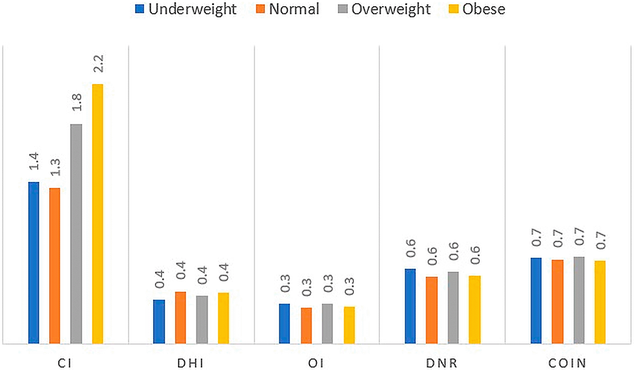
To evaluate dose distribution, parameters were extracted from dose–volume histograms (DVHs). Tumor coverage was examined by measuring the HR-CTV and calculating the proportions of the target that received 100%, 150%, and 200% of the prescribed dose (V100, V150, and V200, respectively). For the OARs, the dose to the most vulnerable regions was recorded as D2 cc, representing the minimum dose to the most exposed 2 cm3 of each organ. In addition, implant quality was judged using several well-established indices. The Coverage Index (CI) describes how completely the HR-CTV was covered by the prescription isodose. The dose homogeneity index (DHI) indicates the extent of uniform dose delivery and the presence of hot spots. The overdose index (OI) is the proportion of the target receiving very high doses (200% or more of the prescription). The dose nonuniformity ratio (DNR) reflected how much of the target received doses above 150%, while the conformity index (COIN) assessed how well the prescription dose conformed to the target shape while sparing adjacent normal tissue. CI reflects target coverage, DHI indicates dose uniformity, OI and DNR quantify hotspots, and COIN reflects conformity between target and prescription volume. The definitions and interpretations of the implant quality indices used in this study are summarized in Table 1.
Radiobiological parameters were also calculated to put the physical doses into the clinical context. The biologically effective dose (BED) was derived using the linear–quadratic model, expressed as BED = D × (1 + d/α/β), where D is the total dose and d is the dose per fraction. The equivalent dose in 2 Gy fractions (EQD2), which allows for comparison across different fractionation schedules, was then obtained as EQD2 = BED/(1 + 2/α/β). For the purpose of this study, an α/β ratio of 10 Gy was assumed for tumor tissue, whereas 3 Gy was used for late-responding organs. As part of the analysis, the influence of patient body habitus was also explored. Height and weight records available at the time of brachytherapy were used to calculate body mass index (BMI) in kilograms per square meter. Patients were then classified according to the World Health Organization (WHO) categories: underweight (<18.5), normal weight (18.5–24.9), overweight (25–29.9), and obese (≥30).
All the collected data were organized in Microsoft Excel and analyzed using SPSS Statistics version 22 (IBM Corp., Armonk, NY) and Epi Info version 7.2.1 (Centers for Disease Control and Prevention, Atlanta, GA). Descriptive statistics were used to summarize the data, with categorical variables presented as frequencies and percentages and continuous variables expressed as the mean values with standard deviation. The distribution of continuous variables was checked using the Kolmogorov–Smirnov and Shapiro–Wilk tests. Group comparisons across BMI categories were carried out using chi-square tests for categorical data and one-way analysis of variance (ANOVA) for continuous data, followed by Tukey’s post hoc test where appropriate. Statistical significance was defined as a p value less than 0.05. Graphical representations, including bar charts, line graphs, scatter plots, and pie charts, were created using Microsoft Excel to aid in data visualization. Since variables were normally distributed, one-way ANOVA was used, followed by Tukey’s post hoc test to adjust for multiple comparisons.
A total of 55 patients with gynecological malignancies who had received definitive external beam radiotherapy (EBRT) followed by interstitial brachytherapy (ISBT) were included in the study. The majority of patients (52, 94.5%) had carcinoma of the cervix, most commonly stage IIB (n = 20, 38.5%) and stage IIIC1 (n = 13, 25%). In addition, two patients were diagnosed with carcinoma of the vagina, and one patient presented with a vaginal vault malignancy following prior hysterectomy. When stratified by body mass index (BMI), 25 patients (45.5%) were classified as having a normal BMI, 11 (20%) were underweight, 9 (16.4%) were overweight, and 10 (18.2%) were obese. Notably, compared with the overall mean age, the mean age of underweight patients was significantly older (59.7 vs. 52.6 years, p = 0.048). Patient demographics, disease characteristics, and BMI distributions are detailed in Table 2.
The mean HR-CTV (V-CTV) for the cohort was 76.05 ± 38.28 cm3, with no statistically significant differences observed across the BMI subgroups. The average values of V100, V150, and V200 for the entire study population were 91.19 ± 9.05%, 53.61 ± 13.73%, and 29.17 ± 9.59%, respectively. Like V-CTV, these dose–volume parameters did not significantly vary across the different BMI categories. Comparisons of dosimetric parameters across BMI categories are presented in Table 3.
The mean coverage index (CI) for the cohort was 1.56 ± 0.93, with higher mean values observed in the obese and overweight groups (2.175 ± 1.354 and 1.844 ± 1.077, respectively). These differences, however, did not reach statistical significance. Across all patients, the mean values of the dose homogeneity index (DHI), overdose index (OI), dose nonuniformity ratio (DNR), and conformity index (COIN) were 0.42 ± 0.12, 0.32 ± 0.09, 0.58 ± 0.12, and 0.71 ± 0.08, respectively. Among the BMI subgroups, the DHI was lowest in the underweight group (0.369), while the OI and DNR were lowest in the normal-weight group (0.301 and 0.559, respectively). COIN values remained relatively consistent across BMI categories, with the lowest value observed in the obese group (0.696). None of these differences were statistically significant. Higher CI values may reflect the densely spaced needle geometry of the Syed–Neblett template, which can increase the prescription isodose cloud. The lower DHI observed in our series likely reflects the large and irregular HR-CT volume typical of patients selected for ISBT. These findings suggest that BMI does not influence implant geometry or HR-CTV coverage. The variation in implant quality indices across BMI groups is illustrated in Figure 1.
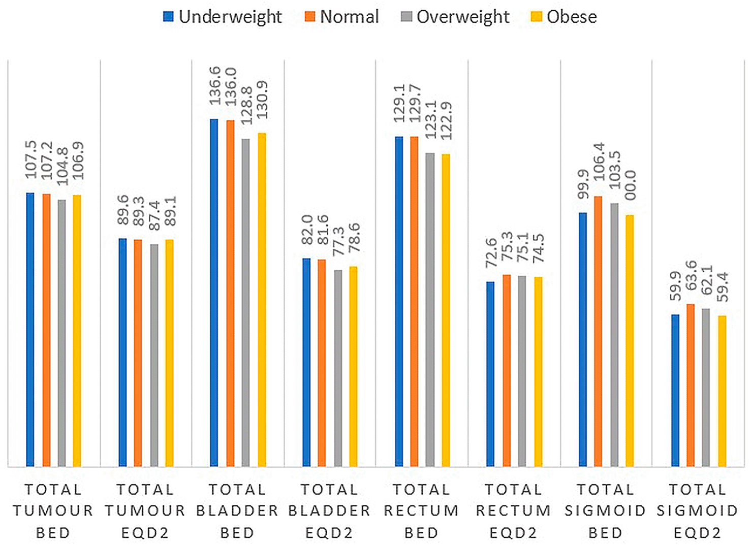
For the study cohort, the mean BED and EQD2 to the primary tumor were 106.79 ± 5.42 Gy10 and 88.99 ± 4.52 Gy10, respectively, with no statistically significant variation across BMI categories. The mean BED to the bladder, rectum, and sigmoid was 134.05 ± 11.67 Gy3, 127.33 ± 7.42 Gy3, and 103.31 ± 16.96 Gy3, respectively. The corresponding EQD2 values were 80.43 ± 7 Gy3, 74.56 ± 4.06 Gy3, and 61.99 ± 10.18 Gy3, respectively.
Rectal BED significantly varied across BMI groups (p = 0.014), with the most pronounced difference observed between patients with a normal BMI and those classified as obese. However, rectal EQD2 did not significantly vary across categories. Both the sigmoid BED and the EQD2 varied significantly with BMI (p = 0.024), with the lowest values recorded in obese patients (99.05 Gy3 and 59.43 Gy3, respectively) and the highest in normal-weight patients (106.45 Gy3 and 63.87 Gy3, respectively). In contrast, bladder BED and EQD2 did not significantly differ across BMI groups. The tumor and organ-at-risk BED and EQD2 values stratified by BMI are shown in Figure 2.
In our cohort, the mean implant quality indices were as follows: CI 1.56 ± 0.93, DHI 0.42 ± 0.12, DNR 0.58 ± 0.12, OI 0.32 ± 0.09, and COIN 0.71 ± 0.08. These values fall within the expected range but differ slightly from those in previously published reports. Sharma et al. reported a mean CI of 0.86 ± 0.03, DHI of 0.69 ± 0.11, DNR of 0.31 ± 0.09, OI of 0.08 ± 0.03, and COIN of 0.79 ± 0.05.10 Kumar et al. reported mean CI values of 0.87, DHI values of 0.58, DNR values of 0.42, OI values of 0.19, and COIN values of 0.74, whereas Poddar et al. reported CI values of 0.95, DHI values of 0.50, OI values of 0.17, DNR values of 0.50, and COIN values of 0.84 among 35 patients with cervical cancer treated with HDR ISBT.11,12 The relatively lower dose homogeneity in our study likely reflects the presence of larger, irregular target volumes where tumor coverage was prioritized during planning.
Although MRI-guided intracavitary-plus-interstitial techniques are commonly reported in EMBRACE I/II, our institution treats patients with bulky, asymmetric, or unfavorable vaginal anatomy using CT-based interstitial HDR with the Syed–Neblett template. This selection explains the predominance of interstitial-only implants in our cohort and should be considered when comparing dosimetric indices to MRI-guided intracavitary series. We add that MRI planning was not consistently available for all cases in the 2019–2023 period and that CT-based planning with GEC-ESTRO contouring was used throughout.
With respect to tumor coverage, our series achieved a mean HR-CTV EQD2 of 89 Gy10. This is higher than the values reported by Sharma et al. (76 Gy) and Poddar et al. (79 Gy)10,12 and aligns with EMBRACE II recommendations, which advocate HR-CTV D90 values of 85–90 Gy and > 90 Gy in patients with bulky or poorly responding tumors.2 “Because CT has lower soft-tissue contrast than MRI does, some degree of interobserver contouring variability is possible.”
For OARs, the mean EQD2 values in our study were 80.3 Gy3 for the bladder, 74.9 Gy3 for the rectum, and 61.9 Gy3 for the sigmoid. In comparison, Kumar et al. reported median bladder, rectum, and sigmoid EQD2 values of 70 Gy3, 64 Gy3, and 48 Gy3, respectively, whereas Sharma et al. reported bladder and rectum EQD2 values of 76 Gy3 and 80 Gy3, respectively.10,13 Although our series recorded slightly higher OAR doses, all values remained within the recommended tolerance limits. In cervical ISBT, central hot spots may be intentionally produced to treat the GTV; therefore, DHI/OI/DNR should be interpreted alongside HR-CTV D90 and OAR constraints.
Recent large-scale image-guided adaptive brachytherapy studies and international consensus guidelines have refined target dose objectives, organ-at-risk constraints, and reporting standards in gynecological brachytherapy.14–30 Although MRI-guided intracavitary-plus-interstitial techniques are commonly reported in EMBRACE I/II, our institution treats patients with bulky or asymmetric disease using CT-based interstitial HDR brachytherapy, which remains widely practiced in resource-limited settings.14,16–18,29,30 To our knowledge, this is the first study to explicitly examine the influence of BMI on implant quality indices and OAR doses in patients with gynecological ISBT. While indices such as CI, DHI, DNR, OI, and COIN did not vary significantly with BMI, we observed meaningful differences in rectal and sigmoid doses. Rectal BED was significantly lower in obese patients than in normal and underweight patients (p = 0.014), and a similar trend was observed for sigmoid EQD2 (p = 0.024). These results suggest that BMI plays a selective role in modulating gastrointestinal OAR exposure. These dose levels are consistent with outcomes reported in image-guided adaptive brachytherapy series demonstrating improved local control with HR-CTV D90 ≥ 85–90 Gy.16–18,29 The protective effect of increased adiposity—likely displacing rectum and bowel loops away from the high-dose region—appears to reduce toxicity risk in obese patients, whereas underweight patients may be at greater vulnerability because of the absence of such protective spacing. Adipose tissue increases perirectal spacing, displacing bowel loops away from high-dose regions, which is consistent with the findings of pelvic organ displacement studies. In contrast, bladder dose was not significantly associated with BMI, possibly owing to the flexibility of interstitial needle loading, which allows individualized sparing regardless of patient habitus. CT-based planning with GEC-ESTRO contouring was used throughout.21–24,28
Our observations are consistent with those of previous reports on pelvic brachytherapy. Boyle et al. and Sabater et al. reported an inverse relationship between BMI and bladder/small bowel doses during vaginal cuff brachytherapy,7,8 whereas Lim et al. reported decreasing rectal doses with increasing BMI in patients with cervical ICBT.9 Collectively, these findings reinforce the interpretation that adipose tissue contributes to the geometric displacement of gastrointestinal organs, thereby reducing high-dose exposure. Dose–volume parameters for the rectum and sigmoid have been shown to correlate with late gastrointestinal toxicity, underscoring the clinical relevance of the BMI-associated dose differences observed in this study.15,17,26 The relatively large HR-CTV in our cohort reflects the selection of bulky tumors for ISBT and the limitation of CT-based delineation compared with MRI; therefore, readers should exercise caution when comparing absolute volume figures with MRI-based series such as EMBRACE. Limitations include a retrospective design, small subgroup sizes, CT-based planning instead of MRI, and the absence of long-term toxicity assessment. Interstitial brachytherapy has been shown to provide improved coverage for bulky and parametrial disease compared with intracavitary techniques, particularly in anatomically unfavorable cases.19,27,28
In summary, our study provides novel evidence that BMI is an important modifier of brachytherapy dosimetry in patients with cervical cancer, particularly with respect to rectal and sigmoid doses. Underweight patients may warrant stricter OAR constraints and closer plan evaluation because of reduced natural tissue spacing. Bladder dose independence from BMI may be related to its superior anterior position and the flexibility of interstitial loading patterns. While implant quality indices remain unaffected, underweight patients may require closer attention during planning and optimization, given their higher risk of gastrointestinal toxicity. BMI may serve as a useful modifier in individualized planning, guiding enhanced OAR-sparing strategies, particularly for underweight patients. Incorporating BMI into individualized risk stratification may enhance treatment personalization and potentially improve patient outcomes. Future studies incorporating MRI-guided adaptive brachytherapy, motion assessment, and prospective toxicity evaluation are warranted to further refine individualized treatment strategies.14,29,30
This study demonstrated that body mass index does not affect implant quality or tumor dose coverage in CT-based interstitial brachytherapy for gynecological malignancies. However, BMI significantly influences gastrointestinal organ doses, with underweight patients experiencing greater rectal and sigmoid exposure and obese patients showing a protective dose reduction. These findings highlight BMI as an important patient-specific modifier in brachytherapy planning. Greater attention to optimization and organ-at-risk constraints may be warranted in underweight patients to minimize toxicity while maintaining effective tumor control.
| Views | Downloads | |
|---|---|---|
| F1000Research | - | - |
|
PubMed Central
Data from PMC are received and updated monthly.
|
- | - |
Provide sufficient details of any financial or non-financial competing interests to enable users to assess whether your comments might lead a reasonable person to question your impartiality. Consider the following examples, but note that this is not an exhaustive list:
Sign up for content alerts and receive a weekly or monthly email with all newly published articles
Already registered? Sign in
The email address should be the one you originally registered with F1000.
You registered with F1000 via Google, so we cannot reset your password.
To sign in, please click here.
If you still need help with your Google account password, please click here.
You registered with F1000 via Facebook, so we cannot reset your password.
To sign in, please click here.
If you still need help with your Facebook account password, please click here.
If your email address is registered with us, we will email you instructions to reset your password.
If you think you should have received this email but it has not arrived, please check your spam filters and/or contact for further assistance.
Comments on this article Comments (0)