Keywords
Artificial Intelligence; Machine learning; Deep learning; Robotics; Neural Networks; Dentistry; Implantology, Diagnosis, Treatment plan
This article is included in the Manipal Academy of Higher Education gateway.
This article is included in the Artificial Intelligence and Machine Learning gateway.
Artificial Intelligence; Machine learning; Deep learning; Robotics; Neural Networks; Dentistry; Implantology, Diagnosis, Treatment plan
The study of computational understanding and the development of computer programs and devices that behave intelligently like humans is known as artificial intelligence (AI) (Tandon and Rajawat, 2020; Suman et al., 2019). AI can perform actions often associated with intelligent human-like behaviour ( Figure 1). AI-based software, apps, machines, and robots are being developed and used in various spheres of life, including the healthcare industry (Park & Park, 2018). They are being employed primarily to record patient history, aid in diagnosis and treatment plans, and manage patient data.
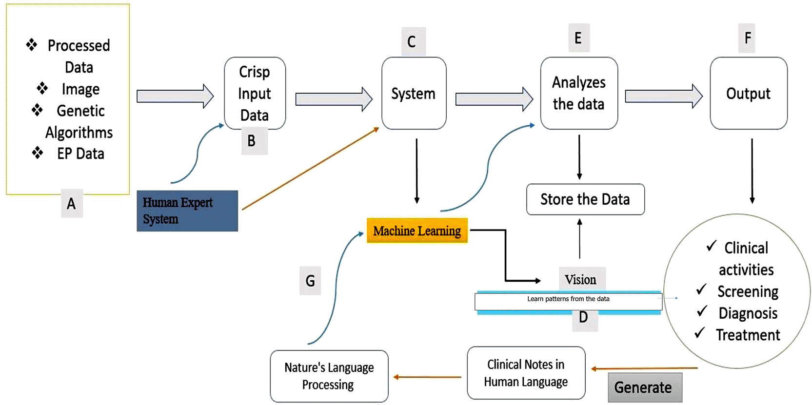
Dentistry is currently using a variety of AI tools and its branches, such as DL, ML, computer vision, cognitive computing, natural language processing (NLP), and decision trees (DT). CNN, ANN, and support vector systems (SVM) are some of the most common AI-based methods used in dentistry (Farzaad et al., 2020) ( Figure 2). AI and robotic systems, along with navigational guidance, are being employed to improve the precision and exactness of dental treatments. Robots have also helped to streamline the work process and acts as dental assistants to provide high-quality patient care. AI and robotics even facilitate dental education; help in diagnoses and treatment plans for various dental conditions like periapical lesion, dental caries, periodontal disease, tumors, cysts, and fractured restorations; designing prosthodontic and orthodontic appliances; digital planning for surgical implant placement; tooth preparation and root canal treatment (Kise et al., 2019; Mcleod and Melder, 2005; Liu et al., 2017) (Supplementary Table 1).
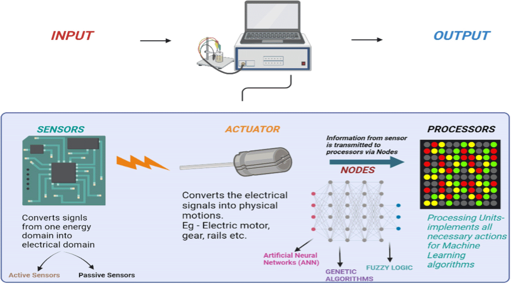
Often, the sensors required for the task are compiled into a single system referred to as an embedded system. With AI, sensor fusion has become easier and more accurate compared to classical algorithms. This allows the ANN to cope with unknown situations in a much better way. In addition, it can detect compensation techniques provided with the training data and potentially increase the value of the delivered result to the customer. The actuator is another component that helps to convert the electric signals into physical events or characteristics. They convert components of machines and energy received into motion. The actuators are only responsible for moving and controlling a system. An actuator can be an electric motor, gears, rails, etc. So, the fine line of demarcation between a sensor and an actuator is that sensors can sense physical events and convert them into electrical signals. Whereas the reverse happens in the case of an actuator. The information is finally transmitted to the processors via nodes. The processing units implement all the required actions for machine learning algorithms.
With rapid development in AI and its manifold use , it is essential to understand how AI and its components are being used in various spheres of dentistry have dentistry. Hence, this review summarizes and provide an overview of the clinical applications of AI and robotics in dentistry.
Six electronic databases, PubMed, Cochrane, Scopus, ClinicalTrials.gov, EBSCO (dentistry and open science access), and Web of Science, were used for selecting relevant articles using the following keywords: (“Artificial Intelligence”[Title/Abstract] OR “Artificial neural network”[Title/Abstract] OR ANN [Title/Abstract] OR “Convolutional Neural Network”[Title/Abstract] OR Robotics [Title/Abstract] OR Robots [Title/Abstract] OR “Deep learning network”[Title/Abstract]) AND (Dentistry [Title/Abstract] OR Dental [Title/Abstract] OR Implants [Title/Abstract]). Only articles written in English were included for extracting data. All observational studies, experimental and clinical studies in humans or animals, case reports/series (including any patient-related data or information), and in-vitro and in-vivo studies (cohort, cross-sectional, case-control) were taken into consideration. All letters, editorials, and reviews were excluded. Duplicates were eliminated after gathering the search results in the Mendeley reference manager (version 1.19.4).
Robotics, ML, DT, CNN, and ANN are some of the common components used in the dentistry he AI-based systems are being used for diagnosis using clinical data and radiographic findings, periodontal probing, patient data management, and minimally invasive surgical procedures like scaling, periodontal surgery, implant placement, extraction of teeth, and orthodontic and orthognathic procedures. AI and robotics are also being used for dental education for students and patients; interpreting dental radiographs for detecting dental caries, periodontal diseases, the pattern of bone destruction, orthodontic wire and appliance fabrication, and crown preparation. An overview of the studies and applications of AI in dentistry is discussed as follows. Various studies on the clinical role of AI and robotic in dentistry and various type of robots used in dentistry are discussed in supplementary Tables 1 and 2.
The use of robots for educating and training dentists was introduced in 1969, when phantom heads were used for training dental students for preclinical exercises (Sun et al., 2014). However, traditionally there were the static mannequin’s jaw and phantom head that did not simulate the clinical situations. Working with these systems did not provide the opportunity for employing communication skills and management of behavioral problems like apprehension, restlessness, anxiety, or managing any unwarranted movements such as coughing, vomiting, and tongue thrusting in patient may arise during treatment (Yu et al., 2015). To overcome such shortcomings, robotic patients with real-life characteristics were developed. Presently, robots with haptic interface technology, virtual reality, and advanced simulation have been developed to provide students with an opportunity to learn clinical skills before treating the real patient. Showa Hanako Tokyo’s Geminoid DK, Simiroids, and Robotutors are the common robotic systems being used for training dental students and performing simulation exercises (Hamura et al., 2011) (Supplementary Table 1). A robot that reproduces jaw movements in a comprehensive and precise manner has also been developed. This robots helps to understand the various jaw movements and their effect on occlusion and the final prosthetic outcome (Ahire et al., 2012). A robotic patient that can move its head in eight directions and can be positioned on a dental chair with the ability to mimic human-like movements was developed and was used for education and training (Liu et al., 2023). These robots even have soft tissue-like structures mimicking the structures in the oral cavity, such as the oral mucosa, tongue, uvula, and salivary secretion. They can converse with the trainee and mimic the movements, allowing students to have real-life scenarios. To mimic a vomiting reflex during clinical examination or dental procedures like impression making, the uvula was additionally equipped with a pressure sensor. In addition to “shaking neck,” “coughing,” “tongue thrusting,” and “mouth fatigue,” this model also simulates blinking, ocular, tongue movements, and mouth motions (Miller, 2007). This robotic patient can be used for practical training sessions like administering oral anesthesia, performing tooth extractions, preparing cavities, and assisting with medical emergencies (Ahmad et al., 2021; Boesecke et al., 2001; Naveen, 2012). A dental humanoid robot named SIMROID®, is another training robot that replicates human behaviour, and was used to teach students how to communicate with patients and to teach them how to manage medical emergencies. Another specific an AI system named LA-VR stimulator has also been developed primarily for learning local anesthetic techniques (Zafar et al., 2021). Kitahara et al. (2024) evaluated operator experiences utilizing a questionnaire survey after hands-on training on the SIMROID® simulator from 112 dental students. They reported that the simulation training was highly appreciated by all participants.
A home dental care model comprising 3D-printed human teeth that provided a realistic image of the dental pulp and better visualization of dental caries was proposed in 2019 for educating patients at home. By evaluating the oral image from the upper and lower quadrants/arches, the system used a DL model to gather information on whether home dental care is needed. Two evaluation criteria, tooth disease and ‘Need for Professional Dental Treatment (NPDT)’ classifications, have been used. Here the AI models using tooth pictures annotated by dentists to make a decision . The results showed that this system can detect tooth disease and provide NPDT classifications with an accuracy greater than 96% and 89%, respectively (Kim et al., 2020; Abe et al., 2018). To train students and clinicians for implant placement , Xiajoung et al. (2019) reported using a virtual reality-enabled haptic feedback system in conjunction with a model software program called CAPPOIS (Computer-assisted Pre-operative Planning for Oral Implant Surgery) (Major L et al., 2021). This specialized software simulates dental implant placement, improving accuracy and reducing risks. It enables virtual implant placement, guided surgical template fabrication, and predictable prosthetic outcomes, particularly benefiting complex cases by allowing precise, tissue-driven planning. It can help in training surgeons to improve skills by allowing them to experience the surgical procedure virtually.
AI is being utilized to plan and perform many oral and maxillofacial surgical procedures (Nieri M et al., 2010). There are many preoperative planning systems that provide surgeons remote access to perform surgery. Here the surgeon can use with an alternative AI software and an image based and an infrared light-based navigation camera to access on the patient's radiograph data supplied by the clinician to plan and perform surgery. AI and robotsare also been used for the detection of anatomic structures like salivary glands, nerves, and blood vessels; detection, segmentation, characterization, classification, and removal of tumours (Dean R et al., 2010; Nathan et al. 2006; DiMaio et al., 2011; Abdolali et al., 2016; Halicek et al., 2017; Zheng Z et al., 2021); therapeutic and selective neck dissection of maligant lesions (Park et al., 2013); and managment of obstructive sleep apnea syndrome (Lin et al., 2013; Cramer et al., 2019). Furthermore, Vicini et al. (2010) reported that transoral surgery using robotics could be performed for managing ‘obstructive sleep apnoea’. An AI-based model is even employed to detect the early malignant changes in the epithelium and connective tissue of the oral cavity (Giorgini et al., 2015). As reported by Vicini et al. (2010), the da Vinci robotics system was the first FDA-approved robotic system for transoral treatment of malignant and non-malignant lesions involving the oropharynx, tongue, and larynx. AI is also used to grade the severity of malignant lesions. For example, Krishnan et al. (2011) used AI system to categorize oral submucous fibrosis lesion into: normal tissue, tissue with ‘oral submucous fibrosis’ (OSMF) without dysplasia, and tissue with OSMF with dysplasia. In 2015, a computer-based quantitative microscope and near-infrared fluorescence, in conjunction with an AI-based system, were employed to identify the amount of keratinized tissue and the presence of ‘keratin pearls’ in histologic images to detect oral cancer with high segmentation accuracy (Das et al., 2015). A multispectral wide-field optical imaging technology and near-infrared fluorescence can differentiate cancerous and non-cancerous cells (Lu et al., 2014; Gao et al., 2018; Jeyaraj & Nadar, 2019). The CNN system, which utilizes a DL image classification system, has been used to diagnose lymph node metastasis on computed tomography images (Ariji et al., 2019). AI-based robots are also used to make incisions and perform many oral surgical procedures. Chang Seow-Wee et al. in 2013, developed an oral cancer prognosis model using a Bootstrap-ANFIS technique and observed that robotic-assisted surgical excision for primary or recurring malignancies in the oral cavity have a better functional recovery rate and recurrence-free survival. The risk of intraoperative or postoperative complications was also less with robotic surgery compared to conventional surgery or radiochemical therapy (White et al., 2010). However, the chances of postoperative hospitalization were higher due to extensive flap reflection compared to open surgery (Gkegkes et al., 2019). The various generations of robots used for surgical procedure are discussed in supplementary Table 1.
Robots have been used for performing various surgical procedures like orthognathic surgery, third molar extraction, implant surgery, and cosmetic surgery like nerve repositioing, canine exposure, lip repositioning, etc. They can avoid mistakes made by surgeons in terms of precise drilling and incisions, apart from monitoring the patient continuously (Danilchenko et al., 2011). The navigation module of the robot can detect and assess the operational performance based on which it can successfully finish the surgery with high accuracy (Grischke et al., 2020). AI-based software is also used for preoperative planning for surgical procedures. CT-Dent is one of the common AI software that uses DL technology to generate an automatic radiological diagnosis of the surgical area. The software was able to locate the position of the inferior alveolar nerve in less than 4.5 seconds and helped to plan the position, placement, and decide the accuracy, speed of implant placement, and minimize the risk of injury to the anatomical structures (Froum et al., 2021). Vinayahalingam et al. (2019) used a bootstrap-ANFIS framework to detect the location of the inferior alveolar nerve during the third molars extraction, implant placement, and maxillofacial surgery (Supplementary Table 2). In addition, a study by Zhang et al. assess facial swelling following the extraction of impacted mandibular third molars by employing an adaptive BP algorithm alongside a conjugate-graded BP method (Zhang et al., 2018). This neural network model accurately established the functional relationship at 98% accuracy between patient-related factors, surgical procedures, and anatomical considerations concerning the degree of facial swelling after the extraction of impacted mandibular third molars. Moghimi et al. (2011) utilized a hybrid genetic algorithm and ANN model to predict the size of unerupted canines and premolars during the mixed dentition phase and to explore the factors influencing treatment options for impacted canines.
AI-based software and robots can be used to design and manufacture various prosthetic solutions for the patients such as cast partial dentures, complete dentures, prepare teeth for receiving fixed denture prosthesis, and fabrication of maxillofacial prosthesis (Wang et al., 2005; Wang HY & Zhang LY, 2005; Jiang et al., 2015; Sreelekshmi et al., 2017). A CRS robot and a MOTOMAN UP6 robot, a single-manipulator and 6-degree-of-freedom, are some of the robots used for the fabrication of complete dentures (Wang et al., 2005). MOTOMAN UP6 robot has a multi-finger hand (TAMFH) comprising three fingers, with each finger having 3 degrees of freedom based on the analysis of motion, and a workplace simulation driven by 84 motors. This robot can recognize any position in space and helps in teeth arrangement (Jiang et al., 2015). However, the manipulation of artificial teeth was found to be challenging with this system due to their complex morphology of the tooth and its alignment. Consequently, a tooth-arrangement robotic system with a slipway mechanism was developed to allow placement and adjustment of teeth along all the three planes. Additionally, AI is being used to fabricate complete dentures. For example, complete denture are being manufactured using three-dimensional animation software known as “Blender” with a robotic hand (Jiang et al., 2015). A virtual 3D tooth arrangement software has also been employed to make or pick files according to the patient's case history and medical records. Based on the data and jaw measurements, 3D virtual tooth arrangements can be facilitated on the screen (Sreelekshmi et al., 2017). AI is also used to classify the morphology of of the arches and plan prosthesis. Using 1184 radiographs (maxilla: 748 images; mandible: 436 images), Takashashi et al. (2020) classified dental arches into four categories: arches with loss of posterior teeth; arches with edentulous space confined; complete edentulous; arches with intact dentition. The study utilized the Keras and TensorFlow DL model to categorize images. Once the learning process was finished, the precision, diagnostic accuracy, recall time, and area under the curve for both the maxilla and mandible were determined. The study found a 99.5% probability of categorizing the image accurately for the maxilla and 99.7% for the mandible, with no differences between the four types of mandibular dental arches. Araie et al. (2018) even developed a ‘robot denture’ for geriatric patients, without any disabilities, to help them chew and swallow their food effectively. Robots have been employed in tooth prepartion for fixed partial dentures. In 2018, a robotic system named the 'Dobot Magician tandem articulated arm' was also used for tooth preparation. This robot worked with a robotic arm having 6 degrees of freedom, a low-heat laser, CAD/CAM software, and an intraoral 3D scanning machine. These components were interconnected to generate a three-dimensional path of motion for the lasers and to plan the required shape needed for individual tooth preparation. The robot was also capable of obtaining three-dimensional information on the teeth to be prepared, the teeth in the opposite arch, and also the adjoining teeth to design, prepare, and mill the prosthesis. Furthermore, robots have been used to observe and mimic the movement of the mastication in the patient and estimate how much force is deployed during opening and closing of jaw, thereby enabling the evaluation and treatment of occlusal load and the possibility of temporomandibular joint dysfunctions (Takanobu et al., 1993). Common examples of such masticatory robots include dental masticatory robots, mouth training robots, WJ (Waseda Jaw) series robots (Alemzadeh and Raabe, 2007), SIMROID (Akiyama et al., 2013), and Dento Munch Robot Simulator (Alemzadeh and Raabe, 2007). The six-degree-of-freedom 'Dento Munch Robot Simulator' is a robotic dental testing tool that includes a compliance module and artificial jaws. It has a prosthetic jaws that are essentially reverse-engineered replicas with artificial teeth implanted that mimic the human maxilla and mandible. They can be used to mimic material wear on teeth, crowns, bridges, or implants. Another similar robotic system called “Bionic Jaw Motion” uses a robotic system with an analyzer that has a camera and an articulator to imitate the movements of the mandible (Carossa et al., 2020). ‘Waseda Asahi oral rehabilitation robot' is another robotic system developed at Waseda University in Japan for massaging the facial muscles of mastication and muscles around the salivary gland and ducts for patients with TMJ disorder, myofascial pain dysfunction, and dry mouth (Koga et al., 2008). AI models have been able to diagnose changes in the masticatory function, jaw movements, osteoarthritic changes in the temporomandibular joint and condyles using biomarkers and ML models (Bianchi et al., 2020).
AI and robotics are being utilized for case history and diagnosis, planning, and determining the progression and prognosis of orthodontic treatment (Woo SY et al., 2017). AI helps in making digital impressions for the fabrication of orthodontic appliances, straightening wires, and the fabrication of brackets, bands, and myofascial appliances (Adel et al., 2021). SureSmile, MOTOMAN UP6, Cartesian type, and LAMDA are some examples of orthodontic arch wire bending robots that use three-dimensional imaging systems and computerized techniques to customize the process of wire bending and appliance fabrication. The Cartesian-type archwire bending robots can even customize the bending process, and analyse the degree of bends can be analyzed (Palanivel et al., 2021; Zhang and Jia, 2009).
Research has confirmed that using AI/ML to detect landmarks and cephalometric points yields higher accuracy results than using conventional manual techniques. AI, CNN, and DL are also used for locating various anatomical landmarks and structures via radiographs for orthodontic planning (Park JH et al., 2019). Some of the common structures and their associated defect that can be detected via AI include the type and morphology of crown and root of the teeth; amount of tooth wear; location of fracture lines; location of CEJ; alveolar bone height and degree of bone resorption/loss; amount of tooth mobility and soft tissue (gingival) profile. AI models use less time and human labor and are more accurate thannon-specialized dentists/clinicians (residents, graduate students, and students) (Muraev et al., 2020; Lindner et al., 2016; Shahidi et al., 2013; Schwendicke et al., 2021). On the contrary, Kunz et al. (2020) in their comparative study reported no difference between AI algorithms and manual/human-based analysis for analyzing the cephalometric radiographs. Muraev et al. (2020) observed that 94% ANNs could predict the requirement of tooth extraction for orthodontic treatment. The determination of the pattern of extraction showed an accuracy of 84.2%, while the determination for the anchorage pattern was found to be 92.8% (Thanathornwong, 2018). AI can aid in the selection of an effective appliance or headgear for correcting the given amount of space and predicting the treatment outcomes (Bahaa et al., 2011). AI system was found to select the appropriate headgear with an accuracy of 95.6% (Akcam and Takada, 2002). ANNs may also anticipate treatment outcomes, evaluate the patient's future profile, and identify whether orthognathic surgery is necessary (Denadai et al., 2019). These outcomes were found to be equivalent to recommendations made by orthodontists (Lahaoud et al., 2021). An AI-driven tooth segmentation system to assist the orthodontists for visualizing, assessing, and predicting the presence of physical growth and development anomalies during craniofacial growth is also available (Lahoud et al., 2021). This model facilitated the determination of reactive sites and the optimal course of action for the treatment of malocclusion. In many clincial situations, such as patients with Class III malocclusion (hyper-mandibular, hyper-divergent, or balanced categories) can have their results predicted by AI-systems (Auconi et al., 2015; Auconi et al., 2011).
ANN is used to predict facial appearance and age perception following orthognathic surgery (Denadai et al., 2019). Research indicates that the application of AI in orthognathic surgery resulted in 66.4% of patients appearing younger following therapy, especially following profile-altering surgery. 74.7% of patients showed comparable gains in their facial attractiveness, particularly following lower jaw surgery (Naran et al., 2018; Meyer and associates (2010). A Bayesian Network and ML algorithm that utilizes the cone-beam computed tomography (CBCT) images have also been used to successfully diagnose the degree of maxillary canine impaction. This enabled the determination of the required amount of palatal expansion and the type of appliance needed (Suri & Taneja, 2008). Additionally, ANN and DL models could predict the skeletal bone age automatically with a mean difference of ±0.8 years between humans. Furthermore, ANN has been used to determine cervical vertebrae maturation and plan the time for orthodontic procedure(Kok et al., 2021).
AI-assisted models are being used to detect and classify dental restorations, carious teeth, and periapical and endodontic lesions; diagnose aesthetically compromised teeth, fractured teeth, and restorations; determine prognosis of teeth; and plan treatment (Al Haidan et al., 2014; Kang et al., 2002; Wenzel, 2001; Devito et al., 2008; Gakenheimer, 2002; Wenzel et al., 2002; Wang et al., 2016; Ekert et al., 2019; Lee JH et al., 2018b; Lee et al., 2020; Aminoshariae et al., 2021; Jokanovic et al., 2022; Revilla-Leon M et al., 2022) (Supplementary Table 2). Computer-based programs and AI-based models have shown to have higher accuracy in detecting caries (39.4%) compared to dental clinicians. Recently, to detect caries an automated caries detection system (BPNN) by using the image texture, intensity, and gradient direction in radiographs was developed (Rad AE et al., 2018). The BPNN system demonstrated 91.33% accuracy in detecting caries, with a 5% level of significance. The ‘Logicon Caries Detector™ program (Logicon Inc., USA) is an automated caries detection model used for detecting and classifying caries on the proximal surface (Kang et al., 2002). This model enabled dentists to identify 20% more cavities extending into the dentin compared to traditional methods. Wenzel et al. (2002), Devito et al. (2008), and Gakenheimer (2002) also developed a more sophisticated caries detection model by using two-dimensional images (intra-oral periapical radiographs) from extracted teeth. Their findings indicated that an AI system could detect caries with higher accuracy. AI based model csn detect caries that even experienced dentists find it challenging to identify. The minor changes in dental X-rays that could signal the initial phases of dental caries using conventional technologies can be detected for early intervention.
AI-based machines/models and robotic systems are also used in cavity preparation for carious teeth, treatment of fractured teeth, and performing root canal treatment (Wenzel et al., 2002). A computer-controlled and supervised microrobot is also used to complete the biomechanical preparation of the root canals (Rawtiya et al., 2014). This helps the practitioner in performing an automated drilling, filling, cleaning, and probing for the canals during endodontic treatment. A compact structure, specialized workspace, and micro millimeter-scale motion of these robots provide a safer and more automated drilling experience (Mittal et al., 2013). CNNs/ANNs are commonly used to understand the root canal anatomy, locate the apical foramen, determine the working length, detect the number and curvature of the roots and canals in the tooth, locate vertical root fracture. It can even predict and understand the 3-dimensional modification after instrumentation, identify the degree and extent of endodontic lesions, and predict if the pulpal stem cells are viable (Bindal et al.,1999; Saghiri MA et al., 2012a, b; Johari et al., 2017; Fukuda et al., 2020; Hiraiwa et al., 2019). Several studies have demonstrated the effectiveness of the DL algorithm in differentiating periapical cysts from other cysts, including dentigerous cysts and keratocysts (Flores et al., 2009; Orhan et al., 2020). AI is also useful for predicting the need for retreatment and determining the chances of failure during retreatment (Rawtiya et al., 2014; Turky M and Dummer PMH, 2025). AI-based models have better accuracy when compared to an experienced endodontist (95% vs. 76%) (Saghiri et al., 2012a, b). Recently, micro-robots have been used for biomechanical preparation, where they have shown catalytic ability to disrupt oral biofilms formed inside the pulpal canals of the teeth (Hwang et al., 2019) (Supplementary Table 2).
AI subsets like ANN, DL, CNN, fuzzy cognitive machines (FCM), fuzzy expert systems (FES), and genetic algorithms are being employed in periodontology for patient education, data collection, and segregation, diagnosis, and treatment planning (Allahverdi et al., 2011; Mago et al., 2014; Lee JH et al., 2018a). CNN and ANN-based models are employed for the diagnosis of periodontal diseases based on clinical findings, radiographic data, and risk factors (Thakur and Akter, 2018). A home dental care system was created by Kim et al. (2023) and included DL models for images of the mandibular and maxillary teeth to categorize the dental conditions in teeth and also to check if a professional dental treatment is necessary. Furthermore, a specialized oral image acquisition tool was created to capture images of the mandibular and maxillary teeth. Around 5251 intraoral images were checked and marked by a dentist with only a bachelor's degree in dentistry and others with a doctoral degree in dental medicine. Both dentists were asked to check for the tooth with disease and classify the teeth where professional dental treatment was needed. The suggested system demonstrated accuracies of over 96% and 89% in the dental disease and professional dental treatment categories, respectively. These findings support our belief that the suggested approach will enable users to efficiently maintain their oral health by identifying dental conditions and informing them when dental care is necessary. AI-based models have been utilized to simulate the ability of clinicians to determine the extent of alveolar bone loss utilizing radiographs with high accuracy. Studies have demonstrated that a CNN-based DL can identify healthy images with an accuracy of 98% and detect the presence of periodontitis. AI-based systems can also detect the depth of periodontal pockets by performing periodontal probing and assessing the severity and nature of the bony defect (Lin et al., 2017). Wavelet transforms and pattern classification techniques have been employed by AI for the evaluation of pocket depth. The 5th generation periodontal probe, which utilizes an ultrasonic wave, has been integrated with AI for both pattern classification and image classification techniques to assess the depth of the periodontal pocket or sulcus (Bertoncini & Hinders, 2010). These AI-based pattern classification methods can accurately identify 86.6% of periodontal pocket depths (Acosta & Hinders, 2020).
AI models are also being used to assess the degree and thicknes (phenotype) of gingival tissue around teeth and implants. Yang et al. (2023), in a pilot animal study, used three-dimensional gingival soft tissue reconstruction and CBCT images to determine the nature of the gingival phenotype. The gingival phenotype (thickness) was clinically assessed via a periodontal probe and digital calliper, and compared with the digital estimation by DL-based CBCT. The authors found a positive correlation between the dentists and the DL model for periodontal pocketassessment. However, a greater agreement was observed between the virtual and clinical measurements for the buccal sites (0.019 ± 0.233 mm) than the lingual sites (0.116 ± 0.215 mm). Chifor et al. (2022) also tried using 3-dimensional ultrasound for periodontal tissue (pocket depth and gingival tissue thickness) analysis using RR-CNN and U-Net-CNN models. CNN could effectively detect both the depth of the periodontal pocket (10%) and the gingiva thickness (75.6%), and this ability to measure the PD was linked to two techniques (wavelet transforms and pattern classification). Studies have shown that with pattern classification, 86.6% of the PD can be predicted effectively, and AI-assisted periodontal probing could provides faster and more reliable pocket charting for dentists. The 5th generation periodontal probe that uses ultrasonic waves to detect PD is also based on the pattern and image classification system of AI (Laugisch et al., 2021). The periodontal probing system has also undergone significant advancement, as evidenced by several studies conducted by Ernst et al. (1997), Lee et al. (2018a), and Yauney et al. (2019) (Supplementary Table 2). A recent clinical study compared the efficacy of a manual periodontal probe to an AI-based periodontal probe (PA-ON Parometer, Germany) and found that the average pocket depth varies significantly between the manual and PA-ON probe (mean difference: 0.38 mm; p < 0.001). The time required for the PA-ON probe was longer (23 ± 11 min) compared to the manual probe (21 ± 11 min), with a p-value < 0.05. The pain score showed statistically significant less for the PA-ON probe compared to the manual probe (Sukegawa et al., 2020).
AI has been used to evaluate various immunological parameters of inflammation, differentiate between different forms of gingivitis and periodontitis, predict the recurrence of oral lesions such as aphthous ulcers, and identify and classify oral malodor or halitosis (Ozden et al., 2015). DT, SVM, and ANN models with propagation algorithms interpret radiographic findings and are also being used for training students to classify periodontal disease (Ozden et al., 2015; Farzaad et al., 2020; Krois et al., 2019). The ANN model demonstrated the lowest accuracy among other AI models, with a score of only 46%. Wen et al. presented a novel AI model using the ‘particle swarm optimization neural network (PSONN)’ and ‘multichannel grey levels occurrence matrix (MGLCM)’ for diagnosing gingivitis. This model was designed to diagnose the disease by differentiating shades of grey using an input data set derived from healthy and inflamed gingiva images. The MGLCM demonstrated a true negative value, true positive value, precision, F1 score, and accuracy of 78.17%, 78.23%, 78.24%, 78.17%, and 78.20%, respectively. ANNs were also employed to classify patients into either aggressive periodontitis or chronic periodontitis with an accuracy rate of between 90% and 98%.The FES has also been employed by AI models to make decisions regarding mobile teeth, based on signs/ symptoms and patient related factors such as pain, infection severity, bone loss, occlusion-related trauma, misalignment, smoking, pregnancy status, oral hygiene status, and hormonal changes in patients (Mago et al., 2011). The software utilized inputs that ranged from 0 to 1, and several functions were employed to produce the expression for each. The ‘IF-THEN’ rule and the ‘Mamdani algorithm-based Fuzzy Inference System’ mechanism were employed. The results demonstrated that the software is capable of accurately predicting periodontal disease with a 94.1% success rate (Tatabatei et al., 2017). CNN models that were trained with a few radiographic images exhibited comparable discrimination abilities to those of dentists for assessing bone loss using orthopantographs. ANN models have been proposed for the diagnosis and classification of aggressive and chronic periodontitis based on blood tests, immunoglobulin levels, and serum antibodies (Papantonopoulos et al., 2014). These models are consisdered to be useful for future studies on periodontal disease diagnosis and classification, provided that other environmental, behavioural, patient-related, personal, clinical, and radiological factors are considered. Immunological markers of periodontitis, such as the CD3, CD4/CD8 ratio, lymphocyte, eosinophil, monocyte, neutrophil count, IL-1, IL-2, IL-4, interferon-gamma, and TNF-α levels have been used in AI tools/models for classification and diagnosis. The antibody levels against Aggregatibacter actinomycetemcomitans and Porphyromonas gingivalis have also been identified using ANN models. As reported by Thakur and Akter (2018), the ANN provided an accurate estimate by considering the levels of CD3, monocyte, eosinophil, neutrophil, and CD4/CD8 ratio. Based on the levels of these immunologic and inflammatory mediators, the severity of the disease can be predicted. Bacterial count in saliva has also been used by AI to predict periodontal disease. A study used salivary bacterial copies from 144 healthy controls and 548 patients with generalized chronic periodontitis were taken to assess the accuracy and dependability of ML models to predict the severity of chronic periodontitis (Kim et al., 2020). After the genomic DNA was isolated, multiplex qPCR was used to measure levels of the following pathogens: Aggregatibacter actinomycetemcomitans, Campylobacter rectus, Eikenella corrodens, Fusobacterium intermedia, Fusobacterium nucleatum, Peptostreptococcus anaerobius, Tannerella forsythia, and Treponema denticola. Based on the red-complex pathogens, the model was able to categorize periodontitis into two groups: healthy, moderate or severe (Ernst et al., 1997; Lee et al, 2018b; Yauney et al., 2019) (Supplementary Table 2). Yauney et al. in 2019 conducted a study using a ‘fluorescent porphyrin biomarker for intra-oral imaging’, along with ML and chair-side periodontal examination. The study correlated the periodontal status with systemic conditions (body mass index, neurological, cardiac rhythm, blood pressure, and evaluation of the tympanic membrane and optic nerve). Gingivitis and periodontitis were found to be associated with abnormalities in the optic nerve. A significant correlation between periodontitis and a family history of eye disease and swollen joints was made. AI-based models are also used for identifying genetic variants for periodontal disease. Ning et al. (2021) analyzed periodontitis-related transcriptomic datasets and immunosuppression genes by using AI models: ‘Dis-Ge-NET and Hisg-Atlas’. The authors also identified differentially expressed genes, transcription factors, and various signaling pathways, which are involved in periodontitis, using bioinformatics analysis. The SVM classifier was used to identify three “master” immunosuppression genes (FOS, FCGR3A, PECAM1). The following transcription factors: FOS, NFKB1, JUN, STAT4, STAT5B, and HIF1A were also identified.
AI is also used to predict the prognosis and calculate the risk for disease. Studies by Sandberg & Fors (2007), Persson et al. (2003), and Page et al. (2005) found that a computerized risk assessment tool for dental diseases was more effective than the judgment of experienced senior dental specialists. For instance, the ‘Periodontal Risk Calculator’ tool can calculate the risk for periodontal disease initiation in healthy patients and is capable of identifying certain variables as potential risk factors for the initiation and progression of periodontal disease. By utilizing algorithms, it can assign a relative weight to these risk factors, which are known to increase the likelihood of developing periodontitis. The variables employed in mathematical algorithms that calculate risk include the patient's age, history of smoking, presence of diabetes mellitus, previous history of periodontal surgery, depth of periodontal pocket, degree of furcation defect, nature of restorations, presence of subgingival calculus, height of bone, and presence of angular bony defects in radiographs (Wen et al., 2020). SVM, ANN, and DL systems have been employed to categorize halitosis based on the microorganisms present in saliva. SVM classifiers have been demonstrated to provide the most accurate classification system among others (Kaloumenou et al., 2022). SVM is employed for the screening of saliva for oral malodor based on the presence of compounds such as methyl or ethyl mercaptan (Supplementary Table 2). AI-based applications even predict the intensity of halitosis by analyzing tongue images and providing the users with a history of their assessments. DL models are also used to measure the bone level, and based on the bone loss. Lee et al. (2022) compared the accuracy of a DL model for detection of bone loss compared with a dentist found no significant difference for the level of bone loss (p = 0.65). The area under the receiver operating characteristic curve of RBL stage assignment for stages I, II, and III was 0.89, 0.90, and 0.90, respectively. The accuracy of the case diagnosis was 0.85. The author concluded that DL model was found to be effective in measuring bone loss measurements and image-based periodontal diagnosis using periapical radiographic images. The accuracy of the model to detect bone loss was found to be 79.54%. Karacaoglu et al. (2023) used DL models to detect radiographic bone level (or CEJ level) as a basic structure for automatically diagnosing and staging bone loss in periodontal disease. They found that, compared to the gold standard, both human observers and ML had significantly different abilities to detect periodontal defects (p = 0.05). However, ML and human observers showed similar performance in detecting these defects, with no significant difference (p = 0.05). This approach could assist dentists in diagnosing and systematically monitoring periodontitis on panoramic X-rays or radiographs, potentially improving the diagnostic accuracy and aiding in the treatment of periodontitis patients.
Micro and nanosized robots have been tested for the removal of oral biofilm and for maintaining effective oral hygiene (Panagakos and Migliorati, 2014). Catalytic Antimicrobial Robots (CARS) is a magnetically driven robot that can accurately kill, degrade, and remove biofilm and bacterial free radicals. These nanorobots metabolize the trapped organic matter, preventing the buildup of calculus, tooth decay, and halitosis (Hwang et al., 2019). Villa et al. (2020) developed a self-propelling microrobot that could break the biofilm and have high antibacterial activity. Such microrobots use low concentrations of fuel for their propulsion, and they achieve an efficient dental biofilm disruption within five minutes. The microrobots are also biocompatible with the epidermal cells and activate the immune response, which helped to combat the microbes. Micro- and nanoscale robots are used as local drug delivery agents for treating dental hypersensitivity and for delivering local anesthetics and antimicrobial agents into periodontal pockets (Jain et al., 2013; Jain et al., 2020; Zhang et al., 2022). Nanorobots measuring 1–10 μm in size could be delivered sub-gingivally, either as a mouthwash or toothpaste (Lang et al., 2014). These nanorobotic dentifrices were found to crawl at a speed of 1–10 μ/s within the oral cavity. They helped in scanning, recognizing, and eliminating pathogenic bacteria present in the plaque and oral cavity by metabolizing the trapped organic matter into odorless vapors. These nanorobots, have been incorporated into dentifrices, named as ‘denti-fobots’ (Lang et al., 2014). A mouthwash containing these smart nanorobots can identify the areas where biofilm was present and to remove the pathogenic bacteria. This allows the commensal bacteria to flourish within the oral ecosystem. Additionally, these robots can remove particles of food, plaque, or calculus from the teeth, interdental areas and restorations as they can penetrate remote surfaces that are difficult to reach with tooth brush bristles/ fibers, or floss. Nanorobots can also deliver a drug, photosensitizer or dye into the periodontal pocket, which may be activated with a light source of a specific wavelength (805 nm) to provide antimicrobial therapy and help control periodontal inflammation. Nanorobots with pulsating laser ablation-like ability are employed for local drug and gene delivery purposes (Zhou et al., 2021; Park et al., 2020).
Robots can be employed in-vitro settings assess the efficacy of a toothbrushing and patient education (Park et al., 2020). AI is also used for plaque removal using AI-based tooth brushes/tools. A robotic hand brushing may replace manual clinical hand brushing for plaque removal. And this may be useful for patients who lack manual dexterity, geriatric, mentally challenged and hospitalized patients. (Grischke et al., 2020). Sakaeda et al. (2017) developed the first robotic toothbrush with an eccentric arm that moves the brushes eccentrically along the row of teeth and helps to automatically clean the tooth surface. Ara, an AI toothbrush, can record the brushing frequency, duration, and effectiveness of plaque removal. This brush can provide a digital readout and weekly emails containing brushing parameters and an analysis of plaque control. Amabrush is another AI-based toothbrush that utilizes the ‘Bass technique’ to simultaneously brush all teeth. It comprises two parts: a motor that provides vibrations and acts as an actuator, and brushes that are like a mouthpiece (Acherkouk et al., 2022). In 2021, another prototype of a robot-assisted brush was developed with the ability to brush in a circular movements (Fones method). The toothbrush's power and ability to brush in multiple directions facilitated effective plaque removal (Liu et al., 2023). Oral-B iO is another commerically available electric toothbrush that employs AI and a linear magnetic drive, along with a smart pressure sensor and enables it to provide pressure in the range of 0.8 N to 2.5 N. This brush utilizes oscillating rotating vibrations that are transmitted to the bristle tip, thereby facilitating a smooth brushing experience and improving patient compliance. The sensor modifies the light according to the force applied, thereby guiding the user to maintain an optimal pressure while brushing (Adam, 2020). Furthermore, users can utilize a compatible ‘Oral-B iO app’ that is connected to their phones via bluetooth and guides them and provides feedback via brushing. The new 'GENIUS X' is another intelligent toothbrush incorporating AI technology to monitor the areas of the oral cavity that are being brushed. It helps generating personalized feedback via the Oral-B app, which in turn educates the patient regarding their brushing practices. Tonetti MS et al., in 2020, evaluated the role of AI-toothbrushes during supportive periodontal care and reported AI users have lower plaque scores at each recall visit. The use of AI toothbrushes improved the overall oral health and hygiene of the patients.
AI and robots have been employed for the diagnosis, planning, and surgical placement of implants (Miller, 2007; Rawal S, 2022; Yan B et al., 2022; Liang SZ et al., 2021; Bai SZ et al., 2021). AI-based software have enabled clinicians to assess the degree of osseointegration, identify type of implant placed, future for peri-implantitis risk, location of implant and type of implant to be used, identify the presence of implant fracture, and even diagnosis the presence of bone loss around implant and the extent of peri-implantitis, and the prognosis of the implant (Miller, 2007). Robotic arms are being used for implant planning and placement ( Figure 3). The preoperative planning system in the robot allows the clinicians to view surgical sites via scanned CT images. Many AI-based tools and robots use surgical navigation sub-system with an infrared navigation camera and ensures that the implant is placed precisely during surgery. The positioning of implants can be accomplished in the specified and planned location with, real-time guided position as the system can check the depth and orientation of the implant drills (Tanzawa et al., 2012). Robots with five degrees of freedom (two rotational and three translational movements) have demonstrated translation accuracy of up to +4 mm and rotational accuracy of ±0.15° for placing the implants (Yuan et al., 2018) (Supplementary Table 2).
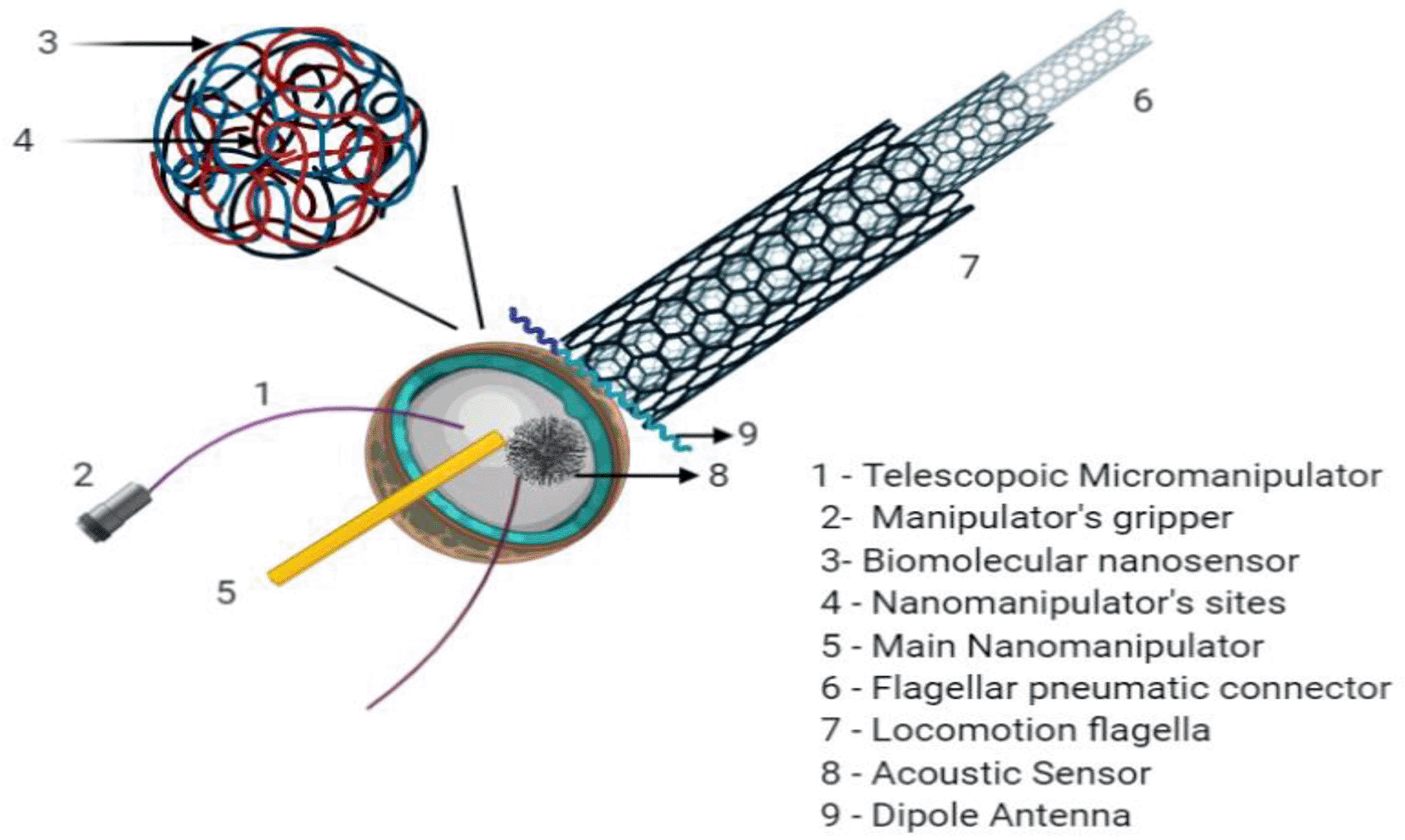
Their design is derived from biological models and may include molecular computers, onboard sensors, actuators, motors, manipulators, and power supplies. The chief elements used to prepare nanoscale gears and other nano-components include carbon, sulphur, hydrogen, oxygen, and fluoride.
Boesecke et al. (2001) were the first to test the role of robotic surgery for implant placement. Initially, a six-degree-of-freedom autonomous system for implanting endosseous dental implants was developed, followed by a three-degree-of-freedom robotic system equipped with a stereo camera. This system was utilized to orient the handpiece and ensure precise implant insertion with the appropriate torque (Miller, 2007). With the introduction of an autonomous implant placement device by Zhao et al. (2018), dental procedures might be carried out without the dentist's involvement. In 2017, the FDA was the first to approve the use of a ‘computerized robotic navigation system called YOMITM (Neocis, Florida, United States of America) for implant surgery (Tanzawa et al., 2012). The Yomi robotic guidance system is another robotic system that employs haptic feedback to provide surgeons with directional and proportional guidance through the robotic guide arm during implant placement. The surgeon can fully control the implant drilling process and can alter the orientation and surgical strategy at any point. The surgeon can also switch from robotic to freehand insertion during surgery. Without the need for additional surgical guides, the Yomi system tracks and directs the drill's depth, orientation, and location, preventing operator error. The navigation system could prepare implant osteotomies with high predictability and precision. It also provided a vibrational feedback and could identify various anatomical structures and landmarks,if encountered during surgery.
Another automatic robot system for dental implants, jointly developed by Beijing University and the Fourth Military Medical University in China, was found to be effective in the placement of zygomatic implants within resorbed alveolar ridges and edentulous maxillae (Wu et al., 2019). A study by Cheng et al. (2021) tested the accuracy of HRCDIS (Human Robotic Collaborative Dental Implant System) in positioning during implant placement. The authors found discrepancies between the intended and actual implant positions: The central deviation at the hex and apex was found to be 0.79 ± 0.17 mm and 1.26 ± 0.27 mm, respectively. The mean horizontal deviation at the hex was found to be 0.61 ± 0.19 mm, and at the apex was 0.91 ± 0.5 mm. The vertical deviation at the hex was 0.38 ± 0.17 mm and at the apex 0.37 ± 0.20 mm. The deviation from the angle of placement was found to be 3.77 ± 1.57 degrees. Mozer et al. (2020) in their study found no difference between the deviations from pre-surgical planning software and static CAD/CAM stereolithographic surgical guides. However, additional variables such as the density of bone, the time when the implant was placed, and the nature of flaps raised determined the accuracy of implant placement. Huang et al. (2021) also evaluated the impact of implant location (maxilla/mandible), protocol for implant placement (delayed/immediate), and surgical technique used for flap reflection (flapless/flap reflection ) during guided implant placement. The authors demonstrated that the accuracy was comparable at the apex, coronal, and angulation levels for the maxilla and mandible. The results were also comparable for both immediate and delayed implants, open flap placement versus the flapless technique. However, a significant discrepancy in depth was observed between the maxillary and mandibular implant sites (p < 0.05). In addition, various factors such as the tooth type, amount of spacing or distance from the adjacent teeth, number of implants placed, implant length, mode of guiding the implant, support taken from the surgical guide, quantity of anchoring pins attached to the guide, and the presence or absence of a reinforcement structure influence the precision of the guided implant placement. Guentsch et al. (2022) investigated the accuracy of guided implant surgery with either closed or open sleeves. Twenty implants were placed using a surgical guide, partially and surgery was partially guided and partially freehand. A comparison was made between the planned position of the implants and the actual position of the implants achieved after the surgery. The results showed that with an increase in the distance between the sleeve of the implant drill and the bone, the fully-guided implant group showed a decline in accuracy, with a more pronounced reduction in accuracy for the open sleeve group compared to the closed sleeve group. The accuracy was minimal for the freehand implant surgery group. The accuracy of the implant surgery that was guided partially was superior to that of freehand placement; however, it was inferior to that of the fully guided groups. The proximity of the implant sleeve to the bone was found to be a critical factor affecting the accuracy and precision of computer-guided implant surgery, for both the closed-system and open sleeve implant surgery. Surgery involving partial guidance with the utilization of a pilot drill with a static guide was found to be less precise than both fully guided and freehand implant surgery protocols.
The prognosis and success rates of implantation have been predicted using AI models. These prediction models are produced by complex correlations of demographic, radiological, and clinical factors (Wua et al., 2021). The utilization of intraoral periapical radiographs and panoramic images enables the application of AI-based CNN and K-nearest neighbor models, which are capable of identifying and classifying dental implants with an accuracy of 93.8% to 98% (Li et al., 2019; Revilla-León et al., 2023). Furthermore, AI models are effective in assessing osseointegration around an implant, with an accuracy of 62.4% to 80.5%. Furthermore, these models can be utilized to select implant designs that enhance osseointegration potential and reduce the degree of stress at the bone-to-implant interface. A study by Révilla-León et al. (2020; 2023) found that the degree of which the implant osseointegrate and its prognosis depend on many variables such the age, gender, lifestyle, physical makeup and conditions of the oral cavity, quality and quantity of the bone receiving the implant, need for bone grafting at the implant site, type of prosthesis provided after placement (Esposito et al., 2010). Papantonopoulos et al. has created an implant map to identify two distinct implant types and assess the risk of peri-implantitis by evaluating three factors related to the implant: length, thread length, and the pitch. They conducted a study and used clinical and radiographic data collected from 72 patients (N=237 implants), where the implant had a mean year of function of 7.4 ± 3.5. The authors used principal component analysis to reduce variables for ensemble selection (ES) and applied SVM for network analysis. They found a 96% of implants were prone to peri-implantitis. The group susceptible to peri-implantitis had an average bone loss of 5.2 mm, and this group included patients in whom implants were placed in the mandibular anterior region and in patients with an average of eight teeth remaining. Factors such as the total number of teeth present in the arch, oral hygiene status as measured by the full-mouth plaque scores, nature of the implant surface, severity of periodontitis, age of the patient, and the glycemic control significantly influenced the outcomes (Papantonopoulos et al., 2017). AI algorithms have also been utilized to estimate the stress at the bone-to-implant contact and for optimizing the implant design (Li et al., 2019). In 2022, Roy et al. used the ANN and genetic algorithms to optimize which type of implant should be placed based on the porosity, length, and diameter of the implant. In a similar study, Zaw et al. (2009) presented an AI method that could precisely determine the implant-bone interface's elastic modulus with micro-robotized dental implant surfaces and found that micro-robotized models enhance the degree of bone-to-implant contact and connection between the bone and implant (Liang et al., 2021).
AI has also been used for the assessment of peri-implant bone loss compared to dentists. Vera et al. (2023) developed an image-processing method using DL techniques to help dental professionals in defining the severity of bone loss around the margins of the implants. A DL (YOLOv3) model was used to identify the prosthesis and implant, along with the changes in the intensity of bone and intersections between the crown and the implant screw. The DL model was able to detect the marginal bone loss around implants with less deviation in pixels. CNN models were found to be precise and accurate in detecting bone loss around a dental implant. The R-CNN model can detect landmarks around dental implants and help assess the severity of peri-implantitis. The study found no statistically significant difference between the modified R-CNN model and dental clinicians (Cha et al., 2021). Wang et al investigated a group of peri-implantitis patients who were planned to undergo regenerative therapy and performed a comprehensive clinical examination along with immunological and microbial profiling using an ML model to check the immune profile. The results showed that an unsupervised ML model can identify individuals with distinct microbial colonization and immune profiles, and predict the outcomes of the regenerative therapy. Individuals with low risk exhibit lower B-cell infiltration and increased levels of M1 to M2 macrophage ratios. The immune profile for low-risk individuals also showed an increase in levels of complement and Th1/Th17 cytokines. The levels of two periodontal pathogens, Prevotella intermedia and Fusobacterium nucleatum, were also found to increase in individuals with high risk. Hence, ML models can be used to predict the signatures of immune and microbiological markers, and this could be useful for predicting peri-implantitis (Wang et al., 2021). A comprehensive evaluation revealed no significant differences between the various ‘computer-aided implant surgery systems. The findings indicated that all dynamic ‘computer-aided implant surgery’ (CAS) systems exhibited superior performance in terms of accuracy when compared with static CAS methods. An increased accuracy for the implant system was observed for dynamic CAS systems in comparison to freehand implant placement, apart from a reduced angular deviation in comparison to static CAS systems (Jorba-García et al. 2021).
AI has been employed to identify the gender of any missing or deceased individual by utilizing a DL Algorithms. Atas I et al. (2002) presented a DL technique that utilizes the pre-trained program known as “DenseNet121.” With the use of panoramic dental X-ray images at various resolutions, this program can distinguish between male and female genders. Additionally, it was found that the mandibular circumference and teeth are significant areas considered when determining an individual's gender. Anic-Milosevic et al. (2023) investigated the anterior bolton ratio and permanent canine dimensions in both sexes. Additionally, a statistical model was developed that can determine the sex of an individual whose sex is unknown. All odontometric variables exhibited sex-specific differences, and a sex prediction ANN model was created with an accuracy of more than 80%.
AI and robotics have revolutionized the various specialties in dentistry. The use of robotics and AI-based software assists dentists in providing quicker, more precise, and specialized dental care. Even though AI and robots are thought to replace humans in multiple professions, robots can never replace the emotional and humane touch of a clinician. There will always be a need for professional human judgment and expertise in creating and designing such algorithms and interpreting reports generated by AI technologies. AI and robots should remain as an adjunct to provide better dental care and never replace the traditional patient-doctor relationship.
| Views | Downloads | |
|---|---|---|
| F1000Research | - | - |
|
PubMed Central
Data from PMC are received and updated monthly.
|
- | - |
Provide sufficient details of any financial or non-financial competing interests to enable users to assess whether your comments might lead a reasonable person to question your impartiality. Consider the following examples, but note that this is not an exhaustive list:
Sign up for content alerts and receive a weekly or monthly email with all newly published articles
Already registered? Sign in
The email address should be the one you originally registered with F1000.
You registered with F1000 via Google, so we cannot reset your password.
To sign in, please click here.
If you still need help with your Google account password, please click here.
You registered with F1000 via Facebook, so we cannot reset your password.
To sign in, please click here.
If you still need help with your Facebook account password, please click here.
If your email address is registered with us, we will email you instructions to reset your password.
If you think you should have received this email but it has not arrived, please check your spam filters and/or contact for further assistance.
Comments on this article Comments (0)