Keywords
Rare, bronchiolar squamous cell metaplasia, tuberculosis
Rare, bronchiolar squamous cell metaplasia, tuberculosis
Squamous cell metaplasia (SCM) is a reversible adaptive process in which the normal respiratory epithelium is replaced by stratified squamous epithelium in response to chronic injury or irritation.1,2 This transformation is characterized by the loss of specialized features inherent to the native epithelium, such as ciliation and mucociliary clearance. While this results in a structurally more resilient lining, it produces a functionally less specialized barrier.3
At the cellular level, SCM reflects epithelial plasticity, a process by which differentiated airway epithelial cells revert to progenitor-like states before redifferentiation into an alternative lineage.4 This capacity for lineage programming facilitates the adaptive response of the airway epithelium to persistent environmental or inflammatory stress.4
The induction of SCM is primarily driven by chronic irritation and persistent inflammatory stimuli, including exposure to environmental toxins, mechanical stress and infectious agents.1
These factors disrupt normal epithelial homeostasis and trigger adaptive remodeling of the airway lining. Sustained inflammatory signaling promotes repetitive cycles of injury and repair, favoring a phenotypic shift toward a protective squamous morphology.1,2 Chronic inflammatory conditions further drive this plasticity through continuous regeneration, facilitating the aberration of airway epithelial cells.1,5 This process is mediated by inflammatory cytokines and alterations in the local microenvironment that disrupt standard differentiation pathways, ultimately resulting in the replacement of the native epithelium.1
In the context of infectious diseases, persistent inflammation plays a central role in inducing such epithelial changes. Chronic respiratory tract infections create a pro-inflammatory milieu that promotes metaplastic transformation as an adaptive response to ongoing tissue injury.1,5 At the molecular level, persistent bronchial inflammation activates TGF-β mediated signaling, which reprograms airway epithelial progenitors away from ciliated cell differentiation and toward a metaplastic, squamous-like phenotype. This is evidenced by the impaired generation of ciliated cells in chronic obstructive airway disease.3
SCM occurs most commonly in regions of the respiratory tract exposed to persistent environmental or inflammatory stimuli, particularly the larger conducting airways such as the bronchi. Repeated exposure to inflammatory triggers disrupts epithelial integrity and promotes the development of stratified squamous epithelium as an adaptive protective mechanism.2,5
Consequently, SCM is characteristically observed in the bronchi rather than the bronchioles or alveolar spaces, reflecting site-specific vulnerability within the respiratory tree.5 Furthermore, SCM is considered a premalignant change in certain contexts, as prolonged exposure to injurious stimuli and chronic inflammation can drive progression to dysplasia and potentially, squamous cell carcinoma.1 At the tissue level, these alterations reflect an adaptive yet potentially maladaptive response, where immediate structural protection is achieved at the expense of specialized physiological functions, leading to both structural and functional compromise of the respiratory system.3,5
Chronic inflammatory conditions, such as pulmonary tuberculosis, establish a persistent microenvironment characterized by chronic immune activation, localized cytokine release and repeated cycles of epithelial injury and repair.5,6 The host immune response to Mycobacterium Tuberculosis (Mtb) triggers granulomatous inflammation, a process heavily mediated by a complex network of pro-inflammatory mediators.5,6 Specifically, Mtb induces the secretion of key cytokines which include INF-γ, IL-1, IL-2, IL-12 and TNF which drive parenchymal inflammation.7,8 Additionally, elevated serum levels of TLR2, IL-6, IL-17, and IL-22 have been documented in patients with both tuberculosis and pulmonary malignancies.6,8 Pro-inflammatory mediators such as TNF and IL-6 may further promote metaplastic transformation by upregulating anti-apoptotic gene expression via NF-κB signaling pathway.8 This sustained inflammatory milieu disrupts homeostatic epithelial maintenance and promotes adaptive remodeling of the airway epithelial lining. The synergy of continuous epithelial insult and altered differentiation cues facilitates cellular plasticity and lineage reprogramming.1,2,5,6
A 37-year-old Caucasian female presented with a progressive, long standing pulmonary nodule in the right upper lobe. Serial PET/CT imaging revealed an enlarging 5 × 4 mm dorsal lesion with associated fibrous tracking and adjacent micronodules, though no hypermetabolic lymphadenopathy was detected. Clinical examination and laboratory profiles, including hematological and biochemical markers were within normal limits. The patient underwent video-assisted thoracoscopic surgery (VATS) for wedge resection of segments S1 and S2, during which a necrotic nodule of less than 1 cm in diameter and multiple subpleural miliary lesions were identified.
Gross examination of the resected lung specimen revealed a subpleural whitish lesion with areas of necrosis measuring approximately 15 × 5 mm, with suspected involvement of the pleura. Additional smaller subpleural lesions were also identified. Histopathological analysis demonstrated lung parenchyma with areas of emphysema and multiple caseating epithelioid cell granulomas. Notably, squamous cell metaplasia of the bronchiolar respiratory epithelium was observed (Figure 1). Additional findings included focal hemosiderosis in the surrounding lung tissue. Examination of the pleural material revealed fibrous tissue and skeletal muscle fragments. The overall morphological features were consistent with pulmonary tuberculosis.
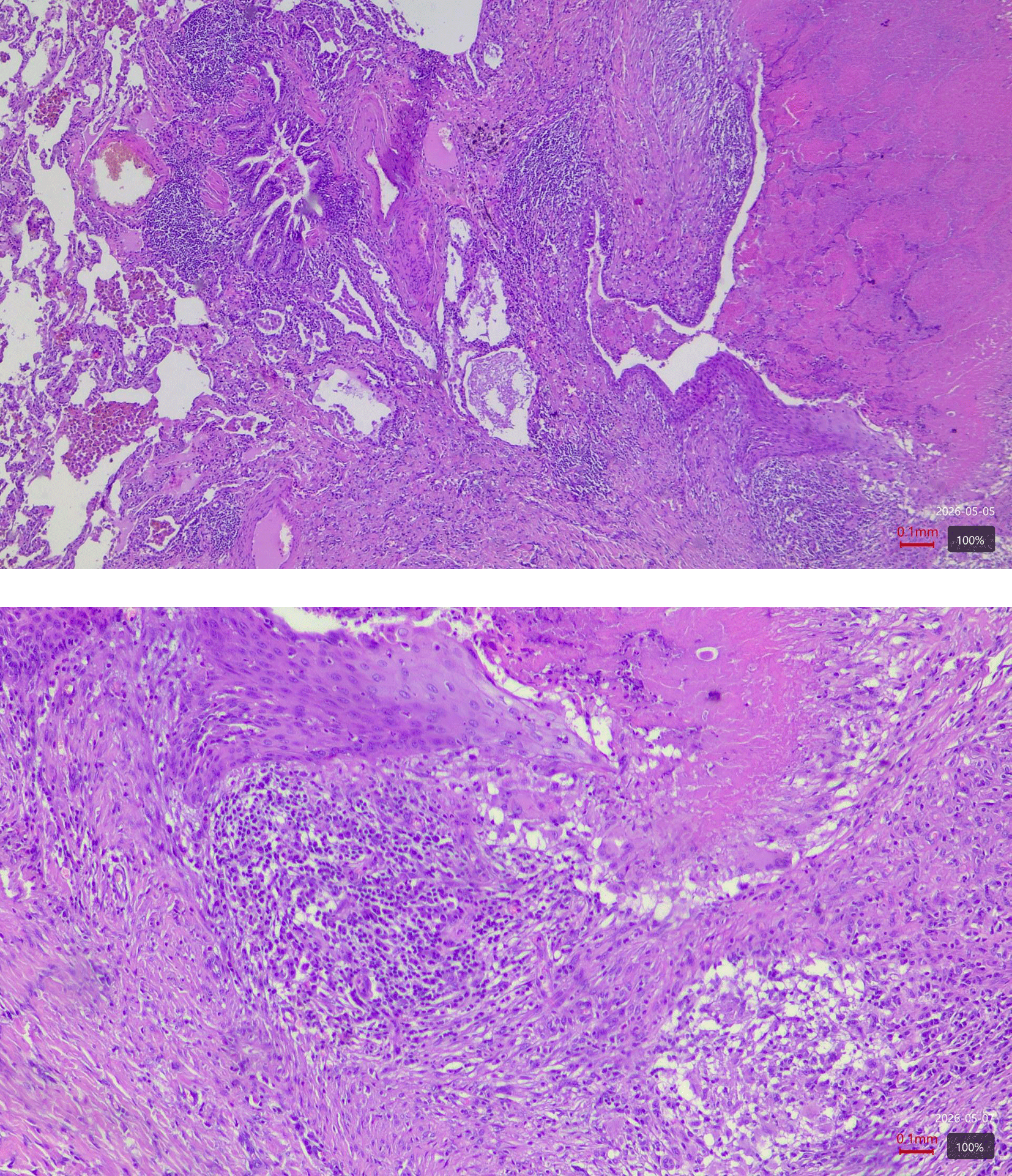
An adjacent bronchiole displays focal squamous metaplasia (***).
Postoperative chest X-rays showed expected changes following a right wedge resection, including a small apical pneumothorax with pleural separation upto 8 mm and subcutaneous emphysema in the right axillary region. Follow-up imaging, demonstrated a reduction in the pneumothorax and subcutaneous emphysema with no new pathological findings.
In this case, we document SCM of the bronchiolar epithelium adjacent to the caseating granuloma in active pulmonary tuberculosis. While SCM classically occurs in proximal, cartilaginous bronchi exposed to chronic irritants, its manifestation in non-cartilaginous bronchioles demonstrates remarkable epithelial plasticity under granulomatous inflammation. The bronchial epithelium composed of club cells and ciliated cells lack the support of bronchial submucosal glands making it uniquely vulnerable to metaplastic transformation under inflammatory stress.9
The subcentimeter growing nodule with fibrous tract extension mimicked early lung adenocarcinoma and prompted a VATS resection under the presumed oncological indication. Intraoperative findings of miliary lesions and caseating epithelioid granulomas confirmed tuberculosis, with SCM metaplasia encircling the necrotic foci. Unlike post-treatment fibrotic scars that may signal premalignancy, SCM in an active Mtb infection suggests the epithelium may adopt this stratified phenotype against mycobacterial destruction.2,5 Pulmonary nodules with interval growth frequently raise the suspicion of malignancy as pulmonary tuberculosis often radiologically mimics neoplastic lesions.10–12 In our case, the subcentimeter size of the lesion limited the utility of PET/CT for FDG-avidity characterization.
Surgical histopathology remains the definitive diagnostic tool, consistent with data showing incidental tuberculosis discovery in 3–8% of resections performed for presumed malignancy.13 The unusual bronchiolar localization of SCM challenges conventional diagnostic patterns, requiring careful histopathological evaluation to avoid a misdiagnosis of dysplasia.2,14,15
Pulmonary tuberculosis presenting as an isolated pulmonary nodule necessitates a multidisciplinary approach involving thoracic surgery, infectious disease and public health coordination. In our case, partial pleurectomy addressed adhesions and a pleural drainage resolved the resulting pneumothorax. Following histopathology results, the clinical team weighed the risks of anti-tuberculosis therapy against the benefits of surgical clearance. Given the negative cultures and localized, unilobar involvement, a strategy of close observation was favored. Surgical resection of localized tuberculosis has been shown to achieve high cure rates with clear margins and in patients with preserved lung function the physiological prognosis is excellent.13
SCM is brought about by a highly regulated form of epithelial programming driven by inflammatory, molecular and epigenetic alterations.16,17 In chronic tuberculosis, necrotizing granulomatous inflammation creates a sustained microenvironment of cytokine release and oxidative stress, which serve as critical drivers for this reprogramming.8,16 At the cellular level, SCM has been shown to arise from airway basal progenitor cells.18 Under chronic stress, these cells, normally responsive for regenerating ciliated and secretory epithelium, undergo hyperplasia and acquire a differentiation program favoring squamous lineage commitment.1,3,18 This shift is marked by the upregulation of cytokeratin CK5, CK6, and CK14, alongside transcriptional regulators such as p63.16,18 Increased expression of CK14 in particular is associated with highly proliferative basal cell populations observed in early epithelial remodeling and carcinogenesis.16,18 Experimental models of chronic lung inflammation also show that CK-14 positive basal cells undergo sustained activation, proliferation and migration, contributing to aberrant epithelial remodeling.16,18
The molecular switch to a squamous phenotype is linked to chronic inflammatory signaling pathway, particularly NF-κB activation.19 Persistent activation of NF-κB signaling promotes transcriptional programs for proliferation and survival, facilitating the transition from pseudostratified respiratory epithelium to stratified squamous epithelium.19 Other pathways such as the Notch and MAPK, further regulate basal cell expansion and differentiation under conditions of chronic injury.1 Elevated levels of TGF-β1 also drive a transition in the bronchiolar epithelium which impairs ciliary cell differentiation and promotes squamous commitment.3 Furthermore, chronic Mtb-induced inflammation also modulates the EGFR pathway, increasing ligand expression and the prevalence of EGFR mutations in cicatricial regions.20,21
Recent studies also highlight the role of epigenetic alterations. Genome-wide methylation analyses show distinct methylome profiles in SCM, suggesting SCM represents an early stage within a continuum of dysregulated epithelial regeneration rather than an isolated morphological phenomenon.17
From a pathogenetic perspective, SCM occupies a critical position in the model of lung squamous cell carcinogenesis. Chronic inflammation induces a sequence of epithelial alterations progressing from basal cell hyperplasia to SCM, followed by varying degrees of dysplasia and ultimately invasive carcinoma.14 While SCM is reversible and distinct from true dysplasia, its presence signifies a field of epithelial instability.14
The relationship between SCM and adenocarcinoma is indirect. While adenocarcinoma arises from glandular or type 2 pneumocyte lineage, both processes can coexist within a shared microenvironment of chronic inflammation.15,20,22 This suggests they may represent parallel outcomes of a common pathogenic milieu. Notably, shared oncogenic drivers, such as BRAF mutations found in lesions with mixed differentiation support the concept of molecular overlap in certain contexts.15
In the present case, the proximity of SCM to a necrotizing granuloma strongly supports a microenvironment-driven mechanism. The tuberculosis-induced milieu, rich in pro-inflammatory cytokines, reactive oxygen species and hypoxia, promotes basal cell activation and lineage reprogramming. This spatial association underscores the role of chronic immune-mediated injury in shaping atypical epithelial differentiation patterns. This coexistence of tuberculosis and SCM reflects a complex epithelial reprogramming within a shared inflammatory environment.22
SCM is a well-recognized adaptive response to chronic inflammation, typically localized in the proximal airways. This case highlights an atypical presentation of SCM within the bronchiolar epithelium in the setting of an active pulmonary tuberculosis, demonstrating the significant phenotypic plasticity of the distal airway under sustained inflammatory trigger. The close spatial proximity between the metaplastic foci and necrotizing granulomas underscores the role of localized cytokine-mediated signaling in driving epithelial remodeling beyond its typical distribution.
In clinical practice, recognizing this distal distribution is critical as bronchiolar SCM may radiologically and histologically mimic dysplastic or neoplastic transformations, particularly in small nodules resected for suspected malignancy. A meticulous correlation with the surrounding granulomatous context is essential to ensure diagnostic accuracy and avoid clinical pitfalls. Ultimately, this case expands the histopathological spectrum of epithelial alterations in pulmonary tuberculosis and emphasizes the need to identify distal airway metaplasia as a benign reactive phenomenon in a chronic infectious setting.
This study was conducted in accordance with institutional ethical standards. Ethical approval was not required for this single case report in line with local institutional policy.
Written informed consent for publication of the patient’s clinical details and histopathological images was obtained from the patient. No identifiable patient information is included in the manuscript or images.
| Views | Downloads | |
|---|---|---|
| F1000Research | - | - |
|
PubMed Central
Data from PMC are received and updated monthly.
|
- | - |
Provide sufficient details of any financial or non-financial competing interests to enable users to assess whether your comments might lead a reasonable person to question your impartiality. Consider the following examples, but note that this is not an exhaustive list:
Sign up for content alerts and receive a weekly or monthly email with all newly published articles
Already registered? Sign in
The email address should be the one you originally registered with F1000.
You registered with F1000 via Google, so we cannot reset your password.
To sign in, please click here.
If you still need help with your Google account password, please click here.
You registered with F1000 via Facebook, so we cannot reset your password.
To sign in, please click here.
If you still need help with your Facebook account password, please click here.
If your email address is registered with us, we will email you instructions to reset your password.
If you think you should have received this email but it has not arrived, please check your spam filters and/or contact for further assistance.
Comments on this article Comments (0)