Case report
A 63-year-old Caucasian man was referred to our hospital for further analysis of slowly progressing pleural effusion with a history of cough and dyspnea. The patient had a long history of epilepsy and meningioma. He was working as head in a department of administration. He was married and had two healthy kids. As a medication he took Pantoprazol, Tegretol and Dorsolamide and had stopped smoking a long time ago.
Our patient was diagnosed with progression of a previous operated (Simpsom3 resection) and postoperatively irradiated (30 x 2 GY with a total doses of 60 Gy within the EORTC 22042 in a study context) atypical left parieto-occipital meningioma (WHO grade-II). Re-resection of the tumor (Simpsom4) was performed and histopathology showed a malignant meningioma (WHO grade-III). In the follow up after re-resection there was an obvious evidence of a residual tumour at the falx cerebri. One year later an asymptomatic re-recurrence was diagnosed (Figure 1A and B), for which conservative follow-up was performed without further surgical intervention. This was given in the form of re-irradiation with a total doses of 130 Gy (60 Gy given for the re-re-recurrent tumor at the resected area + additional 70 GY applied as an integrated boost with IMRT-technic for the residual tumor at the falx cerebri). This decision was taken due to the higher degree of aggressiveness of the malignant meningioma, as further surgical intervention would harm the patient rather than curing him.
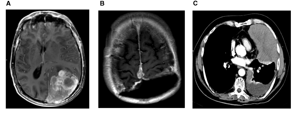
Figure 1. MRI and CT scanning of the original meninigioma intra-cerebral and at distance metastasis intra-pleural.
A: T1 weighted image after administration of Gadolinium based contrast fluid shows a large extra-axial enhancing lesion in the left parieto-occipital region with local mass effect. Note that the sagittal sinus seems to be invaded. B: Contrast enhanced T1 weighted image after administration of Gadolinium based contrast fluid showing a large resection cavity after the second operation and recurrent disease at the most upper margin of the resection plane with enhancing areas surrounding the sagittal sinus. C: Midthoracal CT slice in the transverse plane. Scan performed after i.v. administration of iodine contrast. The lesion is easily distinguished at the left ventral thoracal intrapleural space, slightly enhanced suggesting solid tissue. Some pleural fluid is also present.
A few months later the patient was hospitalized with dyspnea, fatigue, productive cough and anorexia. Multiple pleural masses were detected at a chest computer-tomography (CT) scan. Histopathology was consistent with malignant meningioma (WHO grade-III, Figure 2A), there was a high expression in the EMA staining, also in the AE1/AE3 staining (Figure 2B). The CD 45 and CD 68 were positive and MIB-1 showed high proliferation. Palliative chemotherapy was offered but refused by the patient. The patient is died one week after discharge from the hospital as a result of voluntary euthanasia as was the will of the patient (valid written declaration).
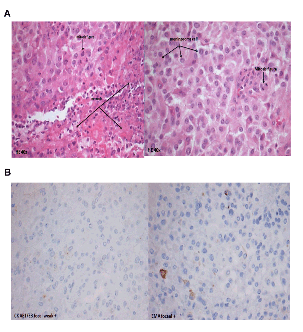
Figure 2. Histology staining of a biopsy of the solid intrathoracal lesion showing the same morphology as the intracranial meningioma.
A: A specimen (HE-stained, 40x) showing histological resemblance between the intrathoracal lesion and the intracranial meningioma. B: Specimen (focal plus and focal weak) showed high expression in the EMA-staining and also in the AE1/AE3-staining.
Discussion
Pulmonary and pleural metastases from an intracranial meningioma are very rare. Distant metastases from meningiomas are infrequently found in clinical practice and mostly associated with atypical or malignant meningiomas. Meningiomas mainly recur loco-regional or adjacent to the radiation treatment fields1,2. There are only isolated case reports regarding pulmonary metastases from meningioma. Most lung metastases were incidentally detected by chest radiography or by CT-scans, because metastatic lesions are usually asymptomatic. The presence of pulmonary metastases appears to negatively affect survival in patients with recurrent meningioma3,4.
Regarding the relationship between the intracranial location and invasion of the sagittal sinus of the tumor and the pleural metastases the route of dissemination is most probably the central venous route to heart and lungs5. In previous case reports the lung was the most common extracranial metastatic site for intracranial meningioma6. Our case was unusual because of the highly rate of recurrences and later the distant metastases. There is no standard treatment in the case of distant metastases.
In this case, histopathologic findings of the primary tumor revealed hypercellularity, wide necrosis, and brain invasion into the normal brain parenchyma. Pathology of the lesion from the left thoracic wall was consistent with malignant meningioma.
Other case studies described that treatment of pulmonary metastasis of malignant meningioma consisted of surgical resection for both the primary or metastatic lesions1,5,7.
Postoperative conventional radiation therapy has been recommended for prevention of local recurrence, especially when resection is subtotal. There are insufficient data regarding radiation therapy by meningiomas with distant metastases, palliative chemotherapy is the only option in the case of distant metastases, however data regarding the efficacy of this systemic treatment are unknown.
Informed consent
Written informed consent for publication of clinical details and clinical images was obtained from the next of kin.
Author contributions
Frits Franssen: supervisor of the respiratory department where patient was admitted. Ryan Accord: the surgeon who did the VATS. Myurgia A. Hamid: histological figures. Shekar Mahesh: radiological figures. Brigitta G. Baumert and Olaf E.M.G. Schijns: literature search and data collection.
Competing interests
No competing interests were disclosed.
Grant information
The author(s) declared that no grants were involved in supporting this work.
Acknowledgments
At the completion of this Case study, I am very thankful to all contributors, who were very helpful and without their support this case study would have never come into its present form.
Faculty Opinions recommendedReferences
- 1.
Figueroa BE, Quint DJ, Mckeever PE:
Extracranial metastatic meningioma.
Br J Radiol.
1999; 72(857): 513–516. PubMed Abstract
| Publisher Full Text
- 2.
Stoller JK, Kavuru M, Mehta AC:
Intracranial meningioma metastatic to the lung.
Cleve Clin J Med.
1987; 54(6): 521–526. PubMed Abstract
| Publisher Full Text
- 3.
Alexandru D, Glantz MJ, Kim L:
Pulmonary metastases in patients with recurrent, treatment-resistant meningioma: prognosis and identification by 111Indium-octreotide imaging.
Cancer.
2011; 117(19): 4506–11. PubMed Abstract
| Publisher Full Text
- 4.
Enam SA, Abdulrauf S, Mehta B:
Metastasis in meningioma.
Acta Neurochir (Wien).
1996; 138(10): 1172–7. PubMed Abstract
| Publisher Full Text
- 5.
Nakano M, Tanaka T, Nakamura A:
Multiple Pulmonary Metastases following total removal of a bilateral parasagittal meningioma with complete occlusion of the superior sagittal sinus: Report of a Case. Hindawi Publishing Corporation.
Case Rep Neurol Med.
2012; 2012: 7. PubMed Abstract
| Publisher Full Text
| Free Full Text
- 6.
Strang RR, Tovi D, Nordenstam H:
Meningioma with intracerebral, cerebellar and visceral metastases.
J Neurosurg.
1964; 21(12): 1098–1102. PubMed Abstract
| Publisher Full Text
- 7.
Kanzaki R, Higashiyama M, Fujiwara A:
Surgical resection of pulmonary metastases from meningioma: report of a case.
Surg Today.
2011; 41(7): 955–8. PubMed Abstract
| Publisher Full Text
Comments on this article Comments (0)