Introduction
Paraovarian cysts occur in the broad ligament between the ovary and the tube, predominantly arising from mesothelium covering the peritoneum (mesothelial cyst) but occasionally also from para mesonephric tissue (paramesonephric cysts or Mullerian cysts) and rarely from mesonephric remnants (mesonephric cysts or Wolffian cysts)1. Paraovarian cysts, constitute 10–20% of all adnexal masses2. Some paraovarian cysts may reach a large size with possible complications like torsion and rupture3. These cysts are usually benign with rare incidence of malignant types4,5. Here, we present a case of unusually extensive proportions.
Case presentation
Consent
Written informed consent for publication of the clinical details and/or clinical images was obtained from the patient.
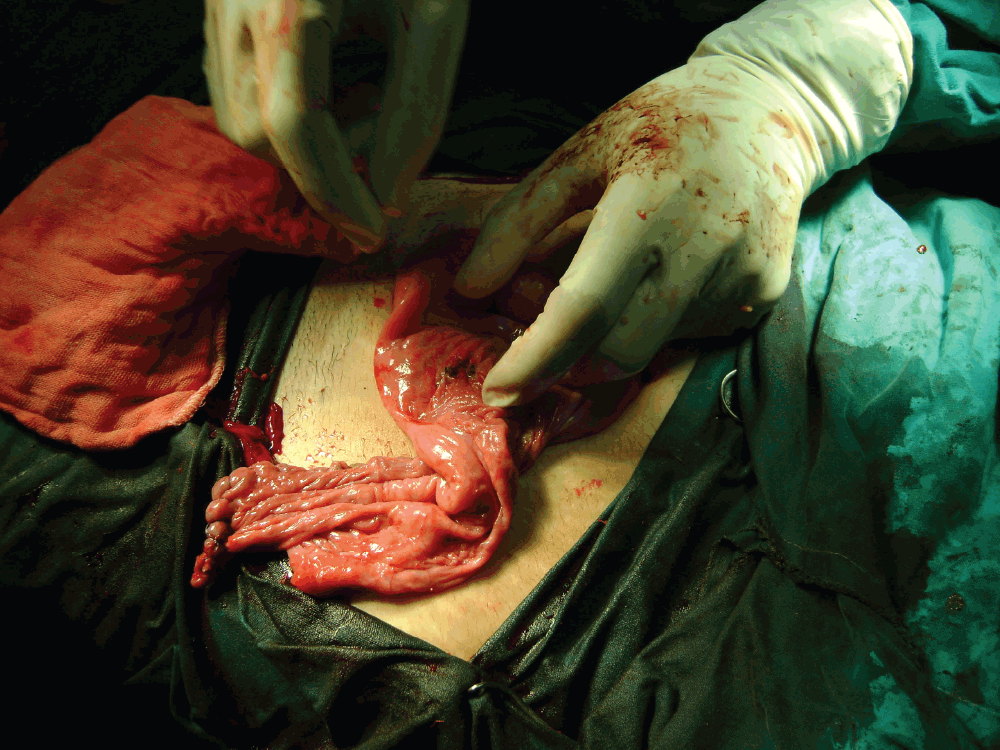
A 17 year old virgin presented with diffuse abdominal pain. History revealed a gradual increase in an abdominal swelling over the preceding 6 months. Physical examination showed a non tender tense cystic pelviabdominal mass of 36 weeks gestational size. Computerised tomography revealed 25×26 cm left ovarian simple cyst with clear contents and no septae. Serum CA125 levels were normal. Other tumor markers were not performed due to financial constraint. Through a subumbilical midline incision, a huge smooth cystic mass overlying the whole abdominal cavity was found. The cyst was isolated from its surroundings with gauze packs. A loose purse string suture was placed in the lowest accessible part of the cyst. A 5 mm laparoscopic trocar with a side track off its main sleeve was connected to a high pressure suction irrigation device via a rubber tube; the trocar was then inserted through the center of the suture which was subsequently stretched to fit around the sleeve. This created a closed system to drain the cyst. The trocar was removed leaving its sleeve in place and suction drained eight liters of clear watery fluid. The collapsed cyst was found to be left paraovarian which was exteriorized and the trocar sleeve was removed. The purse string suture was tightened to close the trocar opening. The left broad ligament was opened and the cyst wall was completely removed from the broad ligament, Figure 1. The redundant ligament peritoneum was excised and subsequently reconstructed with preservation of the tubal integrity as seen in Figure 2. The patient had an uneventful postoperative recovery.

Figure 1. Appearance of the aspirated cyst wall after opening the broad ligament.

Figure 2. The final shape of the left broad ligament after its reconstruction with intact tube.
Postoperative histology reported simple benign serous cyst of mesothelial origin. The peritoneal fluid showed proteinaceous material entangling few lymphocytes and mesothelial cells with no evidence of malignancy.
Discussion
Huge paraovarian cysts are uncommonly reported in the literature. On revising the literature, there were three case reports which had addressed comparable large paraovarian cysts but with implementation of larger incisions extending over the umbilicus for cyst extraction and excision without a policy to decrease its size before its exteriorization6–8. However, a case report for three adolescents with large paraovarian cysts had addressed decompression technique before cyst externalization and excision but in a different way9. In our case report we had dealt with such a huge cyst in a way not only to avoid morbidity of extending surgical incision but to guard against the risk of spillage of cyst contents as well. Concerning the endoscopic role, Darwish et al. reported a series of paraovarian cysts which had been excised laproscopically but were smaller in size with the largest not more than 13 cm10. However, there were two reports of large paraovarian cysts removed laparoscopically where in the first one, it was associated with acute lower abdominal pain while in the second it was associated with pregnancy11,12. We think that in all these laparoscopically operated cases, the implemented cyst decompression procedure before its removal had less control and precautions during it and in turn more risk of cyst spillage than our mentioned maneuver. It was thought that laparoscopy would be technically difficult in this case due to huge size of the cyst reaching close to xiphesternum. Direct abdominal entry with a Veress needle or trocar may have traumatized the cyst leading to risk of spillage of its content. Through laparotomy we employed a closed drainage system and safely aspirated the cyst without spillage of its content.
Conclusion
Open surgery remains the gold standard route to deal with giant paraovarian cysts. Aspiration of the cyst using a closed system followed by excision is a safe and effective treatment.
Author contributions
MK prepared the framework of the case report, introduction, discussion and references. TS was the surgeon and prepared the presentation of the case in the manuscript. MZ assisted during the surgery and helped preparing the presentation of the case in the manuscript.
Competing interests
No relevant competing interests were disclosed.
Grant information
The author(s) declared that no grants were involved in supporting this work.
Faculty Opinions recommendedReferences
- 1.
Stenback F, Kauppila A:
Development and classification of paraovarian cysts: an ultrastructural study.
Gynecol Obstet Invest.
1981; 12(1): 1–10. PubMed Abstract
- 2.
Alpern MB, Sandler MA, Madrazo BL, et al.:
Sonographic features of parovarian cysts and their complications.
AJR Am J Roentgenol.
1984; 143(1): 157–160. PubMed Abstract
- 3.
Lurie S, Golan A, Glezerman M, et al.:
Adnexal torsion with a paraovarian cyst in a teenage girl.
J Am Assoc Gynecol Laparosc.
2001; 8(4): 597–9. PubMed Abstract
- 4.
Altaras MM, Jaffe R, Corduba M, et al.:
Primary parovarian cystadenocarcinoma: clinical and management aspects and literature review.
Gynecol Oncol.
1990; 38(2): 268–72. PubMed Abstract
| Publisher Full Text
- 5.
Kaur K, Gopalan S, Gupta SK, et al.:
Parovarian cystadenocarcinoma: a case report.
Asia Oceania J Obst Gynaecol.
1990; 16(2): 131–5. PubMed Abstract
- 6.
Azzena A, Quintieri F, Salmaso R:
A voluminous paraovarian cyst. Case report.
Clin Exp Obstet Gynecol.
1994; 21(4): 249–52. PubMed Abstract
- 7.
Lazarov N, Lazarov L, Angelova M:
Paraovarian cyst in an 18 year patient.
Akush Ginekol (Sofiia).
2000; 40(4): 50. PubMed Abstract
- 8.
Mukhopadhyay S:
Giant paraovarian cyst.
J Obstet Gyncol India.
2006; 56(4): 352–353. Reference Source
- 9.
Damle LF, Gomez-Lobo V:
Giant paraovarian cysts in young adolescents: a report of three cases.
J Reprod Med.
2012; 57(1–2): 65–7. PubMed Abstract
- 10.
Darwish AM, Amin AF, Mohammad SA:
Laparoscopic management of paratubal and paraovarian cysts.
JSLS.
2003; 7(2): 101–106. PubMed Abstract
| Free Full Text
- 11.
Sindos M, Pisal N, Mellon C, et al.:
Laparoscopic excision of a large paraovarian cyst presenting with acute lower abdominal pain.
J Obstet Gynecol.
2004; 24(6): 717–718. PubMed Abstract
| Publisher Full Text
- 12.
Rouzi AA:
Operative laparoscopy in pregnancy for a large paraovarian cyst.
Saudi Med J.
2011; 32(7): 735–7. PubMed Abstract


Comments on this article Comments (0)