Keywords
Aerosols, Aerobiology, Quantitative Growth Kinetics, Environmental Monitoring
Aerosols, Aerobiology, Quantitative Growth Kinetics, Environmental Monitoring
The virtual colony count (VCC) microbiological assay has been utilized for over a decade to measure the antimicrobial activity of peptides such as defensins. The initial VCC publication (Ericksen et al., 2005) used two methods of transferring cells to microplates using a 20–200 microliter multichannel pipettor: 22.2 microliters added to 200 microliters of media in calibration experiments and 50 microliters added to 50 microliters of solutions in phosphate buffer. Further experimentation has demonstrated that only the former method safely and effectively transfers cells to the intended wells, and the latter method can result in cross-contamination.
The reason for this difference is that adding cells suspended in 50 microliters directly to a like volume caused unacceptable froth, bubbles and background turbidity that is incompatible with the VCC method of measuring growth kinetics by an increase in optical density using a 96-well plate in a plate reader. This problem, which affects optical density readings in turbidimetric assays, was initially solved by holding pipette tips just above the liquid but below the rims of the wells and adding cell suspensions as droplets. Accurately holding the multichannel pipettor within this narrow range seemed to require placing one’s eyes as close as possible to the 96-well plate, but further experiments using biosafety cabinets have proven that the method can be done by a well-trained operator looking through the glass. Assays conducted in 2012 and 2013 within a biosafety cabinet at the University of Maryland Baltimore (UMB) resulted in frequent cross-contamination of the 36 contamination control edge wells. Light microscopy revealed adhesive and cohesive clumps and biofilms formed by Escherichia coli ATCC 25922 and Staphycococcus aureus ATCC 29213. Changes in particle size distribution and adhesive properties due to clumping apparently resulted in increased aerosol formation, which made cross-contamination far more common than in the initial studies in 2003–2004 preceding the 2005 publication of VCC. Using this procedure for hazardous microorganisms outside a biosafety cabinet would pose a safety risk.
The VCC plate configuration as initially published in 2005 used the 36 wells around the edge of the 96-well plate (rows A and H and columns 1 and 12) as contamination control wells. Turbidity in these wells could have been the result of either environmental contamination or cross-contamination, but sampling wells over the course of many experiments revealed colony morphologies that were almost invariably consistent with the bacterial strain studied that day. Six alternating VCC experiments using Escherichia coli ATCC 25922 and Staphylococcus aureus confirmed this conclusion by producing colonies only consistent with the strain studied that day, not the strain studied in the previous experiment or an environmental isolate with a colony morphology matching neither strain.
Two hypotheses regarding the origin of cross-contamination were pursued: cells emanating from the pipette tips as they were passed directly over the contamination control wells or cells ejected up out of the wells as aerosols when the cell suspension was expelled. To distinguish between these possibilities, 13 experiments were conducted not with a single ring of 36 contamination control wells around the edge, but with an additional ring (columns 2 and 11 and rows B and G), totaling 64 uninoculated wells. In these experiments, quadruplicate 8-point 10-fold calibration dilutions were made by adding 22.2 microliters beneath 200 microliters of media, pipetting up and down 15 times, expelling tips, transferring 22.2 microliters to the next column of four wells, etc. None of the 832 uninoculated wells turned turbid after overnight incubation at 37 degrees shaking in a Tecan Infinite M1000 plate reader, indicating a lack of cross-contamination or environmental contamination that is viable in rich media originating from the laboratory, reagents, operator or reader. Next, several VCC experiments were conducted using eight cross-contamination control wells in column 12 with controls lacking antimicrobial agents in column 11 as described in the initial 2005 paper, during which all 24 cross-contamination control wells in column 12 turned turbid in all three experiments. Four changes were made to the procedure in an attempt to remove possible sources of contamination that may have caused cells to become more adhesive and cohesive, which in turn would have caused cross-contamination to become far more likely: 1. using a small HEPA-filtered air purifier, 2. replacing in-house deionized Milli-Q water with purchased molecular biology grade water, 3. replacing 2XMHB prepared and autoclaved in-house using reusable jars with Teknova 2X cation-adjusted MHB, and 4. filter-sterilizing phosphate buffers made near the portable air purifier, rather than autoclaving in reusable jars. After those changes, 25 mL TSB cultures of Escherichia coli ATCC 25922 grown simultaneously as a biosensor no longer produced macroscopic clumps with diameters on the scale of millimeters. However, cross-contamination in VCC experiments persisted. In several of these experiments, a separate 96-well plate containing media only was interposed between the reagent reservoir containing the cell suspension and the experimental 96-well plate, and in no case did any well in these additional plates turn turbid. Had cells been transiently adhering to the outsides of the tips or trailing from the liquid held by capillary action at the openings of the tips, many or all of the 96 wells of the cross-contamination plates would have turned turbid, since all cross-contamination wells in column 12 on the right edges of experimental plates turned turbid. Therefore, contamination caused by passing the tips over these wells without expelling was ruled out. The next simplest explanation is that, while the plunger of the multichannel pipettor was depressed to deliver cells as droplets below the rims but above the liquid in the wells, the tips expelled viable aerosols that travelled in an upward trajectory and escaped the intended wells in such great numbers that the cross-contamination of adjacent wells was probable to the point of inevitability.
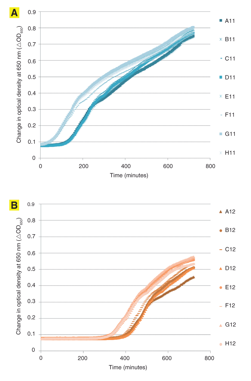
In Experiment 1, all eight wells in column 12 turned turbid and produced growth curves with the same growth rate and doubling times as the other growth curves on the same microplate (Figure 1). Colony morphologies of samples from these wells also matched E. coli ATCC 25922. A comparison of threshold times indicated almost the same difference between input and output controls in columns 11 and 12 (Table 1). There was a roughly 70-minute difference in input and output threshold times in the input and output control wells in Experiment 1, which agreed closely with another roughly 70-minute difference in the threshold times of the adjacent wells. Contamination caused by environmental factors would have been expected to produce widely varying threshold times, if not visible differences in the appearance of the turbid wells. Therefore, the 70-minute difference indicated that the cross-contamination occurred at the same time that cells were transferred.

Uncorrected growth kinetics of columns 11 (panel A) and 12 (panel B) of the 96-well plate in Experiment 1. In these two columns (n=16), the threshold ΔOD650 value of 0.02 corresponded to a mean ± standard deviation uncorrected OD650 of 0.0989±0.0043, which corresponds to a %RSD of 4.4. The line marked “0.1” is approximately at the position of the threshold ΔOD650 of 0.02.
| Columns | |||
|---|---|---|---|
| 11 | 12 | ||
| Rows | A | 121.0 | 393.9 |
| B | 124.3 | 398.8 | |
| C | 120.8 | 385.8 | |
| D | 122.2 | 403.7 | |
| E | 48.4 | 322.8 | |
| F | 50.4 | 333.0 | |
| G | 47.9 | 318.2 | |
| H | 48.2 | 325.4 | |
| Mean, A-D | 122.1 | 396.1 | |
| Mean, E-H | 48.7 | 324.9 | |
| Mean, output minus Mean, input | 73.3 | 71.2 | |
A11-D11 are the “input” control wells and E11-H11 are “output” control wells. Cells were added to those two wells two hours apart, resulting in a 73.3 minute difference in Tt values. Cross-contaminated wells gave a corresponding Tt difference of 71.2 minutes, indicating that A12-D12 were inoculated as cells were being expelled over A11-D11, and E12-H12 were inoculated as cells were being expelled over E11-H11.
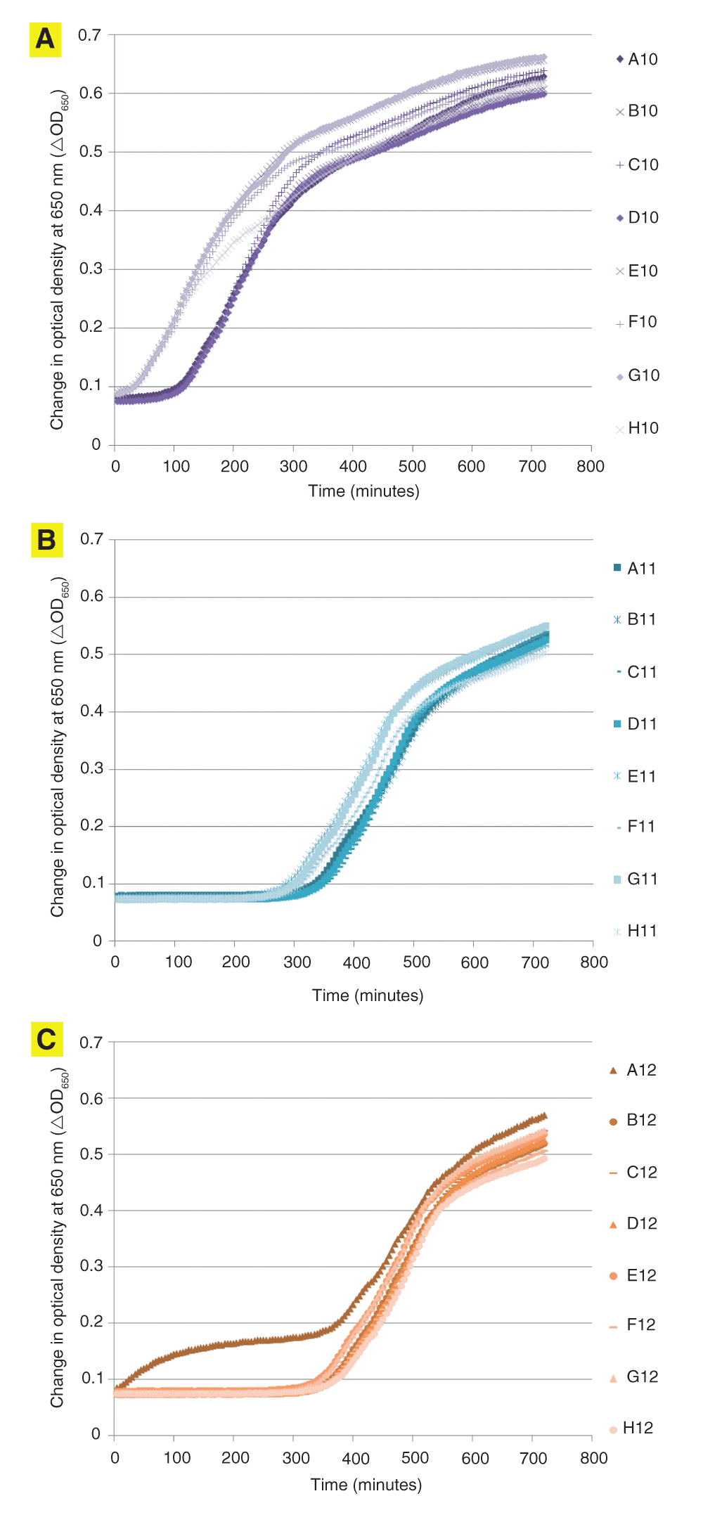
In Experiment 2, the threshold times again reflected a roughly 70-minute difference between input and output controls. (Figure 2 and Table 2) However, this difference was not reflected in threshold times of the cells growing in column 12, indicating that the contamination of those wells was the result of a second contamination event unrelated to the timing of the transfer of cells into the wells in column 10. The only reasonable explanation of this agreement in threshold time differences between columns 10 and 11 and the far larger Tt values resulting from column 12 is that cross-contamination occurred while cells were expelled, and the aerosols thus formed travelled to the adjacent wells but not the wells in column 12 or the intervening 96 contamination control wells in the contamination control plate, none of which turned turbid after overnight incubation at 37 degrees. These results indicate that 96-well plates and threshold times are useful for detecting contamination, and that cross-contamination occurs in experiments where cells are added as droplets from above.

Uncorrected growth kinetics of columns 10 (panel A), 11 (panel B) and 12 (panel C) of the 96-well plate in Experiment 2. In these three columns excluding well A12 (n=23), the threshold ΔOD650 value of 0.02 corresponded to a mean ± standard deviation uncorrected OD650 of 0.0988±0.0053, which corresponds to a %RSD of 5.4. The biphasic curve in well A12 was unique among the 96 wells analyzed in this assay, and is caused by an initial phase of optical density increase caused by condensation on the lid followed by a second phase caused by increased turbidity due to cell growth within the well.
A10-D10 are the “input” control wells and E10-H10 are “output” control wells. Cells were added to those two wells two hours apart, resulting in a 68.2 minute difference in Tt values. Cross-contaminated wells gave a corresponding Tt difference of 42.4 minutes. The difference between these two values, 25.8 minutes, could be accounted for by the growth of additional cells added in a second contamination event reflected by wells B12-H12 Tt values that also caused media in the reservoir to turn turbid when collected and incubated overnight. Thus, Tt values detect cross-contamination in adjacent wells and can distinguish between separate contamination events.
The method of enumeration of cells in a VCC assay is confounded if the cells form clumps, because that clumping and biofilm formation affects optical density readings. Other experiments revealed macroscopic clumps and biofilms visible to the unaided eye. In addition, microscopic clumps were revealed by light microscopy. Cohesion, adhesion, clumps and biofilms affect not only threshold times but also the particle size distribution of the cell suspension and the degree of adhesion as the cells are expelled through the pipette tips. Therefore, both clumps suspended in solution formed by cells adhering to each other but not surfaces and adhesive cells could affect the physical properties of the liquid as it is transformed to an emulsion that generates aerosols. Cross-contamination was far more common in the experiments I conducted in 2012–2013 compared to experiments I conducted in 2003–2004 in an adjacent room, suggesting that some change in environmental factors between those times or locations caused greater cell clumping and adhesion, which in turn greatly increased the probability that a cross-contamination control well would become turbid. It is postulated that one or more clumping environmental factor (CEF) is responsible for the change in cross-contamination and a 23-fold fluctuation in virtual lethal dose values reported by the HNP1 positive controls of the assay throughout E. coli ATCC 25922 experiments in 2013.
In 2011, a modified VCC procedure (Welkos et al., 2011) was published for use with the BSL-3 pathogen Bacillus anthracis, based on the procedure originally developed at UCLA in the laboratory of Robert I. Lehrer. The 50 microliter cell transfer step mentioned in the 2005 publication and used at the University of Maryland was replaced with the addition of cells suspended in a smaller volume of liquid, 10 microliters, added to 90 microliters of buffer. This procedure, similar to the calibration experiments detailed in the original VCC publication, did not generate unacceptable turbidity when cell suspensions were added with the tips placed at the bases of the wells beneath the buffer when it was tested in 2013 in the Institute of Human Virology building at IHV. Adding cell suspensions under liquid apparently greatly reduces the probability of aerosol formation, which is of concern not only for safety reasons, but also because the aerosol cloud within the well can alter experimental results by generating cells that adhere to the sides of the well during the exposure to the antimicrobial agent, then drop down to inoculate the outgrowth media after the antimicrobial peptides have been neutralized by broth during 12 hours of vigorous shaking within the plate reader. VCC users are cautioned to use the 2011 procedure, not the 2005 procedure, to add experimental cell suspensions. Following the 2005 procedure to add Staphylococcus aureus cell suspensions in droplets above the liquid in the wells rather than injecting the cell suspension beneath the liquid in the wells could expose the eyes to aerosols containing a biosafety level 2 pathogen that could cause blepharitis, corneal stromal microabscess, stromal edema, uveitis, ocular necrotizing fascitis, and blindness. (Boto-de-Los-Bueis et al., 2014; Shield et al., 2013) Biosafety level 2 precautions such as those recommended by the Centers for Disease Control in Biosafety in Microbiological and Biomedical Laboratories, 5th Edition (Miller et al., 2012) should be taken for any study of Stapylococcus aureus, including the safer 2011 VCC procedure.
These results highlight an advantage of using the VCC data analysis procedure of enumerating cells (Brewster, 2003), termed quantitative growth kinetics (QGK) by analogy to quantitative polymerase chain reaction (QPCR). (Heid et al., 1996) QGK and QPCR use a mathematically identical procedure for quantifying the initial number of cells or amplicons that were present at the start of the assay. The QGK threshold time Tt is equivalent to the PCR cycle time Ct. Calculating Tt values in the two experiments reported here unequivocally identified the time of the contamination event, gave quantitative batch culture growth kinetic data that suggested that the contamination was cross-contamination, and distinguished between two cross-contamination events. These features of QGK would greatly improve the quality of environmental monitoring data when used to detect contamination by aerosols or ambient viable microorganisms compared to turbidity measurements in the absence of a plate reader or observing the appearance of colonies on agar plates, neither of which provides kinetic data.
Finally, it should be emphasized that the simple improvement of adding cells beneath liquid simultaneously achieves two useful changes at once, reducing the probability that cells inoculate wells other than the ones intended while simultaneously also limiting the probability that cells escape the 96-well plate entirely. Although the reason why the addition of 50 microliters of cells beneath 50 microliters of liquid was unacceptable in VCC experiments stemmed from the turbidimetric nature of the assay, this method of preventing cross-contamination is far from trivial or confined to VCC assays. It teaches a technical lesson limited not just to environments where airborne CEFs are present, but broadly applicable to all experiments where microbes are transferred using pipette tips, thereby potentially improving the usefulness of a wide range of laboratory procedures that might otherwise generate aerosols. Any change in a procedure that improves its safety and efficacy also improves its utility ad oculos.
VCC assays were conducted as described (Ericksen et al., 2005) and modified (Zhao et al., 2013). Twice-concentrated cation-adjusted Meuller Hinton Broth was purchased from Teknova, Inc. Phosphate buffers were made using Sigma monobasic and dibasic sodium phosphate dissolved in molecular biology grade water or equivalent purchased from multiple sources. Rainin GreenPak LTS 200 microliter filter tips were used with an eight-channel 20–200 microliter pipettor. Costar 3595 96-well plates were analyzed in a Tecan Infinite M1000 plate reader at 37°C.
Two experiments were conducted using Escherichia coli ATCC 25922. In Experiment 1, four each of “input” and “output” controls were placed in column 11 of the 96-well plate, with eight cross-contamination control wells in column 12. Wells A11-D11 contained controls in wells added at the time the cells were exposed to antimicrobial agents, termed the “output” controls, and equivalent to the controls mentioned in the initial 2005 publication. In addition, wells E11-H11 contained identical controls that had been stored on ice during the two-hour exposure to antimicrobial agents in phosphate buffer, termed the “input” controls because their Tt values represent the concentration of cells that were present when they were put into the assay at the start of the two-hour incubation. Since the antimicrobial assay is beyond the scope of this report, which focuses only on aerosol cross-contamination, columns 1–10 and the antimicrobial agents therein will not be discussed here.
Next, in Experiment 2, the controls lacking antimicrobial agents were moved from column 11 to column 10, and columns 11 and 12 contained 16 uninoculated contamination control wells. Wells A10-D10 contained controls in wells added at the time the cells were exposed to antimicrobial agents, termed the “output” controls, and equivalent to the controls mentioned in the initial 2005 publication. In addition, wells E10-H10 contained identical controls that had been stored on ice during the two-hour exposure to antimicrobial agents in phosphate buffer, termed the “input” controls. These controls are designed such that comparing the difference in threshold times between the input and output controls, relating that difference to the calibration curve elsewhere on the same 96-well plate, and assuming that adhesion or cohesion and lag phases in exponential growth were the same for all cells, the growth of the cells during the two hour incubation on the plate could be quantified. Cells grow during the two hour incubation step, so enumerating the change in cell concentration during that step would allow the calculation of the difference in virtual survival values that would correspond to bacteriostatic activity.
F1000Research: Dataset 1. Growth kinetics optical density readings for Experiments 1 and 2, 10.5256/f1000research.5659.d38055 (Ericksen, 2014).
I thank Peprotech, Inc. for funding.
The funders had no role in study design, data collection and analysis, decision to publish, or preparation of the manuscript.
| Views | Downloads | |
|---|---|---|
| F1000Research | - | - |
|
PubMed Central
Data from PMC are received and updated monthly.
|
- | - |
Competing Interests: No competing interests were disclosed.
Competing Interests: No competing interests were disclosed.
Alongside their report, reviewers assign a status to the article:
| Invited Reviewers | ||
|---|---|---|
| 1 | 2 | |
|
Version 2 (revision) 14 Jan 15 |
read | |
|
Version 1 06 Nov 14 |
read | read |
Click here to access the data.
Spreadsheet data files may not format correctly if your computer is using different default delimiters (symbols used to separate values into separate cells) - a spreadsheet created in one region is sometimes misinterpreted by computers in other regions. You can change the regional settings on your computer so that the spreadsheet can be interpreted correctly.
Provide sufficient details of any financial or non-financial competing interests to enable users to assess whether your comments might lead a reasonable person to question your impartiality. Consider the following examples, but note that this is not an exhaustive list:
Sign up for content alerts and receive a weekly or monthly email with all newly published articles
Already registered? Sign in
The email address should be the one you originally registered with F1000.
You registered with F1000 via Google, so we cannot reset your password.
To sign in, please click here.
If you still need help with your Google account password, please click here.
You registered with F1000 via Facebook, so we cannot reset your password.
To sign in, please click here.
If you still need help with your Facebook account password, please click here.
If your email address is registered with us, we will email you instructions to reset your password.
If you think you should have received this email but it has not arrived, please check your spam filters and/or contact for further assistance.
Comments on this article Comments (0)