Keywords
GTPS, gluteal tendinopathy, tendon repair, tenotomy, ultrasound imaging
GTPS, gluteal tendinopathy, tendon repair, tenotomy, ultrasound imaging
Greater trochanteric pain syndrome (GTPS) is a common cause of posterolateral hip pain typically seen in peri and post menopausal women. Coexistent obesity, ilio-tibial band (ITB) syndrome, low back pain, osteoarthritis are added risk factors1. Evaluation using imaging and histology techniques revealed degenerative tendinosis with tears of the gluteal tendons2–4. The primary underlying pathology of GTPS is a gluteal tendinopathy with or without tears of the tendon5. Although GTPS is widely referred to as greater trochanteric ‘bursitis’, there is no inflammation of the greater trochanteric bursa on histological evaluation of surgical specimens6.
The natural history of GTPS is favourable in most cases and responds to physical therapy, weight loss, non-steroidal anti-inflammatory drugs and behaviour modification1. However in some patients the condition causes significant disability and necessitates intervention. Corticosteroid injection into the bursa and surgical repair of any torn gluteal tendons are the current common treatment options. Corticosteroid injections are controversial in a degenerating tendinopathy7. Corticosteroid injections carry the risk of a dampening effect and progressive worsening of tendon pathology8. A pioneering percutaneous treatment for all tendoligamentous and cartilage tears using autologous platelet rich plasma (PRP) tenotomy under high resolution imaging control was routine clinical management in the author’s practice. The same treatment was performed in a patient with GTPS. This report is the 1-year follow up on the clinical outcome and imaging appearance of this patient.
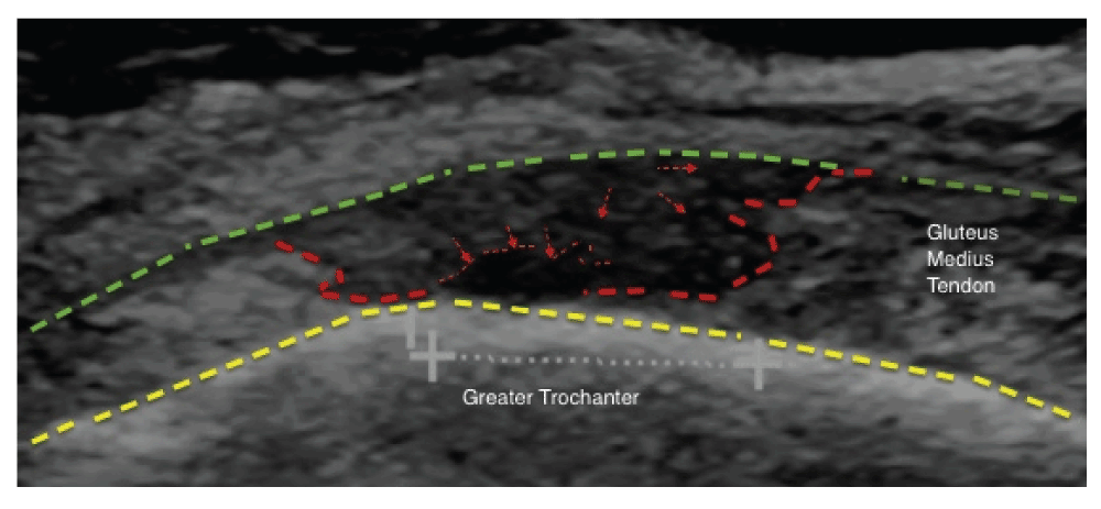
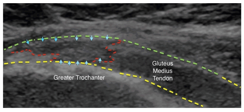
A 56 year old female Caucasian patient presented with 6 months of progressive left sided greater trochanteric pain syndrome. She complained of pain every day for many months, was unable to climb stairs and experienced moderate stiffness of the outer hip during early morning awakening. Pre-procedural clinical examination revealed an overweight individual with a BMI of 27, valgus knees, gynacoid pelvis, localised tenderness of the gluteus minimus and medius insertions into the facets of the greater trochanter, painful limitation of passive and active hip abduction and provocation to resisted abduction due to gluteal tendon dysfunction. She wished to be very active, commence a walking holiday and reduce her body weight. She refused both corticosteroid injection therapy and surgery. Full written informed consent to treat and publish the data was obtained from the patient. Ultrasound scan showed a 10 mm × 12 mm high grade full thickness tear of the degenerating gluteus medius tendon insertion (Figure 1) and gluteus minimus tendinopathy with partial split tears (image not shown). Under ultrasound imaging (GE Logic 9, 9MHz probe) control, 1% lignocaine 5 cc was infiltrated through a 22 G needle into and around the minimus tendon. Percutaneous tenotomy was performed into the foot print and the adjacent gluteal cuff under real time imaging guidance. 4–5 cc of autologous PRP (REGEN Switzerland, Adistem Hong Kong) was infiltrated. Repeated percutaneous tenotomy with PRP of the medius tendon was performed in a similar manner 12 days later. Routine rehabilitation with range of motion exercises, graded activity and a strengthening program was commenced. She took no time off work. Her symptoms improved within 4 weeks. At 6 months post treatment, she enjoyed a walking holiday in the hilly terrains of the Kimberly, Western Australia without limitation. Clinical and ultrasound exam follow-up at 12 months revealed a weight reduction of 6 kg with a near normal BMI of 25.1. Her daily pain had resolved, with moderate pain only on ascending stairs. There was minimal greater trochanteric tenderness and no limitation of hip abduction. Ultrasound of the gluteal tendons revealed that neotendon tissue had replaced the degenerative tendinotic tissue with obliteration of the previously known tear defect (Figure 2).
Tendon pathologies in an ageing population contribute to a significant bulk of musculoskeletal pain syndromes resulting in a major health burden worldwide. They represent the third highest health care expenditure, costing AUD$517 million per annum in Australia9. To date there are no effective treatment strategies that uniformly address the pathology of degeneration in tendinopathy7.
For those patients who do not respond to conservative measures, the current options for treating tendinopathy by corticosteroid injections are ineffective7. Corticosteroid injections may result in progression of a catabolic tendon environment with worsening of tendon degeneration, dampening of protective pain sensory feedback resulting in over-activity and tendon rupture. Surgical repair is reserved as the very last option and data on efficacy is limited to retrospective case series10–12. Data from 2008 suggest that direct surgical costs and indirect costs from bone and joint disorders amount to USD 915 billion in the United States13. This is likely to escalate in the next few decades13. Therefore there is a medical and economic need to treat tendon disorders with innovative methods that will address these issues. The ideal procedure should aim to repair the tendon with minimum direct and indirect expenditure, be repeatable in a single course of treatment, reproducible across health care systems and widely available.
The search for new options should begin with an understanding of the basic anatomy, physiology and pathophysiology of tendon disorders. Tendons do not possess adequate vascular or nerve supply14. The ability of the human body to repair tendons is therefore inherently limited. The paratendon structures surrounding tendon fibrils contain neurovascular tissue that enables neuronal mediation and reparative tendon homeostasis via immunomodulatory and inflammatory molecular pathways. Dysfunctional neuronal mediation and an inadequate tendon homeostasis are the causes of chronic tendon disorders. An understanding of this inadequacy is the key to treating chronic painful dysfunction in tendon disorders14.
It is intuitive to suggest that the initial step should aim for the correction of limitation of neurovascular supply of tendons. Such a correction may facilitate neuronal mediation and improve immunomodulatory and inflammatory molecular pathways. This may pave the way for an adequate, albeit prolonged normal healing response. On this basis, biologicals that possess an anti-inflammatory/immunomodulatory effect that promote neo-tissue regeneration and neo-angiogenesis may offer a solution. The optimal biological material should facilitate neuronal mediation and tendon homeostasis.
The second step is the understanding that the macrostructure of the musculotendinous junction, the full extent of the tendon and the enthesis at the tendon bone fibrocartilage interface are implicated in chronic tendinopathy. Therefore any treatment should be aimed at correction of the entire extent of this anatomy where possible. Delivery of therapeutic material into the affected tendon should fulfill the requirement of being able to penetrate 1) through splits and tears within the substance of the tendon, 2) surrounding paratendon and the 3) fibrocartilage footprint of the insertional portion of the tendon, ensuring that as much affected tendon is treated as possible.
In addition, there is regional attrition of more than one tendon in any given region and any treatment should address augmentation of these regional tendons to ensure additional ‘scaffolding’ of a progressively weakened attritional environment. Placement of therapy through the neighbouring attritional tissues may contribute to additional scaffolding via ‘neotissue’ regeneration around a weakened tendon environment. For example, in gluteal minimus/medius tears, the iliotibial band, gluteus maximus interface should be augmented. As far as the author is aware, this concept of percutaneous tendon augmentation via biologicals has not been previously described in the literature.
Autologous PRP is a supra physiologic concentration of platelets containing various growth factors secreted by the alpha granules in platelets. PRP has gained increasing and controversial popularity in the past few years. Application of PRP in treating degenerative rotator cuff lesions is made on the basis of its role in the regulation of matrix gene expression and cell proliferation15. The application of PRP into the enthesis is based on the regenerative effects on meniscal cells that share similar fibrocartilage histology16. Previous reports of autologous PRP are mixed, with some showing improvement, failure of intervention and reflect the various preparations of therapeutic material and the mode of delivery of PRP usually as an injection17,18. The role of high resolution imaging control in the delivery of therapeutic material is either absent or unclear in these prior reports.
This case report is unique in the simultaneous correlation of clinical and imaging findings as part of routine clinical practice. This ensured an accurate diagnosis of the afflicted tendons and was followed by delivery of the percutaneous treatment precisely into the culprit tendon and the augmentation of surrounding tissues using high resolution imaging control. This procedure represents a unique delivery of care model where clinical evaluation, imaging confirmation and percutaneous treatment were all performed by a single treating specialist dedicated to image-guided orthopaedic intervention. On this basis, the results here shown are incomparable to other existing published studies that use a different service delivery model.
In this patient, there was a combination of clinical improvement together with imaging follow-up demonstrating neotissue tendon regeneration within the gluteus medius tendon tear. The reduction in body weight and the improved BMI are important markers for improved outcome in lower limb tendon disorders. This implies improved mobility and overall health due to life style modifications subsequent to the treatment.
The author has previously published data on neotissue regeneration in a full thickness rotator cuff tear with complete abolition of pain that lasted nearly two years post percutaneous repair in an elderly patient19. As far as the author is aware this is the second report on neotissue tendon regeneration following ultrasound guided percutaneous liquid PRP tenotomy and the first report of percutaneous repair of a gluteus medius tendon tear.
This single report cannot form the basis of routine clinical application in other dissimilar service delivery models. Clinical use of this technique should be subject to regular clinical follow-up and outcome evaluation. Further research is needed prior to routine clinical use outside these parameters.
In conclusion, the treatment of degenerative tendon tears has entered a new paradigm. In this patient, percutaneous tendon repair under imaging guidance with autologous PRP tenotomy resulted in neotissue tendon regeneration of a gluteal tendon tear. This correlated with improvement in the clinical syndrome, implementation of life style modification, reduced BMI with very minimal loss of revenue due to time off work. This suggests that percutaneous repair may efficiently address the issues of the pathology and help in minimizing a sedentary life style in an increasingly overweight population with very minimal or no loss of time from work. This in turn should reduce healthcare costs to a stretched health system, loss of revenue to the patient and the indirect costs to the community. There is an economical and medical need for more research on this new paradigm shift in tendon repair.
Written informed consent for medical treatment and publication of this anonymized report was obtained from the patient.
| Views | Downloads | |
|---|---|---|
| F1000Research | - | - |
|
PubMed Central
Data from PMC are received and updated monthly.
|
- | - |
Provide sufficient details of any financial or non-financial competing interests to enable users to assess whether your comments might lead a reasonable person to question your impartiality. Consider the following examples, but note that this is not an exhaustive list:
Sign up for content alerts and receive a weekly or monthly email with all newly published articles
Already registered? Sign in
The email address should be the one you originally registered with F1000.
You registered with F1000 via Google, so we cannot reset your password.
To sign in, please click here.
If you still need help with your Google account password, please click here.
You registered with F1000 via Facebook, so we cannot reset your password.
To sign in, please click here.
If you still need help with your Facebook account password, please click here.
If your email address is registered with us, we will email you instructions to reset your password.
If you think you should have received this email but it has not arrived, please check your spam filters and/or contact for further assistance.
Comments on this article Comments (0)