Wolf Horrell E and D'Orazio J. UV-independent induction of beta defensin 3 in neonatal human skin explants [version 1; peer review: 1 approved, 2 approved with reservations]. F1000Research 2014, 3:288 (https://doi.org/10.12688/f1000research.5794.1)
NOTE: If applicable, it is important to ensure the information in square brackets after the title is included in all citations of this article.
1The Markey Cancer Center and the Department of Physiology, University of Kentucky College of Medicine, Lexington, KY 40536, USA 2The Markey Cancer Center and the Department of Pediatrics, University of Kentucky College of Medicine, Lexington, KY 40536, USA
OPEN PEER REVIEW
REVIEWER STATUS
Abstract
In order to determine the effect of UV radiation on β-defensin 3 (BD3) expression in human skin, freshly-isolated UV-naïve skin was obtained from newborn male infants undergoing planned circumcision. Skin explants sustained ex vivo dermis side down on RPMI media were exposed to 0.5 kJ/m2 UVB, and biopsies were taken from the explant through 72 hours after radiation. mRNA expression was measured by qRTPCR and normalized to TATA-binding protein. BD3 expression at each time point was compared with an untreated control taken at time 0 within each skin sample. Extensive variability in both the timing and magnitude of BD3 induction across individuals was noted and was not predicted by skin pigment phenotype, suggesting that BD3 induction was not influenced by epidermal melanization. However, a mock-irradiated time course demonstrated UV-independent BD3 mRNA increases across multiple donors which was not further augmented by treatment with UV radiation, suggesting that factors other than UV damage promoted increased BD3 expression in the skin explants. We conclude that BD3 expression is induced in a UV-independent manner in human skin explants processed and maintained in standard culture conditions, and that neonatal skin explants are an inappropriate model with which to study the effects of UV on BD3 induction in whole human skin.
Corresponding author:
John D'Orazio
Competing interests:
No competing interests were disclosed.
Grant information:
This work was funded by the National Cancer Institute (R01 CA131075) awarded to J.D., as well as T32CA165990 which supported E.M.W.H.
The funders had no role in study design, data collection and analysis, decision to publish, or preparation of the manuscript.
The melanocortin 1 receptor (MC1R) is a Gs-protein-coupled receptor expressed on melanocytes that regulates several key aspects of cutaneous UV responses. When bound by agonistic ligands, most notably α-melanocyte stimulating hormone (MSH)1, MC1R initiates a cascade of UV-protective events mediated by activation of adenylyl cyclase and generation of cAMP that result in melanin production and enhanced genome stability via enhancement of DNA repair2. In addition to MSH, MC1R signaling is regulated by other soluble ligands, most notably agouti signaling protein (ASIP) which antagonizes MC1R signaling, decreases cAMP levels, and diminishes downstream melanocyte responses such as pigment induction3,4. Recently, it has become clear that β-defensin 3 (BD3), known for its role in innate antimicrobial immunity, binds and influences MC1R signaling as a neutral MC1R agonist that blunts MSH-mediated MC1R activation as well as ASP-mediated MC1R antagonism5–8. Thus, BD3 may be an important regulator of MC1R-dependent melanocyte responses in the skin.
Because UV radiation is a major causative agent for melanoma and other skin cancers and because MC1R signaling mediates critical UV-protective responses such as melanization of the skin and melanocytic resistance to UV mutagenesis, it is important to understand how UV affects expression of MC1R ligands in the skin. MSH levels increase in response to UV exposure of the skin. Cui and coworkers reported that UV promoted transcriptional increases in pro-opiomelanocortin (POMC), the protein precursor for MSH, in a cell damage and p53-dependent manner in epidermal keratinocytes9, supporting the hypothesis that melanocytic MC1R responses are modified by intracutaneous UV-regulated mechanisms. Similarly, recent studies reported that UVB radiation caused an increase BD3 mRNA and protein levels both in vivo and in vitro10, either in a cell-autonomous, damage-dependent manner or in response to inflammatory mediators such as interleukin-1 (IL-1β) and tumor necrosis factor (TNFα) known to promote its induction11,12. In an effort to understand the mechanisms of how BD3 production may be influenced by UV radiation, we determined its expression in freshly isolated human skin explants. Here we report that BD3 expression increases in a UV-independent manner in neonatal human skin explants in response to processing and culturing of tissues ex vivo.
Methods
Neonatal foreskin explants. Freshly-isolated, de-identified neonatal foreskins were collected from normal newborn infants undergoing planned circumcision from the University of Kentucky Birthing Center under an IRB-exempted protocol. Foreskins were collected only from patients who were consented prior to delivery. Samples were placed into 30 ml of Roswell Park Memorial Institute (RPMI) media (Life Technologies) and stored at room temperature for a maximum of four hours before processing. Samples were rinsed in phosphate buffered saline (PBS) + 1% penicillin streptomycin (Life Technologies), and dermal fat was manually removed by forceps to the point that explants would lie completely flat. Explants were placed in 3 cm cell culture dishes and floated dermal side down on 3 mL of RPMI media with 10% fetal bovine serum for 16–18 hours at 4°C until use.
Skin color measurement. Skin reflective colorimetry was assessed with a CR-400 Colorimeter (Minolta Corporation, Japan) calibrated against a white background. Degree of melanization (darkness) was quantified as the colorimetric measurement on the *L axis (white-black axis) of the CIE standard color axis13. The degree of pigmentation was determined by three independent measurements for each sample.
UV exposure. Skin explants were exposed (epidermal side up) to an overhead double bank of UVB lamps (UV Products, Upland, CA) to receive 0.5 kJ/m2 UVB, a dose similar to that reported previously with respect to cutaneous BD3 induction in vivo10,14. UV emittance was measured with a Model IL1400A handheld flash measurement photometer (International Light, Newburyport, MA) equipped with separate UVB (measuring wavelengths from 265–332 nm; peak response at 290 nm) and UVA (measuring wavelengths from 315–390 nm; peak response at 355 nm) filters corresponding to International Light product numbers TD# 26532 and TD# 27108 respectively. Spectral output of the lamps was determined to be roughly 75% UV-B and 25% UV-A.
mRNA isolation. Total RNA was harvested from skin using TRIzol (Invitrogen). 25 mg of sample were placed in 500 ul of TRIzol and ground to a fine consistency using a dounce homogenizer. RNA was isolated in the aqueous phase and dissolved in RNase free water. cDNA was reverse transcribed in a Mastercycler epgradient thermocycler (Eppendorf International) utilizing random hexamers and M-MLV reverse transcriptase (Promega).
qPCR. Quantitative real-time PCR (qRTPCR) analysis was performed using an Applied Biosystems 7500 Real Time PCR System (Life Technologies) (10 ng cDNA/reaction) utilizing TATA-binding protein (TBP) as a reference gene. Primer sets for TBP were 5´-CAGCGTGACTGTGAGTTGCT (left) and 5´-TGGTTCATGGGGAAAAACAT (right) and for BD3 were 5´-TAGGGAGCTCTGCCTTACCA (left) and 5´-CACGCTGAGACTGGATGAAA (right) (Integrated DNA Technologies).
Statistics and data analysis. Correlation and linear regression analysis were performed using GraphPad Prism 5.0 (GraphPad Software, CA). Data were considered statistically significant if p values were less than 0.05 from multiple independent experiments.
Results
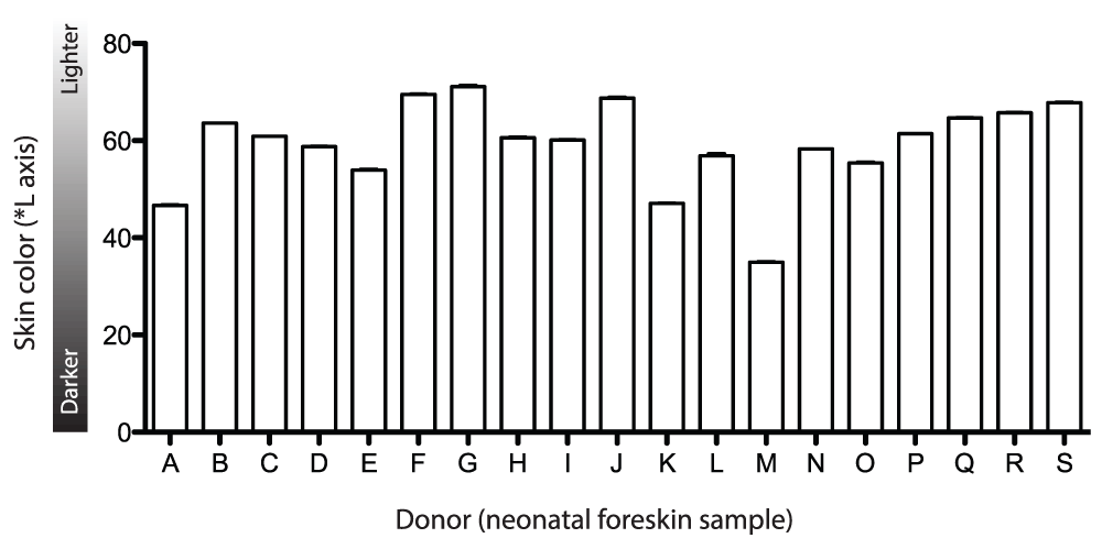
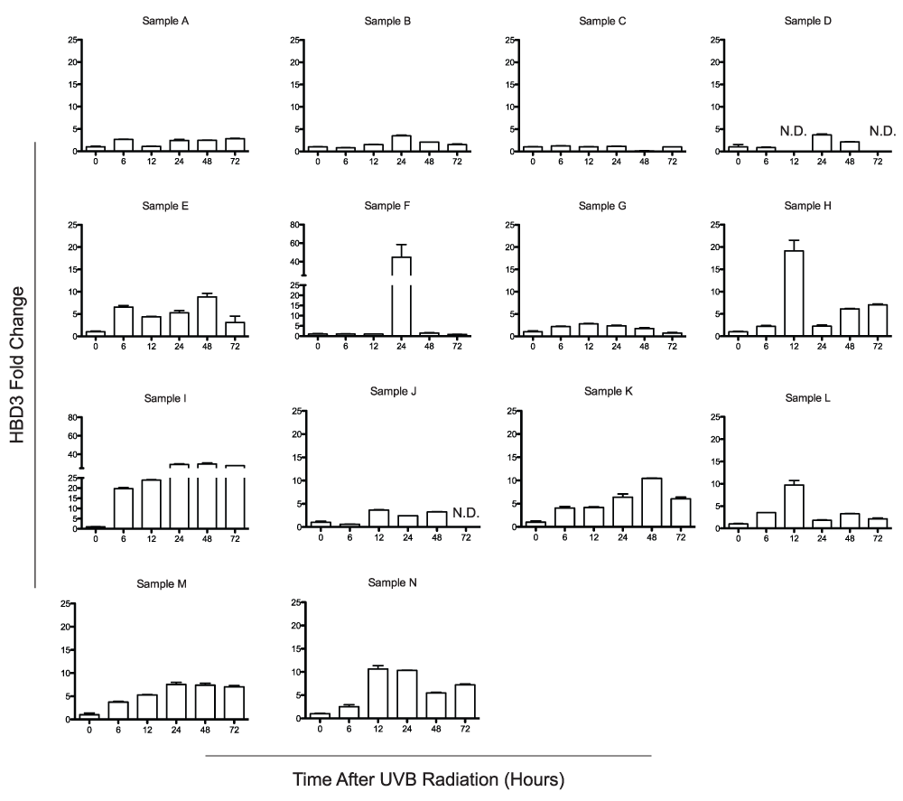
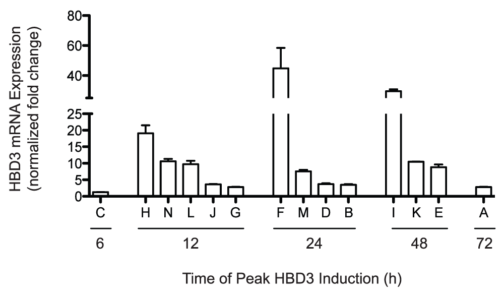
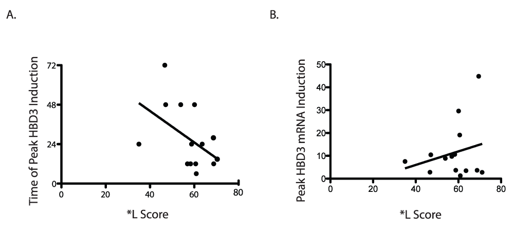
To understand the effects of UV radiation on BD3 expression in human skin, freshly-isolated foreskins were exposed to 0.5 kJ/m2 UVB. Fourteen de-identified samples were obtained from normal healthy male infants undergoing elective circumcision before discharge from the neonatal nursery. Skin pigmentation was measured for each sample by reflective colorimetry in order to estimate melanin content of the epidermis. The skin samples exhibited a range of melanization as determined by the *L score which quantifies color on a black-white color axis (a lower *L score is indicative of a blacker/darker color and correlates with epidermal eumelanin content15). The majority of the samples were derived from light-skinned infants, however at least 3 samples were darker in color (Figure 1). Skin explants were exposed to 0.5 kJ/m2 UVB, and biopsies were taken from the explants at 6, 12, 24, 48, and 72 hours following UV exposure. BD3 mRNA expression was measured by qRTPCR at 6, 12, 24, 48 and 72h after radiation, normalized to TBP, and compared to an unirradiated control taken at time 0. Due to the small size of the skin explants (roughly 1 cm2), it was not possible to have a time-matched mock-irradiated control at each time point, therefore values were normalized to unirradiated controls from each skin sample. We noted extensive variability in both the timing and magnitude of BD3 induction across individuals (Figure 2). Normalized BD3 fold induction ranged from 1.3-fold to 44.8-fold, and peak induction ranged from 6–72 hours depending on the sample (Figure 3). We tested whether the amount of BD3 expression correlated with skin pigmentation, hypothesizing that more melanin in the skin might inhibit UV penetration into the skin and therefore blunt UV effects on BD3 expression. In fact, BD3 expression did not appear to be influenced by pigment phenotype, as manifested by a positive trend between higher BD3 expression and darker skin samples (Figure 4A; r2 = 0.057, p = 0.41). Similarly, a negative trend between skin color and time of peak BD3 expression was observed, although this too did not reach statistical significance (Figure 4B; r2 = 0.234, p = 0.08).
Figure 1. Degree of skin pigmentation from each donor.
Skin color determination is shown for each sample. *L Score is measured by reflective colorimetry and represents color of the skin on a black-white axis. Lower *L score is indicative of a more darkly pigmented phenotype. Data represent the average *L score ± SEM for three measurements per skin sample.
Figure 2. BD3 expression over time among 14 distinct donor after UV radiation.
Fourteen independent human skin explants were treated ex vivo with 0.5 kJ/m2 UVB radiation. BD3 mRNA expression was determined at 6, 12, 24, 48, and 72 hours following UV treatment and compared to an untreated control. qRTPCR was performed in duplicate for each sample, and results are expressed as mean fold change over control ± SEM.
Figure 3. Time of maximal BD3 expression after UV radiation.
Peak BD3 mRNA expression for human skin explants (n=14) is arranged by time of maximal induction for each individual donor. qRTPCR was performed in duplicate for each sample, and results are expressed as mean fold change over control ± SEM.
Figure 4. Relationship between donor skin color and BD3 expression.
A) *L score versus peak BD3 mRNA induction. qRTPCR was performed in duplicate for each sample, and data represent mean BD3 induction for 14 human skin explants. There was no correlation between donor *L score and amplitude of BD3 induction (r2 = 0.057, p = 0.41). B) *L score versus time of peak BD3 mRNA induction. qRTPCR was performed in duplicate for each sample, and data represent mean BD3 induction for 14 human skin explants. Although a weak negative trend existed between donor *L score and time of BD3 induction, the correlation was not statistically significant (r2 = 0.234, p = 0.08).
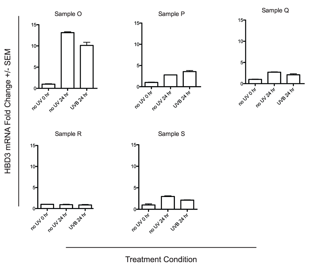
Figure 5. UV-independent BD3 expression in human skin explants cultured ex vivo.
Five additional human skin explants were treated ex vivo with 0.5 J/m2 UVB radiation. BD3 mRNA expression for the UV treated biopsy and an untreated time matched control was compared to an untreated control taken at the time of UV treatment. qRTPCR was performed in duplicate for each sample, and data represent the mean fold change over the untreated control taken at the time of UV treatment ± SEM. BD3 induction in the human skin samples was independent of UVB treatment.
We then considered the possibility that BD3 expression might be affected simply by time in culture and measured BD3 expression over time in samples exposed to 0 or 0.5 kJ/m2 UV exposure. Each of five explants were divided into three sections and sampled either at time 0 (no UV) or at 24h following exposure to either 0 or 0.5 kJ/m2 UVB. Similar to prior experiments, BD3 expression was measured by qRTPCR and normalized to TBP, however values could also be compared with mock-irradiated, time-matched conditions. We observed clear induction of BD3 expression in each of the mock-irradiated samples over time (Figure 5), and exposure to 0.5 kJ/m2 UV did not substantially alter BD3 mRNA expression when compared to individual mock-irradiated time-matched controls. These data suggest that either tissue removal or the process of culturing skin explants ex vivo is sufficient to enhance BD3 expression in whole human skin and that the addition of 0.5 kJ/m2 UV does not impact BD3 expression in this setting.
Dataset 1.Colorimetry measurements from each donor.
Skin pigmentation was determined via reflective colorimetry and is represented by an *L score. The *L score was measured three times for each sample. The “A” column represents each donor. The “B”, “C”, and “D” columns represent the first, second, and third measured *L score respectively.
Dataset 2.Cycle threshold values for qRTPCR.
Cycle threshold (CT) values were calculated for BD3 and TBP (housekeeping genes) for each donor. CT values were determined in duplicate for each sample. The “A” column represents the donor. The “B” column represents the sample treatment for the donor. The “C” column represents the target mRNA measured. The “D” column represents the CT value determined for that sample.
Conclusions/discussions
In an effort to develop a model in which to study UV induction of cutaneous BD3, we measured its expression over time in UV-naïve human skin explants. Although there was a high degree of variability in the magnitude and kinetics of BD3 induction between samples harvested from different donors, we observed BD3 up-regulation in each case. To control for the possibility that tissue processing and/or ex vivo culture conditions might impact BD3 expression in the explants, we compared BD3 mRNA levels between mock-irradiated versus UV-treated sections of skin samples harvested from the same donor. This experiment, which included samples from five donors, indicated that BD3 expression increased over time irrespective of UV exposure (at 0.5 kJ/m2), suggesting that BD3 expression is induced in human skin explants in a UV-independent manner.
BD3 expression is reported to be up-regulated in wound healing processes16, therefore it might be plausible that its increase over time in skin explants may be related to normal wound physiologic processes activated by surgical excision of the skin and/or its processing after harvest. Indeed, the small size of the skin samples isolated from neonatal circumcision (on average 1–1.5 cm2) implies that the majority of the tissue in the explant will be in close proximity to at least one cut surface, raising the possibility of local trauma-induced factors contributing to BD3 expression in the samples. It may also be possible that harvesting skin biopsies from the sample over the experimental time course may have promoted wounding responses in the explants and fueled BD3 expression. Alternatively, it is possible that one or more factors involved in sustaining the skins in culture (media, temperature, oxygen tension, pH, etc.) may have promoted BD3 expression in the explants. We do not as yet understand the mechanisms underlying variability of BD3 induction amplitude or kinetics observed between samples, however it is possible that wounding or inflammatory responses induced by tissue removal may vary between normal individuals. We conclude that because of confounding variables involved in their generation and maintenance, neonatal foreskin explants may not be an appropriate model to isolate the effects of UV on BD3 expression in the skin, however other models may still be appropriate.
Consent
De-identified neonatal foreskin samples were obtained from the University of Kentucky’s Chandler Medical Center Newborn Nursery without accompanying clinical information under an institutionally-reviewed IRB-exempted status.
EWH and JD conceived the study and designed the experiments. EWH carried out the research and data analysis. JD and EWH wrote the manuscript. All authors agree to the final content.
Competing interests
No competing interests were disclosed.
Grant information
This work was funded by the National Cancer Institute (R01 CA131075) awarded to JD, as well as T32CA165990 which supported EWH.
The funders had no role in study design, data collection and analysis, decision to publish, or preparation of the manuscript.
Acknowledgements
The authors wish to thank the staff of the University of Kentucky Obstetrics and Pediatric clinical services for their help in obtaining freshly-isolated neonatal foreskin samples and alerting us to their collection in a timely manner.
Faculty Opinions recommended
References
1.
Suzuki I, Cone RD, Im S, et al.:
Binding of melanotropic hormones to the melanocortin receptor MC1R on human melanocytes stimulates proliferation and melanogenesis.
Endocrinology.
1996; 137(5): 1627–33. PubMed Abstract
| Publisher Full Text
2.
Kadekaro AL, Kavanagh R, Kanto H, et al.:
alpha-Melanocortin and endothelin-1 activate antiapoptotic pathways and reduce DNA damage in human melanocytes.
Cancer Res.
2005; 65(10): 4292–9. PubMed Abstract
| Publisher Full Text
3.
Millar SE, Miller MW, Stevens ME, et al.:
Expression and transgenic studies of the mouse agouti gene provide insight into the mechanisms by which mammalian coat color patterns are generated.
Development.
1995; 121(10): 3223–32. PubMed Abstract
4.
Suzuki I, Tada A, Ollmann MM, et al.:
Agouti signaling protein inhibits melanogenesis and the response of human melanocytes to alpha-melanotropin.
J Invest Dermatol.
1997; 108(6): 838–42. PubMed Abstract
| Publisher Full Text
6.
Kaelin CB, Candille SI, Yu B, et al.:
New ligands for melanocortin receptors.
Int J Obes (Lond).
2008; 32(Suppl 7): S19–27. PubMed Abstract
| Publisher Full Text
7.
Beaumont KA, Smit DJ, Liu YY, et al.:
Melanocortin-1 receptor-mediated signalling pathways activated by NDP-MSH and HBD3 ligands.
Pigment Cell Melanoma Res.
2012; 25(3): 370–4. PubMed Abstract
| Publisher Full Text
| Free Full Text
8.
Swope VB, Jameson JA, McFarland KL, et al.:
Defining MC1R regulation in human melanocytes by its agonist alpha-melanocortin and antagonists agouti signaling protein and beta-defensin 3.
J Invest Dermatol.
2012; 132(9): 2255–62. PubMed Abstract
| Publisher Full Text
| Free Full Text
9.
Cui R, Widlund HR, Feige E, et al.:
Central role of p53 in the suntan response and pathologic hyperpigmentation.
Cell.
2007; 128(5): 853–64. PubMed Abstract
| Publisher Full Text
10.
Glaser R, Navid F, Schuller W, et al.:
UV-B radiation induces the expression of antimicrobial peptides in human keratinocytes in vitro and in vivo.
J Allergy Clin Immunol.
2009; 123(5): 1117–23. PubMed Abstract
| Publisher Full Text
11.
Jia HP, Schutte BC, Schudy A, et al.:
Discovery of new human beta-defensins using a genomics-based approach.
Gene.
2001; 263(1–2): 211–8. PubMed Abstract
| Publisher Full Text
12.
Harder J, Bartels J, Christophers E, et al.:
Isolation and characterization of human beta -defensin-3, a novel human inducible peptide antibiotic.
J Biol Chem.
2001; 276(8): 5707–13. PubMed Abstract
| Publisher Full Text
13.
Wagner JK, Jovel C, Norton HL, et al.:
Comparing quantitative measures of erythema, pigmentation and skin response using reflectometry.
Pigment Cell Res.
2002; 15(5): 379–84. PubMed Abstract
| Publisher Full Text
14.
Hong SP, Kim MJ, Jung MY, et al.:
Biopositive effects of low-dose UVB on epidermis: coordinate upregulation of antimicrobial peptides and permeability barrier reinforcement.
J Invest Dermatol.
2008; 128(12): 2880–7. PubMed Abstract
| Publisher Full Text
15.
D'Orazio JA, Nobuhisa T, Cui R, et al.:
Topical drug rescue strategy and skin protection based on the role of Mc1r in UV-induced tanning.
Nature.
2006; 443(7109): 340–4. PubMed Abstract
| Publisher Full Text
16.
Kesting MR, Stoeckelhuber M, Hölzle F, et al.:
Expression of antimicrobial peptides in cutaneous infections after skin surgery.
Br J Dermatol.
2010; 163(1): 121–7. PubMed Abstract
| Publisher Full Text
17.
Wolf Horrell EM, D’Orazio JA:
Colorimetry measurements from each donor.
F1000Research.
2014. Data Source
18.
Wolf Horrell EM, D’Orazio JA:
Cycle threshold values for qRTPCR.
F1000Research.
2014. Data Source
1
The Markey Cancer Center and the Department of Physiology, University of Kentucky College of Medicine, Lexington, KY 40536, USA 2
The Markey Cancer Center and the Department of Pediatrics, University of Kentucky College of Medicine, Lexington, KY 40536, USA
This work was funded by the National Cancer Institute (R01 CA131075) awarded to J.D., as well as T32CA165990 which supported E.M.W.H.
The funders had no role in study design, data collection and analysis, decision to publish, or preparation of the manuscript.
Wolf Horrell E and D'Orazio J. UV-independent induction of beta defensin 3 in neonatal human skin explants [version 1; peer review: 1 approved, 2 approved with reservations]. F1000Research 2014, 3:288 (https://doi.org/10.12688/f1000research.5794.1)
NOTE: If applicable, it is important to ensure the information in square brackets after the title is included in all citations of this article.
track
receive updates on this article
Track an article to receive email alerts on any updates to this article.
Share
Open Peer Review
Current Reviewer Status:
?
Key to Reviewer Statuses
VIEWHIDE
ApprovedThe paper is scientifically sound in its current form and only minor, if any, improvements are suggested
Approved with reservations
A number of small changes, sometimes more significant revisions are required to address specific details and improve the papers academic merit.
Not approvedFundamental flaws in the paper seriously undermine the findings and conclusions
The authors describe their studies of the effects of UV-irradiation on the expression of beta defensin on human neonatal foreskins treated and cultured in vivo. After finding variable responses between donors which did not correlate with skin phototype, they investigated
... Continue reading
The authors describe their studies of the effects of UV-irradiation on the expression of beta defensin on human neonatal foreskins treated and cultured in vivo. After finding variable responses between donors which did not correlate with skin phototype, they investigated the possibility that beta defensin expression was affected by culture conditions. They found that there were no differences between mock- and UV-irradiated samples. We have seen similar changes in genes of interest in the mock-treated tissues using ex vivo skin cultures. This study is valuable in that it highlights the need to include proper controls in this system. I have several additional minor comments to make about the manuscript and data:
The protocol for mRNA isolation notes that tissue is ground in trizol and RNA is purified from the aqueous layer. There will not be two layers until chloroform is added and this should be included in the protocol.
Figure 1 claims to represent degree of skin pigmentation +/- SEM but there are no error bars.
The qPCR data shows Ct values as high as 32.9 for beta defensin, with almost all of the time=0 samples above 29. Many qPCR assays are not linear in this range. Did the authors do an examination of the performance of their primers in this range? Although these high Ct values make me a little skeptical about the absolute values of the fold change reported in figures 2 and 5, I am nevertheless persuaded that the differences between mock- and UV-treated samples is negligible as the authors conclude.
Competing Interests: No competing interests were disclosed.
I confirm that I have read this submission and believe that I have an appropriate level of expertise to confirm that it is of an acceptable scientific standard.
John D'Orazio, The Markey Cancer Center and the Department of Pediatrics, University of Kentucky College of Medicine, Lexington, KY 40536, USA
19 Feb 2015
Author Response
We sincerely thank you for your thorough reading and insightful comments. We have provided a point-by-point response to each of your concerns.
Methods: The protocol for mRNA isolation via Trizol is
...
Continue readingWe sincerely thank you for your thorough reading and insightful comments. We have provided a point-by-point response to each of your concerns.
Methods: The protocol for mRNA isolation via Trizol is not complete.
We have expanded our methods to include these details.
General Concern: Figure 1 claims to represent degrees of skin pigmentation +/- SEM but there are no error bars.
We have expanded our methods to be more specific with how the measurements were determined. The degree of pigmentation for each skin sample was determined three times by colorimetry measurement. Neonatal skin samples were, as a rule, fairly uniform in their pigmentation. Their homogeneity between measurements therefore resulted in very small standard error of the mean values.
We sincerely thank you for your thorough reading and insightful comments. We have provided a point-by-point response to each of your concerns.
Methods: The protocol for mRNA isolation via Trizol is not complete.
We have expanded our methods to include these details.
General Concern: Figure 1 claims to represent degrees of skin pigmentation +/- SEM but there are no error bars.
We have expanded our methods to be more specific with how the measurements were determined. The degree of pigmentation for each skin sample was determined three times by colorimetry measurement. Neonatal skin samples were, as a rule, fairly uniform in their pigmentation. Their homogeneity between measurements therefore resulted in very small standard error of the mean values.
Competing Interests:No competing interests were disclosed.Close
The authors attempt to establish a skin explant model to investigate the effect of UV exposure on the expression levels of b-defensin 3, an antimicrobial peptide that also binds to Melanocortin 1 receptor and attenuates downstream signaling. They find extensive
... Continue reading
The authors attempt to establish a skin explant model to investigate the effect of UV exposure on the expression levels of b-defensin 3, an antimicrobial peptide that also binds to Melanocortin 1 receptor and attenuates downstream signaling. They find extensive variability in the expression levels of b-defensin 3 in the UV-induced samples and cannot demonstrate that is in in fact up-regulated. They do show some minor increase in b-defensin 3 expression over time that is independent of UV irradiation and suggest that this may be due to a wound healing response to the skin excision process. The authors should have included in their analysis a few more genes known to be upregulated after UV exposure (for example, MC1R) both in keratinocytes and melanocytes. This way, they would have been able to define whether their culture system was improper to study b-defensin3 regulation specifically. In the mock irradiated experiment they should have also looked at levels of IL1 and TNFalpha to support their hypothesis that b-defensin 3 increases as a result of a wound healing response. In the paper by Glaser et al (2009) where b-defensin 3 was reported to be up-regulated after UV exposure, the authors used explants of adult skin. Is it possible that newborn and adult skin (perhaps based on the anatomy or level of cellular differentiation) respond differently to UV exposure that could explain the disparate results?
Competing Interests: No competing interests were disclosed.
I confirm that I have read this submission and believe that I have an appropriate level of expertise to confirm that it is of an acceptable scientific standard, however I have significant reservations, as outlined above.
John D'Orazio, The Markey Cancer Center and the Department of Pediatrics, University of Kentucky College of Medicine, Lexington, KY 40536, USA
19 Feb 2015
Author Response
We sincerely thank you for your thorough reading and insightful comments. We have provided a point-by-point response to each of your concerns.
General comment: The authors should have included a few
...
Continue readingWe sincerely thank you for your thorough reading and insightful comments. We have provided a point-by-point response to each of your concerns.
General comment: The authors should have included a few more genes known to upregulated after UV exposure in both keratinocytes and melanocytes.
We have included the induction of additional genes including tyrosinase in four human neonatal skin samples. We determined that tyrosinase gene expression was induced following UV radiation in half of the skin samples suggesting the model with our culture conditions may be appropriate to study other genes.
General Comment: The authors should have looked at levels of IL1 and TNF alpha to support their hypothesis that BD3 increases as a result of wound healing.
We have included the gene expression data for TNF alpha following UV radiation and determined that its expression correlates with BD3 expression.
General Comment: Is it possible that the neonatal skin explants behave differently than the adult skin explants?
Yes, this is a possibility, however as our study was limited to neonatal explants, we cannot directly address it with data. However, we thank the reviewer for this comment and have raised this caveat in our revised discussion.
We sincerely thank you for your thorough reading and insightful comments. We have provided a point-by-point response to each of your concerns.
General comment: The authors should have included a few more genes known to upregulated after UV exposure in both keratinocytes and melanocytes.
We have included the induction of additional genes including tyrosinase in four human neonatal skin samples. We determined that tyrosinase gene expression was induced following UV radiation in half of the skin samples suggesting the model with our culture conditions may be appropriate to study other genes.
General Comment: The authors should have looked at levels of IL1 and TNF alpha to support their hypothesis that BD3 increases as a result of wound healing.
We have included the gene expression data for TNF alpha following UV radiation and determined that its expression correlates with BD3 expression.
General Comment: Is it possible that the neonatal skin explants behave differently than the adult skin explants?
Yes, this is a possibility, however as our study was limited to neonatal explants, we cannot directly address it with data. However, we thank the reviewer for this comment and have raised this caveat in our revised discussion.
Competing Interests:No competing interests were disclosed.Close
The study by Horell and D'Orazio aimed at addressing the production of BD3, a MC1R antagonist, in neonatal human foreskins, using skin explants, and its possible regulation by UV irradiation. The authors did not find a correlation between the pigmentary status
... Continue reading
The study by Horell and D'Orazio aimed at addressing the production of BD3, a MC1R antagonist, in neonatal human foreskins, using skin explants, and its possible regulation by UV irradiation. The authors did not find a correlation between the pigmentary status of the skin and BD3 production, or a consistent effect of UV radiation. However, they noted altered production of BD3 over time in the skin explants, which they attributed to a wound repair-like reaction, or to inadequate culture conditions for the skin explants. They concluded that the use of foreskins in organotypic culture is not an appropriate model to assess BD3 production.
It is not clear from the report exactly how the skin explants were maintained for the duration of the experiments. Were they maintained in a humidified incubator at 37°C? Was the culture medium changed daily? Obviously, the culture conditions can affect the viability of the explants. Did the authors check on the viability of the skin, especially at the end of the experiment (e.g. by examining the histology after H&E staining)? This is important, and the "health" of the skin can explain the erratic production of BD3. If the viability of the skin is compromised, this will have a generalized effect on its metabolic state.
Since IL-1 is known to stimulate BD3 expression, did the authors check the levels of IL-1, particularly after UV exposure?
If the change in BD3 production is due to a wound healing-like response, the authors might have to consider cutting the foreskins into equal parts at the beginning of the experiment, to avoid taking biopsies at different time points.
It could very well be that the experimental conditions are responsible for the unexpected results that were obtained. So the conclusion that using foreskin explants is not an appropriate model might not be necessarily true.
I suggest that the authors submit this report as a "methodology" report, after investigating the concerns raised above.
Competing Interests: No competing interests were disclosed.
I confirm that I have read this submission and believe that I have an appropriate level of expertise to confirm that it is of an acceptable scientific standard, however I have significant reservations, as outlined above.
John D'Orazio, The Markey Cancer Center and the Department of Pediatrics, University of Kentucky College of Medicine, Lexington, KY 40536, USA
19 Feb 2015
Author Response
We sincerely thank you for your thorough reading of our manuscript and insightful comments. We have provided a point-by-point response to each of your concerns.
Methods: It is unclear as to
...
Continue readingWe sincerely thank you for your thorough reading of our manuscript and insightful comments. We have provided a point-by-point response to each of your concerns.
Methods: It is unclear as to how the skin explants were maintained.
We have expanded the methods to include these details.
General Comment: Did the authors check the viability of skin samples?
We have included hematoxylin and eosin stained histological slides for the skin explants at 0 and 24 hours following culture conditions to assess the viability of the samples and have determined the samples were still viable. Furthermore induction of TNF alpha suggests viability of the tissues over the time course of the experiment.
General Comment: Did the authors check the level of IL-1B following UV radiation?
We have determined the levels of TNF alpha which is also known to induce beta-defensin 3 expression. TNF induction appears to correlate with the beta-defensin 3 induction suggesting they are related.
Methods: The authors may consider cutting the foreskins into equal parts at the beginning of the experiment to determine whether the change is due to a wound healing response.
The samples were divided into equal parts at the beginning of the experiment prior to UV radiation. We apologize for the confusion in the methods and have updated the methods to include this information.
General Comment: The conclusion that the use of human neonatal foreskins may not be an appropriate model to study BD3 induction may not be true and the results may be due to the experimental conditions.
We thank the reviewer for this comment and have adjusted our discussion to accommodate it.
General Comment: The authors should submit this report as a “methodology report.”
Given the fact that our data raise important caveats about the use of neonatal human foreskins to study BD3 induction, we feel that our findings may be better reported as a research article rather than as a methodology report.
We sincerely thank you for your thorough reading of our manuscript and insightful comments. We have provided a point-by-point response to each of your concerns.
Methods: It is unclear as to how the skin explants were maintained.
We have expanded the methods to include these details.
General Comment: Did the authors check the viability of skin samples?
We have included hematoxylin and eosin stained histological slides for the skin explants at 0 and 24 hours following culture conditions to assess the viability of the samples and have determined the samples were still viable. Furthermore induction of TNF alpha suggests viability of the tissues over the time course of the experiment.
General Comment: Did the authors check the level of IL-1B following UV radiation?
We have determined the levels of TNF alpha which is also known to induce beta-defensin 3 expression. TNF induction appears to correlate with the beta-defensin 3 induction suggesting they are related.
Methods: The authors may consider cutting the foreskins into equal parts at the beginning of the experiment to determine whether the change is due to a wound healing response.
The samples were divided into equal parts at the beginning of the experiment prior to UV radiation. We apologize for the confusion in the methods and have updated the methods to include this information.
General Comment: The conclusion that the use of human neonatal foreskins may not be an appropriate model to study BD3 induction may not be true and the results may be due to the experimental conditions.
We thank the reviewer for this comment and have adjusted our discussion to accommodate it.
General Comment: The authors should submit this report as a “methodology report.”
Given the fact that our data raise important caveats about the use of neonatal human foreskins to study BD3 induction, we feel that our findings may be better reported as a research article rather than as a methodology report.
Competing Interests:No competing interests were disclosed.Close
Alongside their report, reviewers assign a status to the article:
Approved - the paper is scientifically sound in its current form and only minor, if any, improvements are suggested
Approved with reservations -
A number of small changes, sometimes more significant revisions are required to address specific details and improve the papers academic merit.
Not approved - fundamental flaws in the paper seriously undermine the findings and conclusions
Spreadsheet data files may not format correctly if your computer is using different default delimiters (symbols used to separate values into separate cells) - a spreadsheet created in one region is sometimes misinterpreted by computers in other regions. You can change the regional settings on your computer so that the spreadsheet can be interpreted correctly.
How to fix it
Save downloaded CSV file
Open spreadsheet program (e.g. Excel)
Click the ‘Data’ tab at the top
Click the ‘From text’ icon (top left)
Browse for downloaded CSV file, click ‘Import’
Ensure ‘Delimited’ radio button is selected, click ‘Next’
Check one of the appropriate delimiter checkboxes (you can visualize the formatting by looking at the data preview below these options)
Wolf Horrell E and D'Orazio J. Dataset 1 in: UV-independent induction of beta defensin 3 in neonatal human skin explants. F1000Research 2014, 3:288 (https://doi.org/10.5256/f1000research.5794.d39057)
Spreadsheet data files may not format correctly if your computer is using different default delimiters (symbols used to separate values into separate cells) - a spreadsheet created in one region is sometimes misinterpreted by computers in other regions. You can change the regional settings on your computer so that the spreadsheet can be interpreted correctly.
How to fix it
Save downloaded CSV file
Open spreadsheet program (e.g. Excel)
Click the ‘Data’ tab at the top
Click the ‘From text’ icon (top left)
Browse for downloaded CSV file, click ‘Import’
Ensure ‘Delimited’ radio button is selected, click ‘Next’
Check one of the appropriate delimiter checkboxes (you can visualize the formatting by looking at the data preview below these options)
Wolf Horrell E and D'Orazio J. Dataset 2 in: UV-independent induction of beta defensin 3 in neonatal human skin explants. F1000Research 2014, 3:288 (https://doi.org/10.5256/f1000research.5794.d39058)
Adjust parameters to alter display
View on desktop for interactive features
Includes Interactive Elements
View on desktop for interactive features
Competing Interests Policy
Provide sufficient details of any financial or non-financial competing interests to enable users to assess whether your comments might lead a reasonable person to question your impartiality. Consider the following examples, but note that this is not an exhaustive list:
Examples of 'Non-Financial Competing Interests'
Within the past 4 years, you have held joint grants, published or collaborated with any of the authors of the selected paper.
You have a close personal relationship (e.g. parent, spouse, sibling, or domestic partner) with any of the authors.
You are a close professional associate of any of the authors (e.g. scientific mentor, recent student).
You work at the same institute as any of the authors.
You hope/expect to benefit (e.g. favour or employment) as a result of your submission.
You are an Editor for the journal in which the article is published.
Examples of 'Financial Competing Interests'
You expect to receive, or in the past 4 years have received, any of the following from any commercial organisation that may gain financially from your submission: a salary, fees, funding, reimbursements.
You expect to receive, or in the past 4 years have received, shared grant support or other funding with any of the authors.
You hold, or are currently applying for, any patents or significant stocks/shares relating to the subject matter of the paper you are commenting on.
Stay Updated
Sign up for content alerts and receive a weekly or monthly email with all newly published articles
Comments on this article Comments (0)