Keywords
Really direct brain-to-brain communications, Mind-to-mind Qualia, Consciousness, Event-related brain potentials (ERPs), P3b-P600, IAPS stimuli
Really direct brain-to-brain communications, Mind-to-mind Qualia, Consciousness, Event-related brain potentials (ERPs), P3b-P600, IAPS stimuli
Colors, sounds and smells do not exist in the outside world. They are the creations of our brain in response to light waves, rhythmic variations of air pressure and inhaled molecules, respectively. External stimulations are responsible for action potentials whose processing in the brain may then produce colors, sounds and smells. These, so-called qualia1,2 are then apparently projected outside around us and constitute our perceptual world, sometimes called the phenomenal world3. Although perceived internally, except in the case of induced out-of-body experiences4, events, such as feelings of meanings, as in the tip of the tongue phenomenon, or such as emotions, conscious intentions to act and sensations of our body can also be seen as qualia.
Understanding consciousness as consisting of self made qualia leads to one of the most enduring philosophical questions: Are qualia the same across individuals? In other words, is the yellow produced by the brain of one person the same as the yellow produced by the brain of another person? Surprisingly, there is no way to know for sure. The fact that the same word is used by all the people speaking a language to designate a qualia merely establishes a correspondence. It does not prevent the qualia it indicates from varying across these people. The yellow qualia for one person could, for instance, be the blue qualia for another person. Nevertheless, such differences across individuals appear unlikely since many use the same associations and agree that red is a warm color and that blue relates to sadness. In the auditory modality, many associate high pitch sounds with sharpness. Moreover, we use the same metaphor and define these sounds as “high” whereas those of longer wavelengths are said to be “low” sounds (for other metaphors see for instance Lakoff and Johnson5). The same relations between qualia thus seem to exist across people whereas if qualia were different across individuals it seems that these relations should differ. It could be agued that metaphors used in a language convey relationships between certain qualia and are thus responsible for building the links between them. However, it seems that new metaphors can be understood at their first occurrence6, which suggests that relations between qualia are, at least partly, independent of language.
In any case, if the qualia produced by our brains in response to a given stimulus were not similar across individuals, one could call the entire human race delusional since we all go through our everyday lives and interact with others as if they perceive the world in pretty much the same way as we do. As a matter of fact, if the phenomenal world of each individual were unique, the most fundamental social consensus would be lost. Sharing feelings by verbalizing emotions would be an illusion and our use of language as if each word designates the same qualia would be incorrect. It thus appears reasonable to hypothesize that qualia are similar across individuals and that we are actually living in similar phenomenal worlds.
At first sight, it is tempting to say that qualia could be similar because of the resemblances existing between the brains of humans. However, this idea is questionable for several reasons. First, when macroscopically comparing the brain of people, one can be stricken by the large differences existing between their shapes (with some extreme, such as the one described by Feuillet et al.7). There are also problems at the microscopic level. For instance, nothing has been found that distinguishes the so-called color-cells of V1 for blue from the V1 color cells for yellow apart from their afferences8. Thus, applied within a person, the neuronal similarity argument would predict that qualia for blue should be similar to the qualia for red or yellow. Another point can be made with the qualia for white, which is generated by the stimulation of the three types of cone cells. Or even by only two complementary colors (e.g., green and red, which stimulate the M and the L cone cells, or blue and yellow). How could the V1 “color cells”, which are processing the output of these cone cells generate the same qualia? There again, similarities between particular neurons and qualia do not work. So the hypothesis of a similarity of qualia creates a problem. How could qualia be similar across individuals when they are said to be, by nature, totally private events not strictly dependent on brain similarities?
Another, apparently unrelated, question is: how can qualia within a given person be so qualitatively different from one another while theoretically originating from the same type of neuronal bioelectrical activity? Sounds appear to be totally orthogonal to colors or smells. Nevertheless, they are induced by the same depolarizations, such as those induced by Penfield and Jasper9 at different places of the cortex. One way to answer this question is to hypothesize that, while dependent on the well-known bioelectrical activities of neurons, the physical nature of qualia is not limited to these activities. The authors of this second hypothesis can grossly be divided into those suggesting, (a) that qualia are also electromagnetic fields (for a recent review, see Jones10) and (b) those developing the even more controverted theory that qualia also include modulations of the wave function described by quantum mechanics (e.g., 11). Each of these two theories thus introduces phenomena, which, by the immense variety of the instances they include, could provide ways to account for the qualitative differences existing between percepts.
Interestingly, thinking about qualia in terms of electromagnetic fields or in terms of modulations of the wave function could also provide a hint as to how qualia are apparently projected to form our perceived environment and also how they could be similar across individuals while being “private events”. Indeed, both physical phenomena propagate. They can thus be projected and travel between individuals. Therefore, some kind of inter-subjective sharing could theoretically occur. In other words, experiencing a qualia might have an impact on the qualia of another person. This means that, at least in some conditions, the brain activity of a person might be influenced by the activity of the brain of another person. No study has reliably12 reported such a direct brain to brain impact but that might be due to the fact that, to the best of our knowledge, no author has yet specifically explored the possibility that qualia propagate.
Testing this possibility was the first aim of the present study. To achieve this goal, we focused on the centro-parietal P600, a late event-related brain potential (ERP) elicited by the presentation of meaningful stimuli, such as, words, objects, faces and scenes. This component belongs to the P3b family of components despite its late maximum, which occurs around 600 ms post stimulus onset when using complex stimuli such as words, objects, faces and even a little later when using scenes. Its amplitude has been reliably related to conscious perception. The greater the amount of information placed in working memory, the larger the amplitude of this potential13,14. Meaningful stimuli presented during attentional blinks that are not consciously perceived elicit no P600, whereas these stimuli do so systematically when they are consciously perceived15,16. On the contrary, the negative component that precedes it, namely, the N400, is evoked by these stimuli even when their processing is only preconscious15–17. Our first goal was thus to measure the amplitude of the P600 elicited by the presentation of a meaningful stimulus to a subject and see if it could depend on whether another person is simultaneously presented with the same stimulus or with a different one (for our purposes, the sameness factor) when one person could not see what the other was presented with. One, potentially greater, impact on the P600s was predicted in the case where different stimuli were presented to each subject, given that the qualia corresponding to each stimulus would be different, and thus that they would generate a greater amount of information in working memory. A different, potentially smaller, impact on the P600s was foreseen in the cases of identical stimuli, since qualia of each person would be similar. An ERP difference between these sameness conditions would support propagation of qualia from the brain of a person to the brain of another person. If this difference pertains to the P600, it would also provide a strong argument for the possibility that the qualia of one person can impact that of another person since qualia are defined here as the building blocks of consciousness and since P600s index consciousness13–16.
However, if the brain activity of one person could have an impact on the brain activity of another person, it seems that this impact should be prevented as much as possible when it is known that the other person is confronted with a different stimulus. Indeed, in these conditions, it seems that the qualia of that person should not interfere. The second aim of the present study was thus to manipulate the beliefs of each pair of participants (for our purposes, the belief factor) by telling them that they would be presented with the same stimuli in some conditions and with different stimuli than their partner in other conditions. These statements were true in half of the blocks and false in the other half, while, again, participants had no way to check and no reason to doubt the statements. Our operational hypothesis was that the hypothesized impact of one participant of a pair on the amplitude of the P600 of the other participant would be minimal when participants are presented with different stimuli and believe it. This was used as our baseline condition.
The third aim of the study had no relation whatsoever to the exploration of the causes of the assumed similarity of qualia across individuals. It was totally separate from the possibility of an impact of one’s activity on the brain of another person. This third goal was to evaluate the impact of social cognition on memory. Indeed, having a mental representation of a partner going through an event (i.e., the presentation of a stimulus), in addition to having a representation of oneself going through the same event, might enrich the encoding in episodic memory and facilitate delayed recognition. Thus, subjects were told to remember each image because there would be a memory test at the end. Our operational hypothesis was that they would have a higher rate of recognition for the stimuli they were presented with when they believed they were seeing the same stimuli as their friend and a lower rate of recognition for the other stimuli.
Thirty-two right-handed participants (25 F, 7 M), pairs of friends, couples, or siblings were recruited because it was assumed, for this first attempt, that testing people in a close relationship could increase the odds of positive findings. The 32 subjects of the 16 pairs underwent exactly the same procedure. All participants learned about the experiment through classified ad websites. They spoke fluent English, were between eighteen and thirty years of age (mean = 23.1, SD = 3.4) and had completed, or were in the process of completing, a university degree. They had normal or glasses-corrected to normal vision. Participants were excluded if they consumed more than twelve drinks of alcoholic beverages per week or if they used recreational drugs, except if they used marijuana less than once per week. Participants were also excluded if they had a history of psychiatric disorder, took medication related to such a disorder, or if one of their first degree relatives had a history of schizophrenia or bipolar disorder. All these inclusion- and exclusion-criteria were checked by an eligibility questionnaire.
The two participants of each pair came to the lab together for approximately three hours. Each participant read and signed an informed consent form accepted by the Douglas Institute Research and Ethics Board. This board, which follows the principles expressed in the declaration of Helsinki, also approved the study itself (Douglas REB #12/12). Data were anonymesed, which did not distort scientific meaning.
Stimuli were images selected from the International Affective Picture System (IAPS,18). Using our own judgment, we chose the 560 most striking pictures of this set to ensure the maintenance of participants’ attention during the tasks. The experiment consisted first of the study phase, which included four blocks, and of a memory test phase. As presented in Table 1, which explains their acronyms, each of the four blocks of the study phase, DBd, SBs, SBd and DBs, corresponded to a particular sameness and belief condition. The order of presentation of these four blocks was randomized across subject pairs using a Latin square. We used four different sets of 70 IAPS stimuli. The allocation of each set to each block was also randomized across subject pairs. In study phase blocks in which different pictures were seen by each member of a pair (i.e., in the DBd and DBs blocks), the picture set seen by one participant in DBd was seen by the other participant in DBs, and vice versa. Therefore, all pictures of the four sets were seen by both participants during the study phase. The memory test phase consisted of a fifth set of pictures that contained, in a random order, all the pictures of the study phase mixed with 280 additional pictures.
The study phase (Table 1) was followed by the memory test phase. As illustrated by Figure 1, each stimulus of the study phase was presented for 1000 ms and was followed by a white screen with a black fixation cross, the duration of which randomly varied between 790 and 1500 ms to prevent the development of a contingent negative variation19. Participants could see their partner in their very peripheral vision field without moving their eyes. Nevertheless, even if they moved their eye or did head movements, they could not see the part of the screen their partner was watching (Figure 2 illustrates this unusual setting). Participants were told to look at each picture for the subsequent memory test phase. Stimuli in that latter phase were presented for 3000 ms in order to allow time for participants to respond.
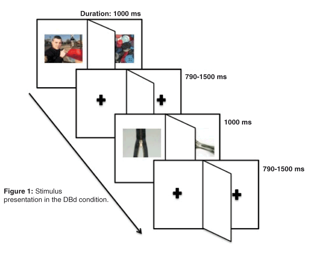
The different-and-believed-different (DBd) condition is used as an example. Note the division of the screen into two halves by the vertical cardboard piece, preventing the two subjects from seeing each other’s stimuli, but not preventing them from feeling close to one another.
During the memory test phase, participants were required to respond by pressing keys on a shared computer keyboard. The participant seated on the left hand side of the keyboard used the typewriter keys and pressed ‘1’ to indicate (s)he believed to have seen the picture previously, and ‘2’ to indicate (s)he believed not to have seen the picture previously. The participant seated on the right hand side of the keyboard used the numeric keypad and pressed ‘4’ to indicate (s)he believed to have seen the picture previously, and ‘5’ to indicate (s)he believed not to have seen the picture previously.
At the end of the memory test phase, there was a debriefing session where participants were asked 4 questions, mainly designed to explore attention differences and whether they detected any deception. The first was: “Did you feel more attentive/distracted seeing the pictures when your friend was present?”. The second was: “Did you feel any different when you knew your friend/partner/relative was looking at the same images that you were seeing?”. The third was: “Did you feel any different when you knew your friend/partner/relative was looking at different images than you were seeing?”. The fourth was: “Did you feel deceived at any point during the experiment?”.
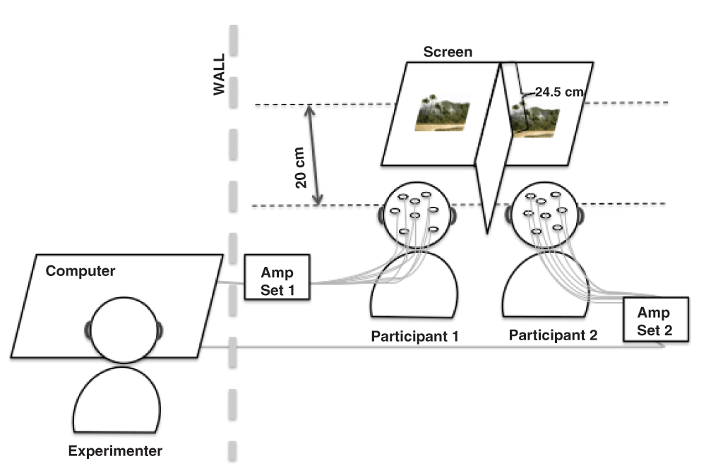
Behavioral key presses were recorded during the memory test phase, as well as the verbatim of the response to the debriefing session’s questions. The electroencephalogram was recorded from 28 electrodes mounted in an elastic cap (Electro-Cap International) during the study phase. Electrodes were placed according to the modified expanded 10–20 system20. For each participant of each pair, these electrodes were grouped into three subsets: sagittal (Fz, Fcz, Cz and Pz), parasagittal (Fp1/2, F3/4, Fc3/4, C3/4, Cp3/4, P3/4, and O1/2), and lateral (F7/8, Ft7/8, T3/4, Tp7/8 and T5/6). There was a separate set of amplifiers for each participant. The right earlobe was used in each subject as the reference for his/her set of amplifiers while the ground of each participant was taken from an electrode two centimeters ahead of Fz. For both sets of amplifiers, high- and low-pass filter half-amplitude cut-offs were set at 0.01 and 100 Hz, respectively, using an additional 60 Hz electronic notch filter. EEG signals were amplified 10,000 times and digitized online at a 256 Hz sampling rate and stored in a single file with 56 (28 × 2) channels.
In each trial, electrodes contaminated by eye movements, excessive myogram, amplifier saturations or analog to digital clipping were removed offline by setting automatic rejection criteria. Electrodes for which analog to digital clipping exceeded a 100 ms duration and electrodes for which amplitude exceeded +/- 100 mV were discarded. Before these rejections, the baseline was set prior to the onset of the stimulus, from -200 to 0 ms. Averages were calculated for each block and each subject in a 1400 ms time window, beginning 200 ms before the onset of the stimulus and lasting for 1200 ms after the stimulus onset. Following averaging, each file was divided into two files, each containing the ERPs of a single subject. The ERPs of each of the 32 subjects were then computed and measured independently of the pair of participants they initially belong to. Based on our a priori hypothesis, we focused on the late positive component (LPC or P600) and computed the mean voltages of ERPs in the 600–900 ms time window for all electrodes, all conditions and all subjects.
Three repeated-measures ANOVAs were run with the version 20 of the IBMSPSS software package to analyze these measures using a multivariate approach. They had sameness (same vs. different stimuli), belief (belief that stimuli were the same vs. belief they differed) and electrodes as within-subject factors. For parasagittal and lateral electrodes, a fourth within-subject factor, hemiscalp (right vs left), was included. Given that there was only one group of 32 subjects, there was not any between-subject factor. Post-hoc analyses were completed for interactions whose p values were smaller than 0.1. The Greenhouse and Geisser21 procedure was used when required to compensate for heterogeneous variances, in which case the original F values and the corrected p values will be given. To provide a priori hypotheses for future studies, we also completed one-way ANOVAs at each electrode to assess each effect found.
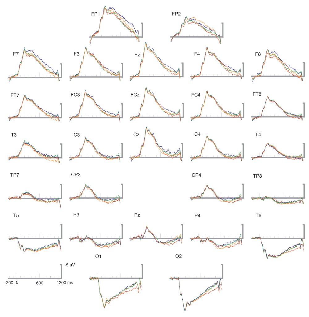
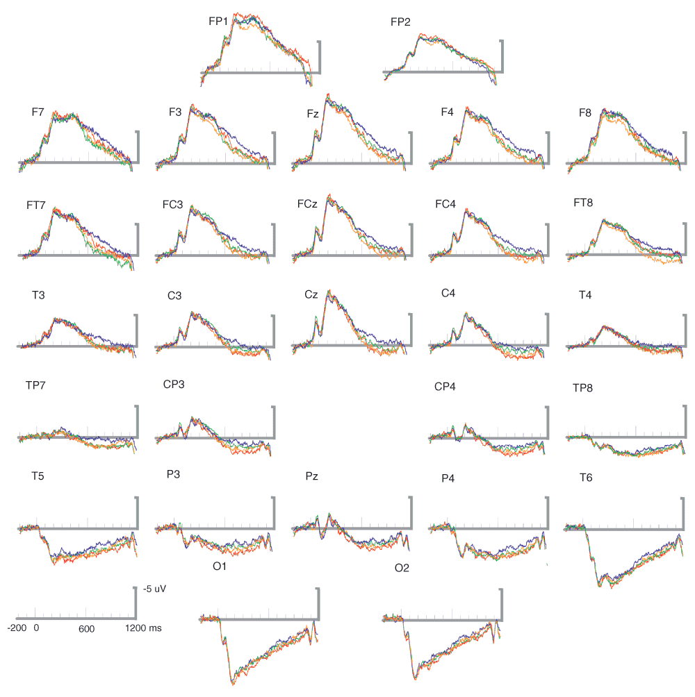
Figure 3 shows the grand averages for the 32 subjects of the 16 pairs tested. Visual inspection of the P600 time window at the electrodes where the amplitude of this ERP component is usually maximal, that is, at the central (Cz) and parietal (Pz) midline sites, reveals that the smallest P600s were obtained for the baseline condition of the study phase where stimuli were different and where participants believe they were seeing an image different from that presented to the other member of their pair (the DBd condition, with the blue waveforms in Figure 3). P600s appear a little bit larger for the Same Believe-same condition (SBs, green waveforms) and maximal for the different believe-same (DBs, orange) and the same believed different condition (SBd, red).
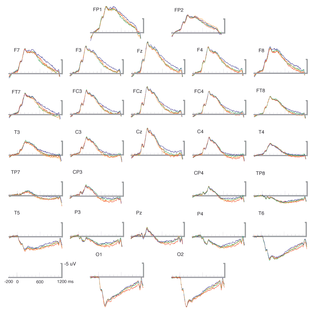
Blue waveforms are for the condition where the two stimuli were different and were believed to be different (DBd); green for when they were, and were believed to be, the same (SBs); orange: different stimuli but believed to be the same (DBs); red, same believed-different (SBd).
Table 2 includes the F and p values of the ANOVAs performed on each subset of electrodes.
Table 3 contains the results of the post-hoc analyses run for each electrode subset to explore the sameness × belief interactions reported in Table 2.
In addition to the findings presented in Table 2 and Table 3, a significant belief × hemiscalp interaction at the lateral electrode set prompted a further analysis, which revealed a marginally significant effect of belief over the left hemiscalp, F(1-31) = 4.24, p = .05.
Effects were then explored relative to the condition where stimuli were different and believed to be different (DBd), as this was the condition where the smallest impact of others’ qualia should occur. Spline interpolated isovoltage scalp maps, including the p values for each electrode (Figure 4), were built to illustrate the scalp distribution of the differences from that baseline condition. These maps were thus made by subtracting the mean voltages of the ERPs in the 600–900 ms time window of that baseline condition (e.g., the DBd condition, blue curves) from those of another condition (e.g., the SBd condition, red curves) at each electrode sites. Although, note that, according to the Bonferroni correction, only 4 stars-sites would be significant when considering electrodes other than Cz and Pz. For the first two maps (Figure 4) the other conditions were SBd and to DBs, respectively. These two maps show that the differences were significant at a large number of scalp sites. In contrast, the 3rd map reveals that the differences between SBs and DBd were more localized at left frontal sites.
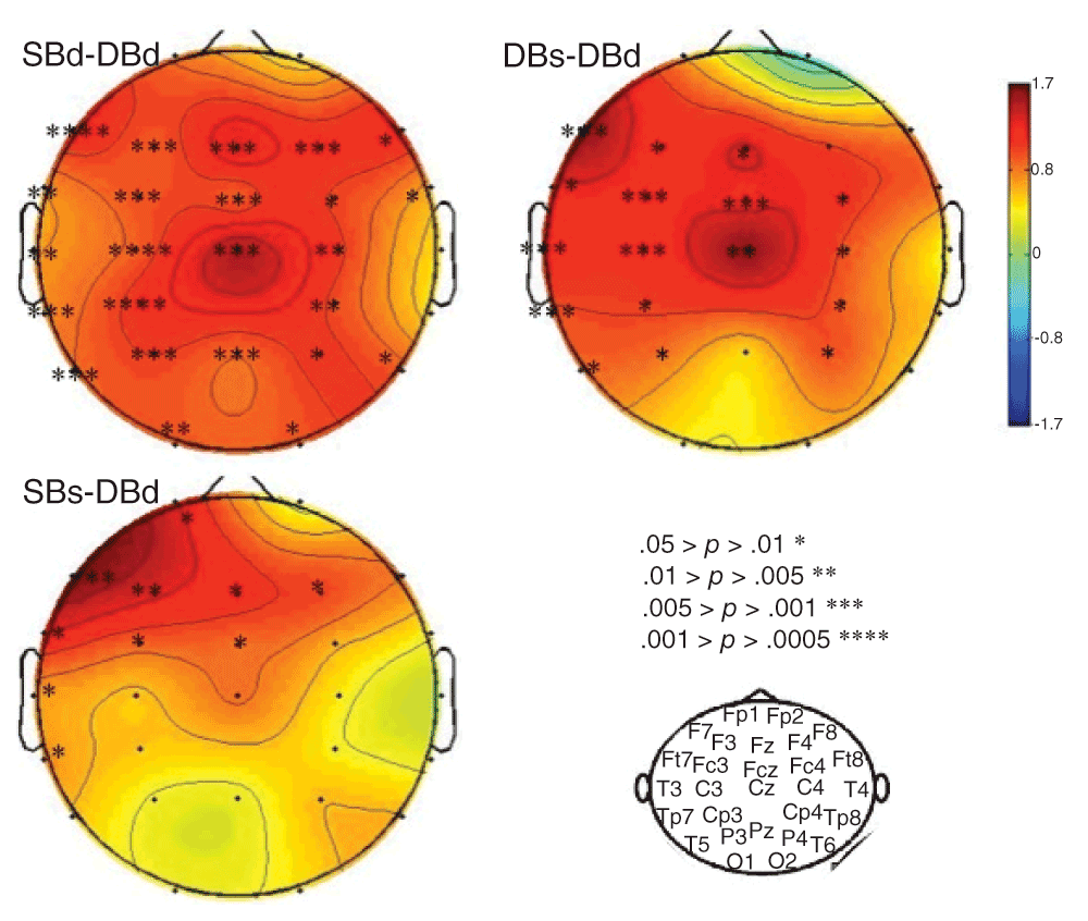
Spline interpolated isovoltage scalp maps computed by subtracting mean voltages of the 600 to 1000 ms time window. P values of the differences are indicated at each electrode site by stars. The baseline condition (i.e., different & believed-different, DBd) was subtracted, in A) from the same & believed-different (SBd) condition, in B) from the different & believed-same (DBs), and in C) from the same & believed-same condition (SBs). Note that, according to the Bonferroni correction, only 4 stars-sites would be significant when considering electrodes other than Cz and Pz since the alpha level would be 0.0018.
On the other hand, the replicability of these findings was explored by computing grand averages of the 16 subjects of the first 8 pairs and the grand averages of the 16 subjects of the last 8 pairs of participants separately. Note that these two sets of subjects went through the exact same procedure and conditions. Figure 5 and Figure 6 display these grand averages. At Cz and Pz, the amplitudes of the P600s for each condition appear to be in the same increasing order, that is from DBd (blue) to SBd (red), via SBs (green) and DBs (orange), as in the grand averages of the 32 participants presented in Figure 3.
Colors as in Figure 3.
We also explored whether large differences in one of the member of the pair were going with large differences in the second member while small differences in one member were going with small differences in the other member. We focused on the conditions that were the most different from each other, namely, SBd-DBd and DBs-SBs and computed the correlations between subjects of each pair for each electrode in order to generate a priori hypotheses for future studies. The significant results that were found are presented in Table 4. Meanwhile, Figure 7 presents scatterplots made by using the data for the electrodes most relevant for the P600, that is, the parietal electrodes P3 and P4 where maximal correlation coefficients were obtained. These correlation coefficients (Table 4) show that, when participants believed they were seeing the same pictures (Bs), the larger the effect of sameness in one participant, the larger this effect in the other. In contrast, when they believed they were seeing different pictures (Bd), these correlations were negative (top of Table 4).
All the correlations having a p value smaller than 0.05 are indicated to generate a priori hypotheses for future studies. However, using a Bonferroni correction for doing 28 analyses (one for each electrode), leads to a corrected alpha level of 0.0018.
| Difference | Electrode | r | p |
|---|---|---|---|
| SBd-DBd | CP4 | -.59 | .03 |
| F8 | -.61 | .02 | |
| FC4 | -.54 | .05 | |
| Fz | -.54 | .05 | |
| P3 | -.61 | .02 | |
| Pz | -.59 | .02 | |
| DBs-SBs | O1 | .55 | .04 |
| O2 | .55 | .04 | |
| P4 | .61 | .02 |
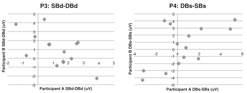
The x coordinate of each point is the size of the P600 effect in one participant of a pair and the y coordinate of the point is that size for the other participant of that pair. At left parietal site (P3) in the believe-different conditions (SBd-DBd), the graph reveals that the greater the size of the effect of sameness on the P600 amplitude in one participant, the smaller this effect in the other member of the pair. In contrast, at right parietal sites (P4) in believe-same conditions (DBs-SBs), the greater the effect in one participant, the greater this effect in the other. The correlation coefficients for these electrodes, as well as for others, are presented in Table 4.
As shown in Table 5, in the memory test phase, there was no difference between study phase conditions in the number of stimuli correctly recognized (hits) or in the number of misses. In sum, participants did not better recall images from any particular condition of the study phase. Similarly, there was no effect of the condition of the study phase on the reaction times of the memory test phase (Table 6).
| Study Phase Condition | Number of Hits (SD) | Number of Misses (SD) |
|---|---|---|
| SBd | 43.3 (10.1) | 26.1 (9.7) |
| DBs | 42.5 (10.9) | 26.6 (11.1) |
| SBs | 43.4 (12.3) | 26.6 (12.2) |
| DBd | 43.3 (10.9) | 26.2 (10.8) |
The results of the debriefing session were as follows. For the question: “Did you feel more attentive/distracted seeing the pictures when you friend was present?”, 8 participants said they were more distracted, 18 said there was no difference, 6 said they were more attentive. To the question: “Did you feel any different when you knew your friend/partner/relative was looking at the same images that you were seeing?”, 17 participants said they felt the same, 14 said they felt different. To the question: “Did you feel any different when you knew your friend/partner/relative was looking at different images than you were seeing?”, 9 said yes, 22 said no. For the fourth question “Did you feel deceived at any point during the experiment?”, 27 said no, 3 misunderstood “deceived”, 2 said yes, but when asked why, they did not suspect the statements of sameness of stimuli. Their suspicion pertained to other aspects (e.g., one said, after the stimulus presentation computer unexpectedly stopped, “I thought that when the computer crashed it was deliberately done so that it was more difficult to remember”).
In each recording session of this study, pairs of related participants were tested together. In each trial, two pictures taken from the international affective picture system (IAPS) were presented simultaneously, one for the first participant, the other for the second participant of the pair. All 32 participants of the 16 pairs tested were asked to remember these pictures during the four different blocks of the study phase. These pictures were then presented again, mixed with new ones, during a subsequent memory test phase.
During both phases, the computer screen was divided in two halves that were separated by a vertical cardboard perpendicular to the screen. Each participant of a pair sat in front of one half of the screen and was presented with one picture at a time. There was no way for a participant to see the picture simultaneously presented to the other participant.
Sameness was manipulated. In two of the four conditions of the study phase, participants were presented with the same picture simultaneously (S conditions). They were presented with two different pictures in the two others conditions (D conditions). The belief (B) pertaining to what the other member of the pair was presented with was also manipulated. Just before the beginning of each condition, or block, of the study phase, participants saw one of two statements on the screen, announcing whether or not the same (Bs vs. Bd conditions) the same picture would appear for both of them on each half of the screen. The four conditions of the study phase were thus: different believed-different (DBd), same believed-same (SBs), different believed-same (DBs) and same believed different (SBd), the latter two thus including deceiving statements.
Event-related brain potentials elicited by the IAPS pictures were recorded during these four conditions of the study phase. There was an interaction of sameness with belief on the amplitudes of the P600s. In the believed-different conditions (Bd), these amplitudes were significantly larger when pictures were actually the same (SBd) than when they were different (DBd), as illustrated by Figure 3 and Figure 4. In contrast, in the believed-same conditions (Bs), P600 amplitudes tended to be larger when pictures were different (DBs) than when they were the same (SBs).
Usual interpretations of these ERPs difference could be ruled out. First, because, in contrast with the third hypothesis, there was no effect of sameness or belief on the recognition scores obtained during the memory test phase. The ERP differences found between the different conditions of the study phase could thus not be related to a Dm effect; that is, to larger P600s at fronto-central electrode sites for stimuli that benefit from a deeper encoding in episodic memory22,23. Second, the ERP differences found were also unlikely to be related to differential allocations of attentional resources. Indeed, all stimuli had the same task relevance since they equally had to be memorized. Moreover, they could not capture attention differentially, since they were identical because their use for each of the conditions of the study phase was counterbalanced across pairs of participants.
The statements seen by participants as to whether or not they would be presented with the same stimuli as the other participant of the pair could have theoretically modulated the allocation of attentional resources and thus P600 amplitudes. Nevertheless, these statements could not have had an effect depending on the actual sameness of the stimuli, since it was something participants had no knowledge of. Third, more preconscious processing does not seem to be useful to account for the greater P600s obtained for the three conditions other than DBd. Indeed, why would more processing have occurred for these SBs, SBd and DBs conditions than for this baseline condition when all stimuli equally had to be memorized?
On the other hand, participants were side by side and could get some auditory and visual input from each other in their very peripheral field (i.e., 90 degrees). Thus, they could in principle influence each other (e.g., through breathing variations, subtle body movements, like postural reactions to aversive stimuli, facial mimicry, eye movements etc). It thus has to be discussed whether or not the present results could be in line with Dumas’ et al. work24 on hyperscanning and inter-brain synchrony mediated through the mirror neuron system. Indeed, direct brain-to-brain propagations do not appear to be the most parsimonious explanation. Given that our participants did not have any task to perform, other than to look at the stimuli, part of their attention could have been allocated to what their friend was doing. Therefore, we have to ask whether the processing of these movements could have been responsible for our results. Nevertheless, for ERPs to differ across conditions in a systematic way, as they did in the present experiment, the movements (or breathing sounds) made by the friend should depend on whether or not the stimuli (s)he was presented with are the same as the one the subject is seeing. This does not seem impossible since, when participants were not seeing the same stimuli, they might not “be moved” in the same way. Their systems might have detected that move difference. However, to account for the results obtained here, the effect of such a detection would also have to depend on whether or not the subject was told that (s)he was presented with the same stimuli as his friend. When (s)he has been told stimuli differ (as in the DBd condition) the move difference detected is congruent with the statement. When the subject was told (s)he will be presented with same stimuli, then, the move difference detected should be further processed since it is contradictory information. However, ERP results are not consistent with this interpretation. Contradiction or incongruence is well known to boost the amplitude of negative going ERPs, such as the N2 and the N400 [e.g., 25, 26]. It thus has an effect on potentials other than the P600 and in the reverse direction. This is completely discordant with the present results. And, even if we hypothesize a very unusual ERP whereby greater P600s would index more processing difficulty, the account would then not explain why the P600s elicited by SBs appears larger than the one elicited by DBd, whereas, in that condition, statement and stimuli were congruent.
Therefore, in accordance with the first two hypotheses, the fact that P600s were larger than DBd in three conditions other than DBd and that P600 amplitudes correlate positively with consciousness13–16 suggest that the two participants of each pair may actually enrich the content of conscious awareness of one another. These effects suggest that the activity of the brain of a participant may have a direct impact on the activity of the brain of the other participant. Given, that the P600 component also indexes conscious perception13–16, these results could thus be related to qualia, the individual instances of subjective conscious experience.
On the other hand, because only phenomena that propagate can account for the impact of one brain on another, these results also suggest that qualia are not limited to the known bioelectrical activity of neurons. They may also include physical phenomena of a different nature, such as electro-magnetic fields, as reviewed by Jones10, or such as modulations of the so-called wave function, studied in quantum mechanics and debatably proposed by Hameroff and Penrose11. The electromagnetic hypothesis can be based on the sensitivity to magnetic fields of at least two molecules: magnetite, whose presence has been demonstrated in the human brain27–29, and cryptochrome30. Furthermore, it is consistent with the fact that mammal behaviors have been shown to depend on magnetic fields, such as that of the earth31,32. However, two properties of magnetic fields are at odds with the idea that the magnetic fields generated by one participant could affect the brain activity of the other participant. First, the magnetic fields generated by the activity of the human brain (only 10 to 103 femto Tesla) are much smaller than the magnetic noise of an urban environment (about 108 femto Tesla). Second, magnetic fields decrease with the square of the distance. The heads of the two subjects of each pair were separated by about 40 cm, a distance much larger than the distance separating the brain from the devices used to capture the magnetic fields it generates in magneto-encephalography (MEG, i.e., less than one cm). Finally, our ERP recording room was not shielded like a MEG recording room. Urban magnetic noise was thus much more important than any field a human brain can generate. These factors make the electromagnetic field explanation appear less likely. In contrast, our experimental conditions and results seem to be more consistent with the theories of consciousness that see qualia as, at least partly, underlain by a modulation of the wave function, and that see direct brain to brain communications possible through quantum entanglement33. Indeed, such modulations do not decrease with distance and could involve many atoms34. Nevertheless, only speculations can be made at this point as to the physical nature of the phenomena by which the activity of a brain could have an impact on the activity of another brain.
The finding of such an impact raises the problem of irrelevant interferences. Indeed, the activity of many brains could then affect the activity of our own. It appears logical to think that filtering exists to prevent such perturbations. One possibility is that, the close relationship existing between the members of each pair in the present study is a prerequisite for the impact to occur, as it may depend on empathy and/or prior common memories. On the other hand, filtering should operate to a greater extent when it is believed that others are processing different stimuli. The results of the present study suggest that this might be the case. When participants were told that they would be presented with different stimuli, the P600s were minimal, which was taken as the baseline condition. However, this happened only when they were actually presented with different stimuli. In the case where the two stimuli were the same (SBd), the P600s were maximal, suggesting that this “belief-based filtering” can operate only when qualia actually differ. P600s at Cz were also maximal when participants believed they were seeing the same stimuli while different ones occurred (DBs). Notably, the scalp distribution of these two additional P600 activities differs from the scalp distribution of the additional P600 activity found when comparing SBs to the baseline condition (DBd) (Figure 4). The latter appeared localized at left frontal sites whereas the former two included that location but were also widespread. This latter fact could suggest that while the “enrichment” of consciousness occurred also in deception conditions, the evaluation of its coherence with the belief might bring up yet additional content in consciousness.
The fact that, at left frontal sites, the 600–900 ms time window used was mainly including the downhill slope of a negativity starting much earlier may be important. Rather than smaller P600s, the significant effects found at these electrode sites might in fact reveal larger late N400s for stimuli that were, and were believed to be, different. This change of perspective might provide an a priori hypothesis for future studies of the filtering mechanism proposed above. Indeed, the N400 has been proposed as an index of an inhibition mechanism whose focus depends on the nature of the inhibited representations [for a brief review see 17].
On the other hand, the nature of the enrichments suggested by the greater midline P600s has to be discussed. The fact that no deception was detected, that is, that no subject realized that (s)he was looking at different stimuli when told (s)he was looking at the same, suggest that the additional content of consciousness was neither verbalizable nor distinguishable from the qualia each participant would have had if (s)he were alone. This strongly supports the mutual enrichment hypothesized in the introduction, where qualia of others would contribute to our own by a merging process occurring without our knowledge.
Interestingly, when participants were told the same stimuli were appearing, the effect of sameness on the size of the P600s in one of the members of a pair positively correlated with that size in the other member (Table 4 & Figure 7). In other terms, the greater the effect in one person, the greater the effect in the other. On the contrary, when participants were told different stimuli were appearing, the correlation was negative, as if the greater the effect in one person, the more its impact was detected and could be prevented in the other.
There is a tradition of research studying the synchronization of EEGs and bold fMRI signals of two persons interacting, imitating each others’ movements [e.g., 35] and of persons going through the same stimulation(s) [e.g., 36, for a review, see 37]. This tradition could be relevant here since, we also recorded the EEG of two participants simultaneously. However, we used ERPs, not EEGs’ synchrony or fMRI, and our participants were not interacting, imitating each other, or being presented with only the same stimulation. Each subject in a pair was going through the experiment on his/her own “despite” the fact that (s)he was sitting side by side with a friend/sibling/spouse. Sameness, and belief in that sameness, were manipulated, which modulated the amplitude of an well-known ERP index of consciousness. To the best of our knowledge, there is thus yet no equivalent to the present study. The hypothesis of a direct sharing of qualia has never been tested. Future studies have to explore whether differential EEG synchrony can also occur within the present design and also test whether qualia sharing could account for part of the EEG synchrony observed in interacting participants, for instance. Indeed, the conscious intention to perform an action, when imitating, can be considered as a qualia and could, according to the present results, impacts the functioning of the brain of the interacting person.
It has to be noted that, if further replicated, these findings could open several avenues of research. For instance, it might be interesting to explore whether young children’s brains learn to produce their qualia with the help of others. It could also be interesting to see if autistic children suffer from a disability of this learning mechanism or whether their tendency to limit contact with others is a strategy that protects them against a deficit of the filtering mechanism.
In any case, the results of the present study provide preliminary data about the mechanisms by which qualia pertaining to the same stimulus could be similar across individuals, something that is assumed in everyday life interactions. Results also suggest that the similarity could be due to an intersubjective impact of brain activities, which could be partially controlled and whose physical bases would remain to be determined.
F1000Research: Dataset 1. Mean voltages of the event-related brain potentials elicited by the IAPS stimuli., 10.5256/f1000research.5977.d4121538
| Views | Downloads | |
|---|---|---|
| F1000Research | - | - |
|
PubMed Central
Data from PMC are received and updated monthly.
|
- | - |
Click here to access the data.
Spreadsheet data files may not format correctly if your computer is using different default delimiters (symbols used to separate values into separate cells) - a spreadsheet created in one region is sometimes misinterpreted by computers in other regions. You can change the regional settings on your computer so that the spreadsheet can be interpreted correctly.
Provide sufficient details of any financial or non-financial competing interests to enable users to assess whether your comments might lead a reasonable person to question your impartiality. Consider the following examples, but note that this is not an exhaustive list:
Sign up for content alerts and receive a weekly or monthly email with all newly published articles
Already registered? Sign in
The email address should be the one you originally registered with F1000.
You registered with F1000 via Google, so we cannot reset your password.
To sign in, please click here.
If you still need help with your Google account password, please click here.
You registered with F1000 via Facebook, so we cannot reset your password.
To sign in, please click here.
If you still need help with your Facebook account password, please click here.
If your email address is registered with us, we will email you instructions to reset your password.
If you think you should have received this email but it has not arrived, please check your spam filters and/or contact for further assistance.
Comments on this article Comments (1)