Keywords
Scedosporium apiospermum, Pseudallescheria boydii, cerebral abscess, kidney transplant
Scedosporium apiospermum, Pseudallescheria boydii, cerebral abscess, kidney transplant
Scedosporium apiospermum is a filamentous fungus causing a rare but serious opportunistic infection. It is the asexual form of Pseudallescheria boydii and is found in many environmental sources including soil and fresh water, but most commonly in stagnant or contaminated water1. The infection may be acquired by inhaling the microorganism or after traumatic inoculation through the skin2. The sites of infection include the lungs, sinuses, bones, skin, joints and notoriously, the central nervous system (CNS)3. S. apiospermum can infect the CNS of both healthy4 and immunocompromised hosts. Cerebral abscess is the most common clinical manifestation of S. apiospermum brain infections, although cases of meningitis and, less frequently, ventriculitis have also been reported5. Brain abscesses may be found as one or multiple lesions6. The overall mortality rate of patients infected with this pathogen is higher than 70%7. Solid organ transplant and its associated immunosuppression are important risk factors for infections with Scedosporium species8. Here we present a case of CNS infection caused by S. apiospermum in a patient who had received a kidney transplant and was treated with dual antifungal therapy and surgical drainage. The patient initially responded well to the therapy.
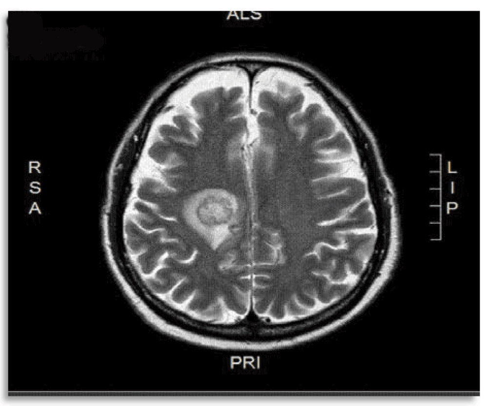
A 64 year-old male patient underwent deceased-donor kidney transplantation following a chronic kidney failure secondary to nephroangiosclerosis. The past medical history was significant for hypertension, hyperlipidemia, peripheral vascular disease, chronic anemia and deep venous thrombosis of right lower extremity. Family history was significant for cardiomyopathy in one brother and diabetes in another. The immunosuppressive medication consisted of tacrolimus 3 mg every 12 hours, prednisone 20 mg daily and mycophenolate mofetil 500 mg every 12 hours. Seventeen days after transplantation, he presented left-sided hemiplegia and dysarthria. A brain MRI was performed, which revealed a hyperintense lesion with ring enhancement at the right paramedian posterior frontal subcortical area with an associated vasogenic edema (Figure 1).

A hyperintense lesion with ring enhancement at the right paramedian posterior frontal subcortical area with an associated vasogenic edema is shown.
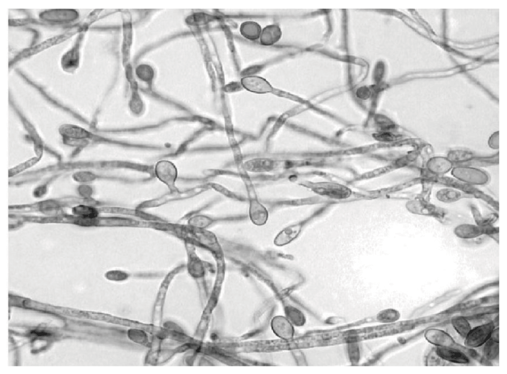
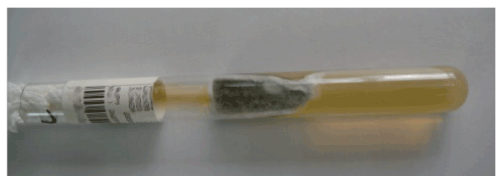
A stereotactic biopsy was performed and tissue examination revealed the presence of a filamentous fungus that was identified as S. apiospermum (Figure 2). The sample was also cultured in Sabouraud’s dextrose agar medium at 25°C for a period of 14 days (Figure 3), the culture turned a dark brown color.

Methylene blue staining showing the morphology of S. apiospermum: unicellular microconidia attached to filaments by conidiophores (original magnification 400×).

The culture grew in Sabouraud’s dextrose agar medium at 25°C and turned a dark brown color on the 14th day.
The patient started a treatment with voriconazole (6 mg/Kg po q12h for two days, then 4 mg/Kg q12h VO) and terbinafine (250 mg po daily) and subsequently was subjected to surgical drainage by craniotomy in order to remove the infected tissue.
The patient was on terbinafine for 4 months and continued to be on voriconazole for almost a year. At a follow up visit he showed significant recovery from the left-sided palsy and also an absence of dysarthria. The brain MRI follow-up images showed an improvement in the brain lesion.
Unfortunately, 8 months later the patient clinical course was complicated and he eventually died of problems unrelated to fungal CNS disease.
Solid organ transplant recipients are highly susceptible to invasive fungal infections9.
During the last few decades there has been a marked increase in the number of immunocompromised patients who have suffered Scedosporium infections, the most frequent cases being infections of the CNS10.
Solid organ transplant patients are susceptible to invasive fungal infections as their immunity might be compromised due to the use of immunosuppressant drugs11. Therefore S. apiospermum should be considered in the differential diagnosis of immunocompromised patients presenting with a brain abscess12.
Within the nervous system, abscesses may be located in brain hemispheres, the cerebellum, the brain stem or the spinal cord, where they may cause alterations of consciousness levels, signs of meningeal irritation or focal neurological deficits13.
If not adequately treated, fungal brain abscesses in immunocompromised patients often result in poor prognosis. The diagnosis of an invasive fungal infection such as S. apiospermum is based on the combination of histopathological, microbiological and clinical findings14. As the clinical and histopathological presentations of S. apiospermum infections are similar to those of other fungi such as Aspergillus and Fusarium spp., a culture is necessary for accurate diagnosis. Furthermore, while most species of Aspergillus (except for Aspergillus terreus) are sensitive to amphotericin, S. apiospermum is usually resistant15. In addition, PCR techniques are important to diagnose as well as to distinguish between different species16.
There are many treatment options described in the literature, but there is an ongoing controversy over which treatment is most suitable for S. apiospermum infections. Voriconazole has emerged as a possible treatment option, since it shows high activity against several species of fungi, including S. apiospermum17. Several reports have shown the successful use of voriconazole synergistically combined with terbinafine against S. apiospermum18 and S. prolificans. Furthermore, many authors recommend the surgical drainage of brain abscesses caused by S. apiospermum19. Therefore, a combined antifungal therapy along with an aggressive surgical approach is recommended for therapeutic success20.
S. apiospermum infection of the CNS is a rare but it is an extremely serious medical condition. Immediate diagnosis in the event of brain abscess in an immunocompromised patient is crucial and the choice of a suitable medical treatment is a priority. Despite the aggressive surgical treatment and the appropriate anti-fungal therapy used, mortality rates continue to be high.
Written informed consent for publication of clinical details and clinical images was obtained from the patient’s family.
MIG, PES and JPC contributed to the design of the study. All the authors contributed to writing the manuscript and agreed to the final contents.
| Views | Downloads | |
|---|---|---|
| F1000Research | - | - |
|
PubMed Central
Data from PMC are received and updated monthly.
|
- | - |
Competing Interests: No competing interests were disclosed.
Competing Interests: No competing interests were disclosed.
Alongside their report, reviewers assign a status to the article:
| Invited Reviewers | ||
|---|---|---|
| 1 | 2 | |
|
Version 1 13 Mar 14 |
read | read |
Provide sufficient details of any financial or non-financial competing interests to enable users to assess whether your comments might lead a reasonable person to question your impartiality. Consider the following examples, but note that this is not an exhaustive list:
Sign up for content alerts and receive a weekly or monthly email with all newly published articles
Already registered? Sign in
The email address should be the one you originally registered with F1000.
You registered with F1000 via Google, so we cannot reset your password.
To sign in, please click here.
If you still need help with your Google account password, please click here.
You registered with F1000 via Facebook, so we cannot reset your password.
To sign in, please click here.
If you still need help with your Facebook account password, please click here.
If your email address is registered with us, we will email you instructions to reset your password.
If you think you should have received this email but it has not arrived, please check your spam filters and/or contact for further assistance.
Comments on this article Comments (0)