Keywords
Oncocytoma, Oncocytic neoplasm, Adrenal neoplasm
Oncocytoma, Oncocytic neoplasm, Adrenal neoplasm
Figure 3 was changed according to the recommendation of the referee.
See the authors' detailed response to the review by Levent Turkeri
See the authors' detailed response to the review by M. Hammad Ather
A 65-year-old Caucasian woman with a history of arterial hypertension underwent an ultrasound examination of the kidneys performed by her general practitioner in 2012. A left-sided suprarenal mass measuring about 5 cm was detected. The physical examination showed no palpable masses in the abdomen, and the peripheral lymph nodes were not enlarged. A 3-T magnetic resonance imaging (MRI) of the adrenal glands presented a heterogeneously enhancing mass measuring 48×48×33 mm in the left apical renal pole in contact with the adrenal gland (Figure 1). The findings seemed to suggest the presence of a renal cell carcinoma. A differential diagnosis of pheochromocytoma had also been considered. Enlarged intra- or retroperitoneal lymph nodes were not detectable in the MRI. Catecholamines and metanephrines, aldosterone-renin ratio and serum cortisol before and after inhibition were within normal range in a 24 hour-urine sample analysis. Thus, a functional adrenal tumor could be excluded.

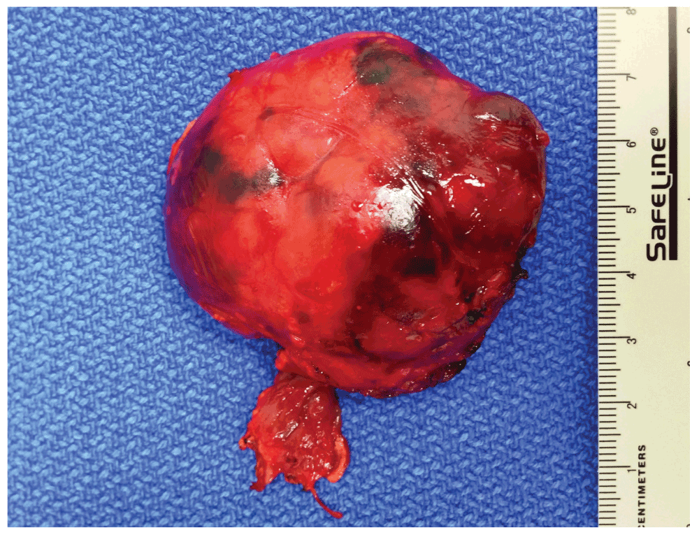
The patient underwent a retroperitoneoscopic exploration. A round shaped 4.5 cm exophytic mass of the upper renal pole was excised retroperitoneoscopically in the fashion of a renal mass enucleation applying the zero-ischemia technique. Intraoperatively, the mass had a very thin, poorly defined pseudo-capsule which was adhering to the renal parenchyma caudally and to the macroscopically inconspicuous adrenal gland medially. The adrenal gland was removed en bloc with the mass (Figure 2). No judgement could be made regarding the origin of the tumor. The postoperative recovery period was uneventful. Histopathological examination (haematoxyline-eosin) of the specimen revealed an oncocytic adrenocortical adenoma that arose from the heterotopic intrarenal adrenal tissue (Figure 3).

The follow-up of this patient (approximately 2 years by the date of article submission) was uneventful. No additional treatment was necessary.
Oncocytic neoplasms of the adrenal glands are extremely rare. A PUBMED search up to December 2013 uncovered only 159 cases of adrenocortical oncocytomas since the first description of this tumor in adrenal in 198617. Most of the papers describe only single patient case reports.
Oncocytic neoplasms or oncocytomas are mostly benign tumors which commonly occur in the kidneys, thyroid, parathyroid, pituitary and salivary glands1,2 and rarely in the respiratory tract3,4, choroid plexus5 and adrenal glands. Oncocytic neoplasms of the adrenal glands, with, to the authors’ knowledge, only 159 previously described cases, are an extremely rare phenomenon. Most of these tumors were discovered as incidental findings on CT or MRI as non-functional adrenal masses. Various malignant potential can be determined in 20% of adrenocortical oncocytic neoplasms6. Oncocytomas arising from the heterotopic adrenocortical tissue have been described in only 10 case reports, seven cases were located intraspinally7–13 and three cases in the retroperitoneum14–16. An oncocytic adenoma arising from heterotopic intrarenal adrenocortical tissue was not suspected in the preoperative assessment of this case because this entity has not been described previously. However, in addition to retroperitoneally located oncocytomas, this should be considered in the future as a differential diagnosis in cases presenting with an intrarenal or retroperitoneal mass.
This paper contains the first report on an oncocytic adenoma arising from the heterotopic intrarenal located adrenal tissue. Heterotopic adrenal tissue or adrenal rest presented mostly by cortical structures is more frequently located in the kidney. Other sites such as the celiac trunk, epididymis, spermatic cord, ovary, broad ligament, liver capsule, gallbladder, pancreas and spleen are rare18.
Oncocytic neoplasms are mostly encapsulated masses with a brown or yellow surface on cut-section. The radial scar can be absent. Oncocytic neoplasms microscopically consist of so called oncocytes, large cells with rich eosinophilic granulations due to the high concentration of mitochondria. The Weiss criteria19, which are commonly used in the histological diagnosis of adrenocortical malignancies, are not applicable to adrenocortical oncocytic neoplasms because all tumors have eosinophilic tumor cytoplasm, diffuse architecture and nuclear atypia. The modified Lin-Weiss-Bisceglia system differentiates between major, minor and definitional criteria for malignancy6. None of these criteria is present in benign adrenal oncocytic neoplasms. Masses with uncertain malignant potential demonstrate the presence of one to four minor criteria (>10 cm or >200g, necrosis, capsular invasion or sinusoidal invasion) in absence of major criteria (mitotic rate >5 mitoses per 50 high-power fields, any atypical mitoses or venous invasion). In adrenal oncocytic carcinomas any of the major criteria could be present.
There are no specific criteria on both computed tomography and MRI and MRI with chemical shift subtraction for adrenal oncocytic neoplasm and its malignant variant20. The bulk size cannot be used as a reliable criterion to estimate the risk of malignancy.
Eighty-three percent of adrenal oncocytic neoplasms are non-functioning masses21. In rare cases, an adrenal oncocytic neoplasm can produce catecholamines, cortisol or testosterone21.
The therapeutic standard is a minimally invasive adrenalectomy. In cases of a large mass, infiltration of surrounding structures, and lymph node bulks, an open surgery approach should be chosen.
Today, there are no recommendations for the follow-up of benign adrenal oncocytic neoplasms. Only one local recurrence of a neoplasm that was originally diagnosed as benign has been described and fulfilled the criteria of uncertain malignant potential21. The oncocytic variant of adrenocortical carcinoma has a poor prognosis, with a postoperative recurrence rate of 75%, and a tumour-related mortality of 40% in a small group of 24 patients with a median follow-up of 21 months (range: 1–180 months)21. An adjuvant or palliative chemotherapy with mitotane can be administered in patients with adrenocortical carcinomas with beneficial effects22, but there is no evidence of efficacy when applied to oncocytic variant.
To the authors’ knowledge, this case report presents the first description of a heterotopic intrarenally located adrenocortical oncocytoma. Although being a rare location, this case is worth mentioning, given the challenging situation with regard to the diagnostics and differentiation from the potentially aggressive malignant lesions. Taking in account the absence of radiological criteria of a benign character, this tumor should be considered and treated as a malignant lesion, although a minimally invasive approach should be chosen when possible. The definitive pathologic diagnosis is in most cases surprising because of the rarity of this type of neoplasm and radiological appearance mimicking renal cell carcinoma or adrenal carcinoma. Benign adrenal oncocytic neoplasms do not require any adjuvant treatment.
Written informed consent for the publication of clinical details and clinical images was obtained from the patient.
| Views | Downloads | |
|---|---|---|
| F1000Research | - | - |
|
PubMed Central
Data from PMC are received and updated monthly.
|
- | - |
Provide sufficient details of any financial or non-financial competing interests to enable users to assess whether your comments might lead a reasonable person to question your impartiality. Consider the following examples, but note that this is not an exhaustive list:
Sign up for content alerts and receive a weekly or monthly email with all newly published articles
Already registered? Sign in
The email address should be the one you originally registered with F1000.
You registered with F1000 via Google, so we cannot reset your password.
To sign in, please click here.
If you still need help with your Google account password, please click here.
You registered with F1000 via Facebook, so we cannot reset your password.
To sign in, please click here.
If you still need help with your Facebook account password, please click here.
If your email address is registered with us, we will email you instructions to reset your password.
If you think you should have received this email but it has not arrived, please check your spam filters and/or contact for further assistance.
Comments on this article Comments (0)