Hypertension is an important manifestation of systemic lupus erythematosus (SLE) but reports of prevalence vary between 20-70% in published reports of adult and pediatric patients. For both children and adults with SLE, the clinical diagnosis and management of hypertension has traditionally been based on guidelines developed for the general population. In clinical trials, the criteria used for defining participants with hypertension are mostly undefined. As a first step towards formally assessing the blood pressure (BP) patterns of children diagnosed with SLE, 24-hr ambulatory BP monitoring data was analyzed on clinic patients who presented with prehypertension or stage I hypertension. In this pediatric SLE cohort, 20% met daytime criteria for a diagnosis of hypertension. Patterns of BP elevation varied widely with white coat, masked, isolated systolic, and diastolic nocturnal hypertension all identified. Nocturnal hypertension was detected in 60% and attenuated nocturnal BP dipping in 90% of both hypertensive and normotensive SLE patients. In SLE patients, the median nighttime systolic and diastolic loads were 25% and 15.5% compared with median daily loads of 12.5% and 11.5%. Daytime and nighttime systolic and diastolic BP load and nocturnal dipping was compared to a control population consisting of 85 non-SLE patients under 21 years old with prehypertension or stage 1 hypertension presenting to hypertension clinic. Median systolic BP dipped 5.3 mmHg in SLE patients compared to 11.9 mmHg in non-lupus (p-value = 0.001). Median diastolic BP dipped 12.9 mmHg versus 18.5 mmHg in non-lupus (p-value = 0.003). Patterns of BP dysregulation in pediatric SLE merit further exploration. Children with or without SLE displaying prehypertensive or stage 1 casual BP measurements had similar rates of hypertension by ambulatory BP monitoring. However, regardless of BP diagnosis, and independent of kidney involvement, there was an increased proportion with attenuated nocturnal dipping and nocturnal hypertension in SLE patients.
Corresponding author:
Scott E. Wenderfer
Competing interests:
No competing interests were disclosed.
Grant information:
This study was funded in part by a Pediatric Pilot Award program, granted to SEW by the Department of Pediatrics at Baylor College of Medicine.
The funders had no role in study design, data collection and analysis, decision to publish, or preparation of the manuscript.
Ambulatory blood pressure monitoring (ABPM) is preferred to casual clinic blood pressure (BP) monitoring in the diagnosis of hypertension (HTN). There are many shortfalls of casual BP readings, including the white coat effect, observer bias/measurement error, regression to the mean with repeated measurements, and variability of blood pressure over time (Flynn, 2011). Additionally, the published normative values for casual BP are based on the auscultation method, yet many clinic measurements are taken with oscillometric devices (Woroniecki & Flynn, 2005). Perhaps the most clinically relevant shortfall is the limited outcome data regarding casual BP measurements and end-organ damage or cardiovascular risk.
There is good evidence supporting the utility of ABPM findings in the prediction of cardiovascular outcomes, both in adults and children (Belsha et al., 1998; Lurbe et al., 2004; Pickering et al., 2006; Singh et al., 2013; Sorof et al., 2002). ABPM can account for the white coat effect as well as measurement and observer errors. Where casual BP measurements account for the magnitude of BP at single points in time, ABPM can define BP loads which measure the proportion of BPs that exceed a defined cutoff, typically the 95th percentile as defined by normative data, over a 24-hr period. Therefore ABPM provides a better appreciation of BP trends which can account for the dynamic nature of BP (such as circadian rhythms, nocturnal BP dipping) (Flynn, 2011).
Although ABPM is a valuable piece of the HTN evaluation, there are potential barriers to its widespread utilization related to both financial and clinical considerations. Insurance companies offer limited reimbursement for ABPM placement and interpretation (Swartz et al., 2008). Although there is vast evidence for normative values in adults; there is more limited normative data for ABPM interpretation in children. The current normative values are based on approximately 950 healthy children with limited variability in ethnicity/race (Wühl et al., 2002). Despite these obstacles, ABPM is considered the gold standard for diagnosis of HTN in both adults and pediatrics. It is useful in predicting cardiovascular risk related to HTN but is also helpful in assessing BP in special pediatric populations such as obesity, sickle cell disease, chronic kidney disease, end stage renal disease, and diabetes (Flynn, 2011). There is limited research on the use of ABPM in the pediatric systemic lupus erythematosus (SLE) population (Canpolat et al., 2013), though HTN occurs in 20–70% of these patients (Bogdanovic et al., 2004; Brunner et al., 2002; Lau et al., 2006; Ruggiero et al., 2013). There is also limited data regarding the management of HTN in the SLE population, with management traditionally based on guidelines developed for the general population (Tselios et al., 2014).
Cardiovascular disease is a leading cause of mortality in adults with SLE and though there are many non-traditional risk factors including altered renal function, impaired endothelial function, chronic inflammation, and an activated renin-angiotensin system (RAS) (Gustafsson et al., 2012; Kiani et al., 2008; Knight & Kaplan, 2013; Pieretti et al., 2007), HTN is still an important risk factor (Contreras et al., 2005; Ginzler et al., 1993; Petrin et al., 1993; Yang et al., 1994). Therefore the use of standardized definitions to define HTN in SLE patients is crucial in better understanding cardiovascular risk and preventing adverse outcomes. Although isolated nocturnal HTN is not considered sufficient for a diagnosis for systemic hypertension in the general population, it is known to associate with increased risk of cardiac outcomes (Yee, 2015). Additionally, attenuated nocturnal dipping, even in the setting of normal 24-hr BP, was noted to be an independent predictor of cardiovascular mortality in a large prospective cohort study in Japan (Ohkubo et al., 2002). Hence, it is also important to use ABPMs to characterize blood pressure patterns in SLE patients so that specific guidelines for ABPM interpretation can be established for this population.
Methods
Study population
BP patterns of the 10 SLE study participants (demographics summarised in Table 1) recruited from a single center were retrospectively reviewed using data from 24-hr ambulatory BP monitoring tests performed between February 2012 and April 2013. ABPM was routinely ordered only on SLE patients seen in a multispecialty pediatric lupus clinic when they presented without kidney disease (non-renal lupus or nephritis in remission) and with casual BP measurements in prehypertensive or stage 1 hypertensive range (National High BP Education Program Working Group on High BP in Children and Adolescents, 2004). The inclusion criteria for the SLE cohort were: (1) diagnosis of SLE by American College of Rheumatology (ACR) criteria, (2) age < 21 years, and (3) ABPM performed. There were 85 patients in the non-SLE cohort (43% female, race and ethnicity unknown). The inclusion criteria for the non-SLE cohort included: (1) age < 21 years and (2) ABPM performed. Exclusion criteria for both cohorts: (1) ABPM uninterpretable due to incomplete/missing data, (2) casual BP measurements all < 90th or all > 99th percentile for age, gender, and height, +5 mmHg (3) end stage renal disease, or (4) kidney transplant recipient. The mean age at ABPM for the non-SLE cohort was 12.4 ± 0.4 years, and the mean BMI was 25.1 ± 0.8 kg/m2 using the Mosteller formula. The study protocol was reviewed and approved by the Institutional Review Board for Baylor College of Medicine (H-32061).
Table 1. Pediatric SLE Cohort Demographics.
Patient Characteristics
Values (n=10)
Age at ABPM (years)
14.6 ± 0.3
Age at SLE Diagnosis (years)
12.4 ± 1.0
BMI (kg/m2)
25.1 ± 1.1
SLE ACR criteria met
5.8 ± 0.4
Gender (% female)
9 (90)
Race
African American (%)
4 (40)
Hispanic (%)
3 (30)
Caucasian (%)
3 (30)
Laboratory Findings (within 3 months of ABPM)
eGFR (ml/min/1.73 m2)
133 ± 6.2
C3 (mg/dL)
83.4 ± 9.5
Sed Rate (mm/hr)
58.7 ± 16.2
Hgb (g/dL)
11.8 ± 0.5
Elevated CRP (%)
1 (10)
Proteinuria (%)
2 (20)
Positive ANA (%)
10 (100)
Positive Anti-DS DNA Antibody (%)
9 (90)
Positive Anti-phospholipid Antibody (%)
9 (90)
Medications (at time of ABPM)
Prednisone dose (mg/kg/day)
0.313 ± 0.07
IV corticosteroids (%)
3 (30)
Hydroxychloroquine (%)
9 (90)
Mycophenolate mofetile (%)
3 (30)
Methotrexate (%)
1 (10)
Azathioprine (%)
1 (10)
Aspirin (%)
7 (70)
ACE inhibitor (%)
1 (10)
Participants. As this study was a retrospective chart review, there were no dropouts.
Sample size. As this was a pilot study, the sample size was not determined by formal power analysis. There were 11 consecutive SLE patients and 100 consecutive non-SLE patients with ABPM data identified, but 1 and 15 patients, respectively did not qualify based on the inclusion and exclusion criteria.
Blinding. All interpretation of ABPM data was performed at the time of clinical testing, prior to inception of the study. There was no formal blinding of ABPM data during the data analysis or sensitivity analysis phases.
Ambulatory Blood Pressure Monitoring
24-hr ABPM was performed using SpaceLabs 90217-1Q or 90217A-1 equipment. SpaceLabs Medical Software (version 90219) was used to evaluate the BP patterns. BP measurements were automatically measured every 20 minutes during the daytime and every 30 minutes during the nighttime over a 24-hr period. Mean diastolic and systolic BPs were calculated for both daytime and nighttime periods and compared to normative data for mean BPs based on age and gender (Wühl et al., 2002). BP loads were calculated for both diastolic and systolic BP, reflecting the percentage of BP measurements above the 95th percentile for gender and age. Blood pressure loads >25% were considered abnormal (Urbina et al., 2008). Additionally, nocturnal dipping of BP was defined as the difference between daytime and nighttime BPs. Nocturnal dipping <10% was considered abnormal (Urbina et al., 2008). American Heart Association (AHA) definitions were used to define normal blood pressure, masked, white-coat and sustained HTN (Flynn, 2011).
Clinical data
Demographic and clinical data was collected from medical records for the SLE cohort. Demographic data included race and age at diagnosis. Clinical data (from within 3 months of ABPM date) included height, weight, BMI, laboratory results, eGFR using the Schwartz formula (Schwartz et al., 2009), presence of proteinuria, medications at the time of ABPM, echocardiogram findings including left ventricular hypertrophy (LVH), left ventricular mass index (LVMI), and relative wall thickness (RWT), ACR criteria for SLE, and SLEDAI score (Systemic Lupus Erythematosus Disease Activity Index). Demographic information for the non-SLE cohort was obtained from the SpaceLabs software, including age at the time of ABPM, gender, height, and weight.
Statistical analysis
Statistical analysis was performed using SigmaPlot software (version 11.0). Patient characteristics and ABPM findings were analyzed using descriptive statistics (medians, and intra-quartile ranges). Fisher exact and Wilcoxon rank sum tests were used to characterize patient demographics and BP patterns. Statistical significance was defined as p-value ≤0.05 (two tailed).
Results
Patient and clinical characteristics
The study population consisted of ten patients, all of whom met ACR diagnostic criteria for SLE (Patient demographics in Table 1). At the time of ABPM, mean age of the SLE cohort was 14.6 years. The mean BMI was 25.1 kg/m2. Nine patients were female. Three SLE patients were Hispanic, three were Caucasian, and four were African American. The non-SLE control population consisted of 85 age- and BMI-matched pediatric patients with casual BP measurements between 90–99th percentile without kidney disease or diabetes.
Case #
BP Diagnosis
ABPM Date
Age at ABPM
SBP load
DBP load
SBP Average
DBP Average
Wake SBP load
Wake DBP load
Wake SBP Average
Wake DBP Average
Sleep SBP load
Sleep DBP load
Sleep SBP Average
Sleep DBP Average
SBP Dip
DBP Dip
Attenuated Dipping
Nocturnal HTN
Wake Sleep Method
Adequate #/% Readings
Age at SLE Diagnosis
SLE Vintage
Race
Gender
Height
Weight
BMI
eGFR (Schwartz formula)
Proteinuria
Hgb
ESR
CRP
total Cholesterol
LDL
HDL
Triglycerides
C3
ANA
Anti-double stranded DNA Ab
Anti-phospholipid antibodies
Steroid dose with IV pulse
Oral Prednisone (mg/kg/day)
AZA
MMF
HCQ
MTX
RTX
ACEI/ARB
aspirin
LVH on echo
LVMI on echo
RWT on echo
Malar Rash
Discoid Rash
Photo-sensitivity
Mouth Sores
Serositis
Arthritis
Renal disease
Neurologic disease
Hematologic disease
Antinuclear antibody
Immunologic disease
# SLEACR Criteria
OSA based on sleep study
1
hypertension
2/4/2012
14
68%
60%
131
82
60%
50%
131
83
100%
100%
128
79
2.3%
4.9%
Yes
Yes
reported
Yes
Based on 95%ile
14
0
African American
Female
1.57
53
21.5
153
Yes
9.1
121
Normal
N/A
N/A
N/A
N/A
47
1280
Yes
Yes
0
0.79
No
No
Yes
No
No
No
No
No
39.2
0.52
Yes
No
Yes
No
Yes
Yes
Yes
No
Yes
Yes
Yes
8
No
2
masked HTN
8/21/2012
14
6%
61%
116
78
0%
61%
120
83
12%
62%
111
70
7.6%
15.7%
Yes
Yes
reported
Yes
Based on 95%ile
5
9
African American
Male
1.45
47
22.4
169
No
13.1
5
Normal
N/A
N/A
N/A
N/A
106
1280
Yes
Yes
0
0.11
No
Yes
Yes
No
No
No
Yes
N/A
N/A
N/A
Yes
No
No
Yes
Yes
Yes
No
No
Yes
Yes
Yes
7
No
3
normotension
2/19/2013
17
18%
6%
122
68
12%
8%
126
72
36%
0%
113
57
10.4%
20.9%
No
Yes
reported
Yes
Based on 95%ile
14
3
African American
Female
1.53
73
31.2
114
No
9.9
79
High
N/A
N/A
N/A
N/A
44
1280
Yes
Yes
x1, 3 weeks prior
0.14
No
Yes
Yes
No
No
No
Yes
N/A
N/A
N/A
No
No
No
No
Yes
Yes
Yes
No
Yes
Yes
Yes
6
No
4
normotension
3/13/2012
15
20%
19%
113
67
9%
15%
116
71
35%
23%
109
62
6.1%
12.7%
Yes
Yes
reported
Yes
Based on 95%ile
14
1
Hispanic
Female
1.58
62
24.8
132
No
12.7
19
Normal
N/A
N/A
N/A
N/A
126
640
Yes
Yes
weekly
0.49
No
No
No
No
No
No
No
N/A
N/A
N/A
No
No
Yes
Yes
No
No
Yes
No
Yes
Yes
Yes
6
No
5
normotension
4/17/2012
15
19%
17%
115
69
0%
3%
113
70
64%
50%
119
68
-5.3%
2.9%
Yes
Yes
default
Yes
Based on 95%ile
14
1
Hispanic
Female
1.54
67
28.3
109
No
13.6
N/A
N/A
174
79
44
253
50
1280
Yes
Yes
0
0.46
No
No
Yes
No
No
No
Yes
N/A
N/A
N/A
Yes
No
No
No
No
Yes
Yes
Yes
No
Yes
Yes
6
No
6
normotension
5/23/2012
15
7%
3%
109
63
0%
3%
110
68
15%
4%
109
58
1.0%
14.8%
Yes
No
reported
Yes
Based on 95%ile
15
0
Caucasian
Female
1.63
76
28.6
137
Yes
10.3
122
Normal
116
66
14
261
90
1280
Yes
No
weekly
0.41
No
Yes
Yes
No
No
No
No
No
35.25
0.40
No
No
No
Yes
No
No
Yes
No
Yes
Yes
Yes
5
No
7
normotension
8/21/2012
14
0%
0%
101
54
0%
0%
103
58
0%
0%
97
48
5.9%
17.3%
Yes
No
reported
Yes
Based on 95%ile
8
6
Caucasian
Female
1.44
41
19.8
131
No
11.4
N/A
Normal
N/A
N/A
N/A
N/A
75
1280
Yes
Yes
0
0.09
No
No
Yes
No
No
No
Yes
N/A
N/A
N/A
Yes
No
Yes
Yes
No
Yes
No
No
No
Yes
Yes
6
No
8
normotension
3/13/2012
14
26%
25%
118
71
12%
17%
119
73
63%
44%
115
67
3.4%
8.3%
Yes
Yes
default
Yes
Based on 95%ile
13
1
Caucasian
Female
1.6
64
25.0
114
No
13.3
25
Normal
N/A
N/A
N/A
N/A
86
1280
Yes
Yes
0
0.31
Yes
No
Yes
No
No
Yes
Yes
N/A
N/A
N/A
Yes
No
No
No
No
Yes
Yes
No
Yes
Yes
Yes
6
No
9
white coat
1/31/2013
13
3%
6%
106
60
0%
4%
109
62
8%
8%
101
55
7.4%
11.3%
Yes
No
default
No
Based on 95%ile
12
1
African American
Female
1.59
65
25.7
129
No
12.6
40
Normal
N/A
N/A
N/A
N/A
106
1280
Yes
Yes
0
0.09
No
No
Yes
No
No
No
Yes
N/A
N/A
N/A
No
No
No
No
No
Yes
No
No
Yes
Yes
Yes
4
No
10
normotension
4/12/2013
15
2%
4%
106
59
0%
3%
107
62
8%
8%
102
54
4.7%
13.0%
Yes
No
reported
Yes
Based on 95%ile
15
0
Hispanic
Female
1.63
63
23.7
142
No
11.9
N/A
N/A
N/A
N/A
N/A
N/A
104
1280
No
No
0
0.24
No
No
Yes
Yes
No
No
Yes
No
42.86
0.39
No
No
No
Yes
No
Yes
No
No
No
Yes
Yes
4
No
Dataset 1.Raw ABPM Data for pSLE Cohort.
File contains the coded ambulatory blood pressure monitoring data for the pediatric SLE cohort abstracted from Space Labs software, using the default 95th percentile cutoff to distinguish normal versus high BP values. ABPM data was matched to demographic, clinical, and laboratory data abstracted from the electronic medical record. N/A (not available) indicates that data was sought but testing was not performed. BMI = body mass index, eGFR = estimated glomerular filtration rate, Hgb = hemoglobin, ESR = erythrocyte sedimentation rate, CRP = Creactive protein, LDL = low density lipoprotein, HDL = high density lipoprotein, ANA = anti-nuclear antibody, AZA = azathioprine, MTX = methotrexate, RTX = rituximab, ACEI = Angiotensin-converting enzyme inhibitor, ARB = angiotensin receptor blocker, LVH = left bentricular hypertrophy, LVMI = left ventricular mass index, RWT = right wall thickness, OSA = obstructive sleep apnea (Dataset 1: Campbell et al., 2015a).
Case #
Age
Diagnosis
SBP load
DBP load
SBP Average
DBP Average
Wake SBP load
Wake DBP load
Wake SBP Average
Wake DBP Average
Sleep SBP load
Sleep DBP load
Sleep SBP Average
Sleep DBP Average
SBP Dip
DBP Dip
Nocturnal HTN
Attenuated?
Wake Sleep Method
Low Success
# Readings
% sucessful
Height
Weight
BMI
Gender
1
11
Hypertension
54%
44%
127
77
67%
56%
135
84
14%
7%
109
58
19.30%
31.00%
No
No
default
No
57
92
1.60
67
26.2
M
2
13
Hypertension
23%
39%
124
73
27%
38%
128
75
7%
33%
112
64
12.60%
14.70%
Yes
No
reported
No
56
90
1.66
54
19.6
M
3
14
White Coat
7%
7%
110
57
0%
6%
110
59
30%
10%
111
52
-0.90%
11.90%
Yes
Yes
reported
Yes
45
32
1.81
110
33.6
M
5
16
White Coat
16%
8%
122
65
17%
11%
130
70
14%
0%
106
54
18.50%
22.90%
No
No
reported
No
49
77
1.75
76
24.8
M
6
9
normotension
9%
11%
111
68
8%
13%
116
74
11%
6%
102
57
12.10%
23.00%
No
No
reported
No
57
92
1.43
54
26.4
F
7
17
White Coat
10%
15%
122
74
0%
14%
124
79
33%
17%
118
65
4.90%
17.80%
Yes
Yes
reported
Yes
41
65
1.92
85
23.1
M
8
16
White Coat
2%
5%
115
66
3%
8%
120
71
0%
0%
103
55
14.20%
22.60%
No
No
reported
No
56
92
1.72
59
19.9
F
9
12
hypertension
26%
4%
120
60
31%
3%
127
64
10%
10%
104
49
18.20%
23.50%
No
No
reported
Yes
46
61
1.59
63
24.9
M
10
5
normotension
2%
32%
108
72
3%
22%
113
76
0%
53%
98
65
13.30%
14.50%
Yes
No
reported
No
53
46
1.11
20
16.2
M
11
19
White Coat
2%
7%
117
78
0%
11%
123
83
6%
0%
107
69
13.10%
16.90%
No
No
reported
No
55
89
1.52
50
21.6
F
13
15
White Coat
9%
21%
116
66
9%
27%
129
75
8%
12%
102
56
21.00%
25.40%
No
No
reported
No
58
42
1.71
58
19.8
M
14
17
Hypertension
42%
37%
130
81
44%
53%
139
92
40%
16%
120
69
13.70%
25.00%
Yes
No
reported
No
57
93
1.54
41
17.3
M
15
11
White Coat
14%
15%
106
64
18%
15%
111
67
0%
15%
94
57
15.40%
15.00%
No
No
reported
No
52
84
1.55
85
35.4
F
16
17
normotension
12%
2%
121
68
13%
3%
127
75
8%
0%
108
54
15.00%
28.00%
No
No
reported
No
51
77
1.83
83
24.8
M
17
11
normotension
30%
16%
116
69
12%
3%
114
68
80%
53%
120
71
-5.20%
-4.40%
Yes
Yes
default
No
57
63
1.73
70
23.4
M
18
15
normotension
14%
14%
123
66
7%
0%
125
68
25%
38%
120
64
4.10%
5.90%
Yes
Yes
default
Yes
44
72
1.82
92
27.8
M
19
12
normotension
12%
35%
113
70
5%
27%
114
70
27%
53%
110
71
3.60%
-1.40%
Yes
Yes
default
No
52
84
1.64
98
36.4
M
20
16
White Coat
0%
4%
112
60
0%
3%
117
64
0%
8%
103
53
12.00%
17.20%
No
No
reported
No
49
79
1.82
67
20.2
M
21
11
normotension
11%
6%
113
63
9%
5%
116
66
15%
10%
105
57
9.50%
13.70%
No
Yes
reported
No
128
91
1.53
71
30.3
F
22
9
Hypertension
32%
20%
116
68
36%
21%
123
74
26%
17%
108
60
12.20%
19.00%
No
No
reported
No
56
90
1.53
63
26.9
M
23
9
Hypertension
44%
28%
119
71
42%
22%
122
74
48%
38%
115
65
5.80%
12.20%
Yes
Yes
reported
No
57
83
1.39
51
26.4
F
24
6
normotension
31%
6%
114
67
25%
3%
117
71
47%
13%
108
61
7.70%
14.10%
Yes
Yes
reported
No
51
82
1.27
25
15.5
F
25
16
White Coat
2%
7%
115
60
0%
7%
123
67
6%
6%
105
50
14.70%
25.40%
No
No
reported
Yes
44
59
1.81
161
49.1
M
26
10
normotension
31%
45%
117
74
21%
17%
121
76
41%
70%
113
72
6.70%
5.30%
Yes
Yes
reported
No
51
82
1.47
74
34.2
M
27
16
White Coat
5%
16%
119
67
3%
9%
126
71
8%
33%
108
59
14.30%
17.00%
Yes
No
reported
Yes
44
70
1.71
80
27.4
M
28
11
Hypertension
67%
32%
128
73
76%
36%
136
79
40%
20%
111
60
18.40%
24.10%
Yes
No
reported
No
57
90
1.58
55
22.0
M
29
6
normotension
0%
4%
99
63
0%
3%
101
67
0%
6%
94
56
7.00%
16.50%
No
Yes
reported
No
50
60
1.21
31
21.2
F
30
15
normotension
2%
11%
116
67
0%
8%
120
73
7%
20%
108
56
10.10%
23.30%
No
No
reported
No
55
86
1.69
66
23.1
M
31
16
hypertension
15%
26%
116
72
18%
30%
122
79
7%
13%
104
58
14.80%
26.60%
No
No
default
No
55
89
1.52
72
31.2
F
32
5
Hypertension
70%
71%
126
85
90%
88%
142
100
32%
41%
105
65
26.10%
35.00%
Yes
No
default
No
63
95
1.01
16
15.7
F
33
6
normotension
6%
6%
102
65
10%
10%
106
70
0%
0%
94
54
11.40%
22.90%
No
No
reported
Yes
47
61
1.14
19
14.6
F
34
7
White Coat
5%
5%
102
59
0%
5%
106
64
9%
5%
99
55
6.70%
14.10%
No
Yes
reported
Yes
42
66
1.28
32
19.5
F
35
15
hypertension
14%
23%
112
71
19%
32%
121
78
5%
5%
96
56
20.70%
28.30%
No
No
reported
No
56
88
1.59
50
19.8
F
37
16
Hypertension
71%
24%
133
70
80%
30%
141
77
47%
7%
114
55
19.20%
28.60%
Yes
No
reported
No
59
94
1.69
68
23.8
F
38
13
Hypertension
38%
2%
122
59
45%
3%
134
68
25%
0%
107
48
20.20%
29.50%
No
No
reported
Yes
45
70
1.75
84
27.4
M
39
6
Hypertension
57%
77%
121
86
66%
89%
126
92
31%
44%
112
71
11.20%
22.90%
Yes
No
reported
No
60
94
1.19
22
15.5
F
42
8
hypertension
29%
12%
115
66
30%
13%
119
70
28%
11%
108
61
9.30%
12.90%
No
Yes
default
Yes
41
65
1.43
67
32.8
M
43
6
Hypertension
44%
33%
118
73
50%
42%
125
79
35%
18%
108
63
13.60%
20.30%
Yes
No
reported
Yes
43
43
1.17
33
24.1
F
44
8
normotension
10%
2%
109
61
13%
3%
113
65
0%
0%
96
46
15.10%
29.30%
No
No
reported
No
49
74
1.42
55
27.3
F
45
8
Hypertension
36%
11%
118
67
39%
13%
120
71
20%
0%
106
50
11.70%
29.60%
No
No
reported
Yes
28
36
1.28
29
17.7
F
46
16
Hypertension
22%
33%
129
73
21%
40%
134
80
27%
13%
119
57
11.20%
28.80%
No
No
reported
No
58
59
1.74
67
22.1
M
48
13
Hypertension
86%
57%
136
78
90%
54%
141
82
77%
65%
126
71
10.70%
13.50%
Yes
No
reported
No
58
91
1.59
62
24.5
F
49
16
White Coat
2%
5%
115
64
2%
5%
120
69
0%
6%
105
56
12.60%
18.90%
No
No
default
No
62
100
1.80
103
31.8
M
50
8
Hypertension
65%
45%
123
76
65%
47%
128
82
67%
40%
114
67
11.00%
18.30%
Yes
No
reported
Yes
49
71
1.40
44
22.4
F
51
6
hypertension
69%
21%
117
64
50%
20%
118
72
79%
21%
116
59
1.70%
18.10%
Yes
Yes
reported
Yes
29
46
1.18
23
16.5
F
52
5
white coat
3%
6%
108
63
4%
4%
112
66
0%
8%
100
58
10.80%
12.20%
No
No
reported
Yes
36
47
1.19
22
15.5
F
53
13
hypertension
38%
23%
127
69
52%
28%
138
77
16%
16%
112
59
18.90%
23.40%
No
No
reported
Yes
48
61
1.60
69
27.0
M
54
17
white coat
2%
2%
114
70
2%
2%
120
77
0%
0%
101
55
15.90%
28.60%
No
No
default
No
56
90
1.65
53
19.5
F
56
17
hypertension
47%
26%
132
78
50%
38%
139
86
40%
0%
118
60
15.20%
30.30%
Yes
No
reported
No
47
77
1.65
72
26.4
F
57
14
normotension
10%
30%
120
72
3%
23%
121
74
30%
50%
119
67
1.70%
9.50%
Yes
Yes
default
Yes
40
60
1.62
104
39.6
M
58
13
white coat
4%
16%
104
64
4%
12%
112
73
6%
20%
100
60
10.80%
17.90%
No
No
reported
No
74
70
1.62
84
32.0
F
60
16
white coat
4%
6%
115
63
2%
5%
119
67
7%
7%
105
55
11.80%
18.00%
No
No
reported
No
55
89
1.79
72
22.5
M
62
16
normotension
9%
30%
122
75
9%
23%
125
78
8%
54%
113
66
9.60%
15.40%
Yes
Yes
reported
No
57
92
1.78
60
18.9
M
64
18
hypertension
49%
33%
130
77
44%
39%
133
81
63%
19%
124
70
6.80%
13.60%
Yes
Yes
default
No
55
82
1.78
115
36.3
M
66
11
hypertension
29%
33%
117
72
34%
30%
122
77
19%
38%
110
64
9.90%
16.90%
Yes
Yes
reported
Yes
49
42
1.54
41
17.3
F
67
14
hypertension
25%
64%
117
78
21%
62%
124
85
30%
67%
110
72
11.30%
15.30%
Yes
No
reported
No
56
81
1.60
68
26.6
F
68
6
normotension
12%
22%
107
69
8%
8%
106
69
20%
50%
108
70
-1.80%
-1.40%
Yes
Yes
reported
No
60
97
1.23
20
13.2
F
69
16
hypertension
35%
43%
134
77
34%
45%
142
84
39%
39%
119
64
16.20%
23.90%
Yes
No
default
No
51
82
1.74
112
37.0
M
71
14
hypertension
59%
23%
127
69
66%
27%
134
75
40%
13%
114
56
15.00%
25.40%
Yes
No
reported
No
56
89
1.70
66
22.8
F
72
8
hypertension
53%
23%
121
69
32%
16%
122
70
87%
33%
119
66
2.50%
5.80%
Yes
Yes
reported
Yes
40
65
1.37
49
26.1
M
73
9
white coat
5%
2%
106
59
4%
2%
110
63
6%
0%
97
51
11.90%
19.10%
No
No
reported
No
64
93
1.38
47
24.7
F
75
12
hypertension
54%
26%
128
70
59%
32%
134
77
39%
8%
115
53
14.20%
31.20%
Yes
No
reported
No
57
86
1.68
86
30.5
M
77
9
white coat
20%
2%
114
60
21%
3%
121
66
18%
0%
105
52
13.30%
21.30%
No
No
reported
No
46
79
1.36
47
25.4
M
78
8
normotension
6%
25%
104
68
10%
26%
108
73
0%
24%
99
61
8.00%
17.00%
No
Yes
default
No
48
79
1.40
19
9.7
M
79
17
hypertension
39%
12%
134
72
44%
18%
145
80
28%
0%
117
59
19.40%
26.30%
No
No
reported
No
52
37
1.67
75
26.9
M
80
17
white coat
23%
2%
122
64
25%
3%
130
69
15%
0%
102
52
21.60%
24.70%
No
No
reported
No
53
85
1.88
119
33.7
M
81
10
hypertension
57%
25%
124
70
61%
37%
130
79
50%
0%
114
54
12.40%
31.70%
Yes
No
reported
No
56
86
1.52
38
16.4
F
82
16
white coat
2%
5%
112
63
4%
7%
118
69
0%
0%
102
51
13.60%
26.10%
No
No
reported
Yes
41
67
1.67
81
29.0
M
83
13
normotension
13%
7%
120
67
13%
8%
127
72
13%
6%
108
56
15.00%
22.30%
No
No
reported
No
55
90
1.52
42
18.2
M
84
16
normotension
39%
22%
134
68
29%
14%
135
71
56%
33%
131
64
3.00%
9.90%
Yes
Yes
reported
Yes
23
37
1.76
116
37.4
M
85
14
hypertension
29%
31%
128
71
18%
32%
132
76
50%
29%
122
62
7.60%
18.50%
Yes
Yes
reported
Yes
42
68
1.75
132
43.1
M
86
9
normotension
3%
7%
102
62
0%
6%
104
66
8%
8%
97
57
6.80%
13.70%
No
Yes
reported
Yes
29
47
1.26
28
17.6
F
87
16
White Coat
4%
21%
119
70
2%
12%
126
75
7%
50%
107
62
15.10%
17.40%
Yes
No
reported
No
56
89
1.72
96
32.4
M
88
17
White Coat
9%
11%
123
77
7%
12%
127
81
20%
10%
111
64
12.60%
21.00%
No
No
reported
No
53
78
1.70
58
20.1
M
89
8
White Coat
30%
22%
114
70
24%
7%
118
74
44%
56%
106
63
10.20%
14.90%
Yes
No
reported
No
60
97
1.42
43
21.3
F
90
14
hypertension
19%
44%
120
73
24%
57%
130
81
6%
18%
103
58
20.80%
28.40%
No
No
reported
No
54
90
1.77
100
31.9
M
91
9
White Coat
7%
9%
109
64
5%
13%
114
70
11%
0%
101
54
11.50%
22.90%
No
No
reported
No
57
89
1.38
46
24.2
F
92
12
hypertension
67%
39%
138
77
67%
44%
141
82
67%
27%
130
68
7.90%
17.10%
Yes
Yes
default
No
54
76
1.67
59
21.2
M
93
13
White Coat
18%
21%
116
71
20%
17%
119
74
9%
36%
106
62
11.00%
16.30%
Yes
No
reported
No
57
84
1.64
75
27.9
F
94
15
hypertension
32%
3%
120
65
38%
4%
125
69
14%
0%
109
56
12.90%
18.90%
No
No
reported
Yes
31
50
1.88
146
41.3
F
95
14
white coat
7%
2%
120
62
7%
0%
124
66
8%
8%
110
54
11.30%
18.20%
No
No
default
Yes
41
73
1.78
77
24.3
M
96
15
White Coat
7%
7%
119
66
9%
7%
128
69
0%
7%
100
59
21.90%
14.50%
No
No
reported
No
60
95
1.67
63
22.6
M
97
11
White Coat
18%
11%
115
64
17%
3%
122
69
19%
19%
109
60
10.70%
13.10%
No
No
reported
No
56
89
1.41
41
20.6
M
98
15
white coat
7%
7%
116
64
0%
5%
117
70
27%
13%
113
55
3.50%
21.50%
No
Yes
reported
No
54
79
1.77
76
24.3
M
Dataset 2.Raw ABPM Data for non-SLE Cohort.
File contains the coded ambulatory blood pressure monitoring data and matched demographic data for the non-SLE pediatric cohort abstracted from Space Labs software, using the default 95th percentile cutoff to distinguish normal versus high BP values. Age is in years, BMI = body mass index (Dataset 2: Campbell et al., 2015b).
All of the SLE patients were antinuclear antibody (ANA) positive, nine were anti-double stranded DNA (anti-dsDNA) antibody positive, and nine tested positive for anti-phospholipid antibodies (aPL). Three SLE patients had echocardiograms and none had sonographic evidence of left ventricular hypertrophy based on adult criteria (LVMI 35.3, 39.2, and 42.9 g/m^2.7, with RWT of 0.40, 0.52, and 0.39). Two patients had proteinuria based on the SLE ACR definition (>0.5 g/day) at the time of ABPM. Among the seven patients with lab values available, the mean ESR was elevated at 58.71 mm/hr (normal <20) and the mean C3 was slightly low at 83.4 mg/dL (normal 90–200).
All SLE patients were prescribed prednisone at the time of ABPM with a mean dose of 0.31 ± 0.08 mg/kg/day. Three patients also received intravenous (IV) pulse steroids within the 3 months prior to ABPM. Two received weekly doses of IV solumedrol (30mg/kg/dose) and the third received a one-time dose in the week prior to ABPM placement. At the time of ABPM, only one was prescribed an ACE-inhibitor, and none were prescribed diuretics or any other anti-hypertensive. No one was treated with rituximab within the 2 years prior to the ABPM, though one patient did receive Rituximab following ABPM. The SLE patients met a median of six SLE ACR criteria for diagnosis of SLE. Five patients met criteria for malar rash; three met criteria for photosensitivity; five had mouth sores; three experienced serositis; eight had arthritis; six had renal involvement; seven met hematologic criteria; nine met immunologic criteria; and one met neurologic criteria. No patients were noted with a documented discoid rash.
ABPM findings
SLE patients tended to have lower daytime systolic blood pressure (SBP) and diastolic blood pressure (DBP) loads and higher nighttime systolic BP loads as compared to the non-SLE patients, and the decreased median SBP load was statistically significant (Table 2). The SLE cohort also showed a significantly higher rate of attenuated nocturnal dipping in both SBP and DBP, when compared to the non-SLE cohort (Figure 1). Ninety percent of SLE patients had attenuated nocturnal dipping compared to only 26% of non-SLE patients. SLE patients also had a higher rate of nocturnal HTN, whether in isolation or in conjunction with daytime HTN.
Table 2. Higher rates of Attenuated Systolic and Diastolic BP Dipping and Nocturnal Hypertension in Children with SLE.
* based on definition of elevated BP as exceeding the 95th%tile for age and gender (Wuhl, 2002)
Figure 1. Attenuated BP dipping and trends toward less daytime and more nighttime HTN in pediatric SLE.
Box plots indicate median, 10th, 25th, 75th, and 90th percentile data, based on Wilcoxon Analysis. Dots indicate the outliers. Differences in %SBP dipping (p = 0.001) and %DBP dipping (p = 0.003) and daytime SBP load (p = 0.01) were statistically significant whereas differences in nighttime SBP load (p = 0.36) and DBP load (p = 0.59) failed to reach significance.
Specifically, only two SLE patients met ABPM criteria for both daytime and nighttime HTN; however four additional patients had isolated nocturnal HTN with normal daytime BPs. Nine of the SLE patients had attenuated nocturnal dipping, regardless of HTN diagnosis. Of the nine SLE patients with attenuated nocturnal dipping, two had proteinuria at the time of ABPM. The one patient with normal dipping had no proteinuria.
Relationships between ABPM findings and clinical characteristics
There were no statistically significant associations between most laboratory measures (complement 3 (C3), ANA, anti-dsDNA antibodies, aPL antibodies) and nocturnal HTN or attenuated nocturnal dipping. The two patients without aPL antibodies did not have nocturnal HTN, though they did have attenuated dipping (Table 3). The one patient who had normal nocturnal dipping was African American, had the highest BMI, low C3 levels, a SLEDAI score of 4, received a dose of pulse steroid within 3 weeks of ABPM, and met more than six SLE ACR criteria. She did have nocturnal and masked HTN. There were no obvious associations between ABPM findings and the presence of specific historical ACR criteria for SLE; however, this study is underpowered to perform formal statistical analysis. Of the six SLE patients who historically met diagnostic criteria for kidney disease, five had nocturnal HTN, while only one of the four patients without a history of nephritis had nocturnal HTN. Moreover, five of the six with a history of nephritis and all four of the non-renal SLE patients had attenuated nocturnal dipping.
Table 3. Individual ABPM and Clinical Laboratory Data for Pediatric SLE Cohort*.
#
BP Diagnosis
Age at ABPM
Wake SBP load
Wake DBP load
Sleep SBP load
Sleep DBP load
SBP Dip
DBP Dip
Attenuated Dipping
Nocturnal HTN
BMI
eGFR
C3
DNA Ab
aPL Ab
SLEDAI Score
# ACR criteria met
1
HTN
14
60%
50%
100%
100%
2%
5%
Yes
Yes
22
153
47
Yes
Yes
15
8
2
masked HTN
14
0%
61%
12%
62%
8%
16%
Yes
Yes
22
169
106
Yes
Yes
4
7
3
normotension
17
12%
8%
36%
0%
10%
21%
No
Yes
31
114
44
Yes
Yes
4
6
4
normotension
15
9%
15%
35%
23%
6%
13%
Yes
Yes
25
132
126
Yes
Yes
4
6
5
normotension
15
0%
3%
64%
50%
-5%
3%
Yes
Yes
28
109
50
Yes
Yes
10
6
6
normotension
15
0%
3%
15%
4%
1%
15%
Yes
No
29
137
90
Yes
No
4
5
7
normotension
14
0%
0%
0%
0%
6%
17%
Yes
No
20
131
75
Yes
Yes
8
6
8
normotension
14
12%
17%
63%
44%
3%
8%
Yes
Yes
25
114
86
Yes
Yes
8
6
10
normotension
15
0%
3%
8%
8%
5%
13%
Yes
No
24
142
104
No
No
6
4
9
white coat
13
0%
4%
8%
8%
7%
11%
Yes
No
26
129
106
Yes
Yes
4
4
* based on definition of elevated BP as exceeding the 95th%tile for age and gender (Wuhl, 2002)
There was also no significant difference in attenuated nocturnal dipping between SLE patients who received pulse corticosteroids within 3 months of ABPM and those who had not. Of the three patients who received pulse steroids, two had nocturnal HTN and one did not. Finally, there were no statistically significant association between use of specific immunosuppressive medication usage (azathioprine, mycophenolate mofetile, hydroxychloroquine, methotrexate) and either nocturnal HTN or attenuated nocturnal dipping. The patient who was on an ACE inhibitor at the time of ABPM did have attenuated nocturnal dipping and nocturnal HTN.
Attenuated dipping was not associated with disease duration. The patient with the longest SLE vintage (9 years from SLE diagnosis to time of ABPM) had both nocturnal HTN and attenuated nocturnal dipping, but the three patients with the shortest disease duration (<1 year from SLE diagnosis to time of ABPM) all had attenuated dipping.
The two SLE patients with daytime HTN also had nocturnal HTN and attenuated nocturnal dipping. They were both African American and one was the only male in the cohort. One patient’s disease duration was 9 years whereas the other was diagnosed in the past year. The patient with disease duration of < 1 year was on a higher oral steroid dose and had proteinuria and low C3 level. They both met more than six ACR diagnostic criteria.
All four of the patients with normal nocturnal BPs still had attenuated nocturnal dipping. Three of these patients’ disease duration was ≤ 1 year while the fourth was 6 years. Their SLEDAI scores ranged from 4–8 at the time of ABPM ± 10 days.
Sensitivity analysis
To determine whether disease-specific BP parameters might be influenced by the thresholds used to define hypertension during analysis of the ABPM data, a sensitivity analysis was performed. Since all patients in our pediatric SLE cohort lacked active nephritis and heart disease, a 95% cutoff was used to distinguish normal versus high BP. To test whether using a 90% cutoff would alter the results, all SLE patient ABPM data was re-interpreted. The data was also reanalyzed using BP loads of >30% (per institutional protocol) rather than 25% (Urbina et al., 2008) to define HTN. In addition, to test if the quality of the ABPM data affected the findings, the comparison between cohorts was repeated after ABPM tests were discarded if either <75% of attempted BP measurements were successful, <50 total measurements were successful, or both. Finally, since 90% of the SLE cohort was female, comparisons were made to the non-SLE controls after eliminating ABPM data from males in the non-SLE cohort. Results showed that decrease in prevalence of daytime SBP load in the SLE cohort lost significance using a 90% cutoff, whereas the increase in incidence of nocturnal HTN became significant using BP loads >30% to define hypertension (Figure 2). The attenuation of nighttime BP dipping in the SLE cohort and all other ABPM findings were not significantly altered by any of the changes.
Figure 2. No major effects of methods for ABPM interpretation on results in SLE cohort.
Boxes represent the range of medians (left) or means (right) obtained from sensitivity analysis. Analyses were repeated comparing non-SLE to SLE cohort, using either 90th or 95th percentiles, and 25% or 30% load, in the definition for hypertension, and by restricting dataset to only include ABPM findings when success rates of measurements were >75%, when >50 total successful measurements were recorded, or both. Ranges of p-values are indicated.
File contains the coded ambulatory blood pressure monitoring data and matched demographic data for the subset of the non-SLE pediatric cohort meeting a more stringent criterion of ABPM data quality, in particular the successful completion of 50 or more BP measurements within the 24-hour monitoring period. ABPM data was abstracted from Space Labs software, using a 90th percentile cutoff to distinguish normal versus high BP values. Age is in years, BMI = body mass index (Dataset 3: Campbell et al., 2015c).
File contains the coded ambulatory blood pressure monitoring data and matched demographic data for the subset of the pediatric SLE cohort meeting a more stringent criterion of ABPM data quality, in particular the successful completion of 75% or greater of the total attempted BP measurements within the 24-hour monitoring period. ABPM data was abstracted from Space Labs software, using a 90th percentile cutoff to distinguish normal versus high BP values. Age is in years, BMI = body mass index (Dataset 4: Campbell et al., 2015d).
File contains the coded ambulatory blood pressure monitoring data and matched demographic data for the subset of the pediatric SLE cohort meeting a more stringent criterion of ABPM data quality, in particular the successful completion of 50 or more BP measurements within the 24-hour monitoring period. ABPM data was abstracted from Space Labs software, using a 90th percentile cutoff to distinguish normal versus high BP values. Age is in years, BMI = body mass index (Dataset 5: Campbell et al., 2015e).
File contains the coded ambulatory blood pressure monitoring data and matched demographic data for the subset of the pediatric SLE cohort meeting a more stringent criterion of ABPM data quality, in particular the successful completion of 75% or greater of the total attempted BP measurements within the 24-hour monitoring period. ABPM data was abstracted from Space Labs software, using a 90th percentile cutoff to distinguish normal versus high BP values. Age is in years, BMI = body mass index (Dataset 6: Campbell et al., 2015f).
Case #
Age
BMI
Gender
HTN
SBP load
DBP load
SBP Average
DBP Average
Wake SBP load
Wake DBP load
Wake SBP Average
Wake DBP Average
Sleep SBP load
Sleep DBP load
Sleep SBP Average
Sleep DBP Average
SBP Dip
DBP Dip
Nocturnal HTN
Attenuated?
Wake Sleep method
Low Success
# Readings
% successful
1
14
21.50
1
Hypertension
77%
70%
131
82
71%
63%
131
83
100%
100%
128
79
2.3%
4.9%
Yes
Yes
reported
No
60
98
Based on 90%ile
2
14
22.35
2
Hypertension
13%
76%
116
78
7%
75%
120
83
19%
77%
111
70
7.6%
15.7%
Yes
Yes
reported
No
54
72
Based on 90%ile
3
17
31.18
1
normotension
24%
11%
122
68
20%
14%
126
72
36%
0%
113
57
10.4%
20.9%
Yes
No
reported
No
63
91
Based on 90%ile
4
15
24.84
1
normotension
25%
27%
113
67
9%
21%
116
71
46%
35%
109
62
6.1%
12.7%
Yes
Yes
reported
No
59
70
Based on 90%ile
5
15
28.25
1
normotension
26%
21%
115
69
3%
3%
113
70
79%
64%
119
68
-5.3%
2.9%
Yes
Yes
default
No
47
76
Based on 90%ile
6
15
28.60
1
normotension
19%
5%
109
63
0%
3%
110
68
41%
7%
109
58
1.0%
14.8%
Yes
Yes
reported
No
58
84
Based on 90%ile
7
14
19.77
1
normotension
4%
0%
101
54
4%
0%
103
58
4%
0%
97
48
5.9%
17.3%
No
Yes
reported
No
51
81
Based on 90%ile
8
14
25.00
1
normotension
33%
33%
118
71
17%
20%
119
73
75%
69%
115
67
3.4%
8.3%
Yes
Yes
default
No
57
92
Based on 90%ile
9
13
25.71
1
white coat
3%
6%
106
60
0%
4%
109
62
8%
8%
101
55
7.4%
11.3%
No
Yes
default
Yes
35
51
Based on 90%ile
10
15
23.71
1
normotension
2%
4%
106
59
0%
3%
107
62
8%
8%
102
54
4.7%
13.0%
No
Yes
reported
No
49
80
Based on 90%ile
Dataset 7.Raw Data: Sensitivity Analysis: pSLE using 90th percentile.
File contains the coded ambulatory blood pressure monitoring data and matched demographic data for the entire pediatric SLE cohort abstracted from Space Labs software, using a looser 90th percentile cutoff to distinguish normal versus high BP values. The 90th percentile cutoff is commonly used to distinguish normal versus high BP values from casual BP measurements in populations at high risk for cardiovascular events, such as in individuals with congestive heart failure, diabetes, and chronic kidney disease. Age is in years, BMI = body mass index (Dataset 7: Campbell et al., 2015g).
Discussion/Conclusions
This study illustrates the potential benefit for further investigation of ABPM use in characterizing BP patterns in SLE patients. Our results show that pediatric SLE patients have a very high rate of attenuated nocturnal SBP and DBP dipping. This was associated with higher rates of nocturnal HTN (whether isolated or in conjunction with daytime HTN), though with standard ABPM-based definitions, this was not statistically significant. A previous study of subclinical cardiovascular disease in pediatric SLE patients reported similar findings with 14 of 21 patients having attenuated nocturnal dipping and higher nocturnal BPs when compared to daytime BPs (Canpolat et al., 2013). However the prior study’s primary focus was cardiovascular risk. There was no control group, and the relationships between ABPM findings and clinical characteristics tested were limited to echocardiogram findings. The small number of studies using ABPMs to characterize BPs in other pediatric chronic illnesses, such as sickle cell disease, has been revealing. This is the first study to investigate the relationship between BP characteristics on ABPM and clinical characteristics in the pediatric SLE population.
SLE patients are at increased risk for death and cardiovascular disease is a leading cause of mortality in this population. This is related to both traditional and non-traditional risk factors in adult patients (Knight & Kaplan, 2013). In a study of 94 adults with SLE, correlations were noted between intima-medial thickness and clinical disease activity scores (Oryoji et al., 2013). Similarly, in a separate study of 64 adults with SLE and nephritis in complete remission, 53% were hypertensive (Shaharir et al., 2015), and the risk factors identified included disease duration (odds ratio (OR) 1.06), longer duration interval to achieving remission (OR 1.10), and the number of disease relapses (OR 2.5). There were no associations between histological classes of nephritis, body mass index, or waist circumference. A study of 51 children with SLE also demonstrated that functional and morphological cardiovascular changes were independent of traditional risk factors such as daytime HTN, hypertriglyceridemia, diabetes, and chronic kidney disease (Sozeri et al., 2013). In SLE, these changes in arterial stiffness, intima-media thickness, and LV mass (Canpolat et al., 2013; Oryoji et al., 2013; Sozeri et al., 2013) are likely to be almost entirely secondary to non-traditional factors, such as disease-related mechanisms like enhanced apoptosis, aPL antibodies, circulating immune complexes, and vasculitis.
Therefore it is important to understand the BP characteristics of these patients, particularly the nocturnal BP patterns, as our study shows that they differ from the general population. There was no effect on this altered ABPM blood pressure pattern in our cohort attributable to medication usage, complement cascade activation and hypocomplementemia, or titers of ANA, aPL antibodies, or anti-dsDNA antibodies. Future studies of ABPM testing in SLE populations can be designed to further assess clinical parameters such as degrees of systemic inflammation, interferon versus neutrophil signatures, or endothelial cell dysfunction, in order to try to understand the possible mechanisms for elevated nocturnal BPs and attenuated nocturnal dipping in SLE patients.
Based on our study, the duration of disease did not seem to play a role in the attenuated dipping, as this pattern was seen even within the first year after SLE diagnosis. Since 90% of SLE patients had attenuated dipping, compared with only 60% of patients meeting criteria for diagnosis of nocturnal hypertension, one might conclude that attenuated nocturnal BP dipping is an earlier change that progresses to nocturnal HTN in the setting of SLE. However, the SLE patient without attenuated dipping did have nocturnal hypertension. Therefore, it is more likely that the disease process in SLE leads to cardiovascular changes sufficient to cause elevated nighttime BP very early in the disease course. If nocturnal HTN or attenuated BP dipping turn out to be pathogenic in SLE, then monitoring for HTN solely with casual daytime clinic measurements may postpone possible interventions that could potentially reduce the increased cardiovascular risk faced by these patients.
One limitation of this study is the small number of patients in the SLE group and the resulting low power. Several findings trended toward significance and might become statistically significant with a larger study population. Although there were a limited number of statistically significant findings, strict inclusion of only SLE patients with prehypertension or stage 1 hypertension, without active nephritis, and most without anti-hypertensive medication, provided for a valid comparison between children with SLE and non-lupus controls. Our study does show that nocturnal HTN and attenuated nocturnal dipping do occur more frequently in pediatric SLE patients than in the non-SLE population. Further research is warranted regarding the association of these findings with other clinical characteristics.
A waiver of consent was obtained from the Institutional Review Board for this study.
Author contributions
JFC and SEW conceived the study. JFC and SEW designed the experiments. JFC and SEW carried out the research. SJS contributed to the design of experiments and provided expertise in analysis of ambulatory BP monitoring data. JFC and SEW prepared the first draft of the manuscript. All authors were involved in the revision of the draft manuscript and have agreed to the final content.
Competing interests
No competing interests were disclosed.
Grant information
This study was funded in part by a Pediatric Pilot Award program, granted to SEW by the Department of Pediatrics at Baylor College of Medicine.
I confirm that the funders had no role in study design, data collection and analysis, decision to publish, or preparation of the manuscript.
Acknowledgements
The authors would like to thank Dr. Michael Braun (BCM) for thoughtful discussions and assistance with reading ambulatory BP monitoring reports, Isenia Medina (BCM) for assistance with performing the ambulatory BP tests, and Debra Canter (BCM) for regulatory support; as well as Dr. Marietta De Guzman and all of the nurses and Pediatric Rheumatologists who see patients in the Texas Children’s Hospital Pediatric Lupus Clinic.
Faculty Opinions recommended
References
Belsha CW, Wells TG, McNiece KL, et al.:
Influence of diurnal blood pressure variations on target organ abnormalities in adolescents with mild essential hypertension.
Am J Hypertens.
1998; 11(4 Pt 1): 410–7. PubMed Abstract
| Publisher Full Text
Bogdanović R, Nikolić V, Pasić S, et al.:
Lupus nephritis in childhood: a review of 53 patients followed at a single center.
Pediatr Nephrol.
2004; 19(1): 36–44. PubMed Abstract
| Publisher Full Text
Brunner HI, Silverman ED, To T, et al.:
Risk factors for damage in childhood-onset systemic lupus erythematosus: cumulative disease activity and medication use predict disease damage.
Arthritis Rheum.
2002; 46(2): 436–44. PubMed Abstract
| Publisher Full Text
Campbell JF, Swartz SJ, Wenderfer SE:
Dataset 1 in: Nocturnal Hypertension and Attenuated Nocturnal Blood Pressure Dipping is Common in Pediatric Lupus.
F1000Research.
2015a. Data Source
Campbell JF, Swartz SJ, Wenderfer SE:
Dataset 2 in: Nocturnal Hypertension and Attenuated Nocturnal Blood Pressure Dipping is Common in Pediatric Lupus.
F1000Research.
2015b. Data Source
Campbell JF, Swartz SJ, Wenderfer SE:
Dataset 3 in: Nocturnal Hypertension and Attenuated Nocturnal Blood Pressure Dipping is Common in Pediatric Lupus.
F1000Research.
2015c. Data Source
Campbell JF, Swartz SJ, Wenderfer SE:
Dataset 4 in: Nocturnal Hypertension and Attenuated Nocturnal Blood Pressure Dipping is Common in Pediatric Lupus.
F1000Research.
2015d. Data Source
Campbell JF, Swartz SJ, Wenderfer SE:
Dataset 5 in: Nocturnal Hypertension and Attenuated Nocturnal Blood Pressure Dipping is Common in Pediatric Lupus.
F1000Research.
2015e. Data Source
Campbell JF, Swartz SJ, Wenderfer SE:
Dataset 6 in: Nocturnal Hypertension and Attenuated Nocturnal Blood Pressure Dipping is Common in Pediatric Lupus.
F1000Research.
2015f. Data Source
Campbell JF, Swartz SJ, Wenderfer SE:
Dataset 7 in: Nocturnal Hypertension and Attenuated Nocturnal Blood Pressure Dipping is Common in Pediatric Lupus.
F1000Research.
2015g. Data Source
Canpolat N, Kasapcopur O, Caliskan S, et al.:
Ambulatory blood pressure and subclinical cardiovascular disease in patients with juvenile-onset systemic lupus erythematosus.
Pediatr Nephrol.
2013; 28(2): 305–13. PubMed Abstract
| Publisher Full Text
Contreras G, Pardo V, Cely C, et al.:
Factors associated with poor outcomes in patients with lupus nephritis.
Lupus.
2005; 14(11): 890–5. PubMed Abstract
| Publisher Full Text
Flynn JT:
Ambulatory blood pressure monitoring in children: imperfect yet essential.
Pediatr Nephrol.
2011; 26(12): 2089–94. PubMed Abstract
| Publisher Full Text
Ginzler EM, Felson DT, Anthony JM, et al.:
Hypertension increases the risk of renal deterioration in systemic lupus erythematosus.
J Rheumatol.
1993; 20(10): 1694–700. PubMed Abstract
Gustafsson JT, Simard JF, Gunnarsson I, et al.:
Risk factors for cardiovascular mortality in patients with systemic lupus erythematosus, a prospective cohort study.
Arthritis Res Ther.
2012; 14(2): R46. PubMed Abstract
| Publisher Full Text
| Free Full Text
Kiani AN, Magder L, Petri M:
Coronary calcium in systemic lupus erythematosus is associated with traditional cardiovascular risk factors, but not with disease activity.
J Rheumatol.
2008; 35(7): 1300–6. PubMed Abstract
Lau KK, Jones DP, Hastings MC, et al.:
Short-term outcomes of severe lupus nephritis in a cohort of predominantly African-American children.
Pediatr Nephrol.
2006; 21(5): 655–62. PubMed Abstract
| Publisher Full Text
Lurbe E, Sorof JM, Daniels SR:
Clinical and research aspects of ambulatory blood pressure monitoring in children.
J Pediatr.
2004; 144(1): 7–16. PubMed Abstract
| Publisher Full Text
National High Blood Pressure Education Program Working Group on High Blood Pressure in Children and Adolescents.
The fourth report on the diagnosis, evaluation, and treatment of high blood pressure in children and adolescents.
Pediatrics.
2004; 114(2 Suppl 4th Report): 555–76. PubMed Abstract
Ohkubo T, Hozawa A, Yamaguchi J, et al.:
Prognostic significance of the nocturnal decline in blood pressure in individuals with and without high 24-h blood pressure: the Ohasama study.
J Hypertens.
2002; 20(11): 2183–9. PubMed Abstract
| Publisher Full Text
Oryoji K, Kiyohara C, Horiuchi T, et al.:
Reduced carotid intima-media thickness in systemic lupus erythematosus patients treated with cyclosporine A.
Mod Rheumatol.
2013; 1–7. Publisher Full Text
Petrin J, Rozman B, Dolenc P, et al.:
The dissociation of arterial hypertension and lupus glomerulonephritis in systemic lupus erythematosus.
Blood Press.
1993; 2(2): 108–12. PubMed Abstract
| Publisher Full Text
Pickering TG, Shimbo D, Haas D:
Ambulatory blood-pressure monitoring.
N Engl J Med.
2006; 354(22): 2368–74. PubMed Abstract
| Publisher Full Text
Pieretti J, Roman MJ, Devereux RB, et al.:
Systemic lupus erythematosus predicts increased left ventricular mass.
Circulation.
2007; 116(4): 419–26. PubMed Abstract
| Publisher Full Text
Ruggiero B, Vivarelli M, Gianviti A, et al.:
Lupus nephritis in children and adolescents: results of the Italian Collaborative Study.
Nephrol Dial Transplant.
2013; 28(6): 1487–96. PubMed Abstract
| Publisher Full Text
Shaharir SS, Mustafar R, Mohd R, et al.:
Persistent hypertension in lupus nephritis and the associated risk factors.
Clin Rheumatol.
2015; 34(1): 93–97. PubMed Abstract
| Publisher Full Text
Singh A, Gianos E, Schwartzbard A, et al.:
Use of ambulatory blood pressure monitoring to guide hypertensive therapy.
Curr Treat Options Cardiovasc Med.
2013; 15(6): 746–60. PubMed Abstract
| Publisher Full Text
Sorof JM, Cardwell G, Franco K, et al.:
Ambulatory blood pressure and left ventricular mass index in hypertensive children.
Hypertension.
2002; 39(4): 903–8. PubMed Abstract
| Publisher Full Text
Sozeri B, Deveci M, Dincel N, et al.:
The early cardiovascular changes in pediatric patients with systemic lupus erythematosus.
Pediatr Nephrol.
2013; 28(3): 471–6. PubMed Abstract
| Publisher Full Text
Swartz SJ, Srivaths PR, Croix B, et al.:
Cost-effectiveness of ambulatory blood pressure monitoring in the initial evaluation of hypertension in children.
Pediatrics.
2008; 122(6): 1177–81. PubMed Abstract
| Publisher Full Text
Tselios K, Koumaras C, Urowitz MB, et al.:
Do current arterial hypertension treatment guidelines apply to systemic lupus erythematosus patients? a critical appraisal.
Semin Arthritis Rheum.
2014; 43(4): 521–5. PubMed Abstract
| Publisher Full Text
Urbina E, Alpert B, Flynn J, et al.:
Ambulatory blood pressure monitoring in children and adolescents: recommendations for standard assessment: a scientific statement from the American Heart Association Atherosclerosis, Hypertension, and Obesity in Youth Committee of the council on cardiovascular disease in the young and the council for high blood pressure research.
Hypertension.
2008; 52(3): 433–51. PubMed Abstract
| Publisher Full Text
Woroniecki RP, Flynn JT:
How are hypertensive children evaluated and managed? A survey of North American pediatric nephrologists.
Pediatr Nephrol.
2005; 20(6): 791–7. PubMed Abstract
| Publisher Full Text
Wühl E, Witte K, Soergel M, et al.:
Distribution of 24-h ambulatory blood pressure in children: normalized reference values and role of body dimensions.
J Hypertens.
2002; 20(10): 1995–2007. PubMed Abstract
| Publisher Full Text
Yang LY, Chen WP, Lin CY:
Lupus nephritis in children--a review of 167 patients.
Pediatrics.
1994; 94(3): 335–40. PubMed Abstract
| Publisher Full Text
This study was funded in part by a Pediatric Pilot Award program, granted to SEW by the Department of Pediatrics at Baylor College of Medicine.
The funders had no role in study design, data collection and analysis, decision to publish, or preparation of the manuscript.
Campbell JF, Swartz SJ and Wenderfer SE. Nocturnal Hypertension and Attenuated Nocturnal Blood Pressure Dipping is Common in Pediatric Lupus [version 1; peer review: 2 approved, 1 approved with reservations]. F1000Research 2015, 4:164 (https://doi.org/10.12688/f1000research.6532.1)
NOTE: If applicable, it is important to ensure the information in square brackets after the title is included in all citations of this article.
track
receive updates on this article
Track an article to receive email alerts on any updates to this article.
Share
Open Peer Review
Current Reviewer Status:
?
Key to Reviewer Statuses
VIEWHIDE
ApprovedThe paper is scientifically sound in its current form and only minor, if any, improvements are suggested
Approved with reservations
A number of small changes, sometimes more significant revisions are required to address specific details and improve the papers academic merit.
Not approvedFundamental flaws in the paper seriously undermine the findings and conclusions
Increased risk for cardiovascular disease is a significant cause of long term morbidity of SLE. In this small retrospective study the authors address one of the potential modifiable risk factors for CV disease in this population, hypertension. In this pilot
... Continue reading
Increased risk for cardiovascular disease is a significant cause of long term morbidity of SLE. In this small retrospective study the authors address one of the potential modifiable risk factors for CV disease in this population, hypertension. In this pilot study the authors focused on children and teens with SLE but no renal disease (or nephritis in remission) with prehypertension or stage 1 HTN who had been evaluated with an ABPM. Although the patient numbers are small, the study is strengthened by the use of the non-SLE control group.
With regard to the methods, findings in the SLE patients were compared to a control group comprised of children undergoing ABPM for evaluation of prehypertension or stage 1 hypertension who did not have SLE (though other details regarding this group are not available). This group was reported to be age matched though it is noted that the mean age of this group was 12.4 years as compared to 14.6 for the SLE group. The mean BMI for the 2 groups was well matched. Unfortunately data for the non-SLE group was limited to information on the ABPM report which was limited to age, sex, height, weight (with BMI calculated from these data). Thus it was not possible to compare race or ethnic group of the control group to the SLE group which was 40% African American and 30% Hispanic.
The authors found that the SLE patients demonstrated a higher prevalence of reduced dipping than the control group (90% vs 26% respectively). Additionally, 60% of SLE patients had nocturnal HTN as compared to 39% of the control group. Due to the small number of patients the authors were not able to demonstrate an association of findings on ABPM with clinical factors such as steroid exposure, disease activity, proteinuria, echocardiograms, etc. Of note all patients with nocturnal hypertension met historical criteria for nephritis even though not active at time of the study. It is possible that these SLE patients had other risk factors for hypertension not addressed here such as positive family history or elevated BMI. Perhaps the authors could comment on the latter point at least. These factors are important given the high prevalence of African American and Hispanic children in their SLE population. Data also showed that SLE patients had a statistically significant lower daytime systolic load as compared to controls while only 50% had elevated nighttime systolic loads and 60% had nocturnal hypertension. Thus it may be that the low daytime systolic load contributed to the finding of attenuated dipping in some of the SLE cohort.
The authors performed various sensitivity analyses that involved using a lower threshold (90th percentile) to define hypertension, use of 30% as cutoff for abnormal load, more stringent requirements for adequacy, and limited non-SLE group to females. These data are somewhat confusing and the authors could consider deleting this section.
The study reported here highlights the need for scrutiny of BPs in SLE patients without active nephritis in whom mild elevations in the office might be attributed to white coat hypertension. Additionally the high prevalence of abnormalities of nocturnal pressures, often occurring in isolation, indicates that this method of evaluation (ABPM) is crucial in the evaluation of this population. These findings should be the basis for further study in a larger group of patients. The study has significant limitations as mentioned above but the findings are compelling and thus warrant reporting.
Competing Interests: No competing interests were disclosed.
I confirm that I have read this submission and believe that I have an appropriate level of expertise to confirm that it is of an acceptable scientific standard.
The authors present the results of a small retrospective case series of children with systemic lupus studied by Ambulatory Blood Pressure Monitoring (ABPM). Their major finding is that these patients have blunted nocturnal BP dipping, which has been associated with adverse
... Continue reading
The authors present the results of a small retrospective case series of children with systemic lupus studied by Ambulatory Blood Pressure Monitoring (ABPM). Their major finding is that these patients have blunted nocturnal BP dipping, which has been associated with adverse cardiovascular risk. This is a novel finding that while needing to be confirmed in a larger, prospective study, could have major implications for the clinical management of children with SLE.
One issue that requires clarification is how the authors characterize the study population. In several places in the paper they state that the SLE patients don't have nephritis, but in reality, about half had a history of nephritis. The authors should restate the description of the SLE patients to clarify that these children were not currently being treated for active nephritis. There are actually some interesting differences in the children with and without a history of nephritis that could be explored in more detail - for example, the ABPM parameters in Table 2 could be further divided into groups of those with and without a history of nephritis. How would that division then affect the results of the other analyses performed?
Another major issue that requires greater exploration is the effect of prednisone treatment on BP dipping. All of the SLE patients were on prednisone and it is likely that this affected the dipping patterns. This needs to be explicitly acknowledged in the discussion, and some discussion of prior studies (if they exist) exploring the effect of corticosteroids on the circadian variation of BP needs to be added.
Minor suggestions:
add the number of subjects to the abstract (it only gives the number of non-SLE controls)
shorten the introduction by 50% (first 2 paragraphs and the first 2/3 of the third paragraph could be deleted)
I don't think the sensitivity analysis adds anything; this could probably be deleted as well
it is unlikely that specific laboratory findings would affect BP outcomes, could some of this information be removed as well?
Competing Interests: No competing interests were disclosed.
I confirm that I have read this submission and believe that I have an appropriate level of expertise to confirm that it is of an acceptable scientific standard, however I have significant reservations, as outlined above.
Scott Wenderfer, Department of Pediatrics, Renal Section, Baylor College of Medicine, Houston, 77030, USA
23 Nov 2015
Author Response
The authors appreciate the detailed and thorough reviews performed by all of the referees, including Dr. Flynn. We have submitted a modified and improved version of the manuscript addressing the comments
...
Continue readingThe authors appreciate the detailed and thorough reviews performed by all of the referees, including Dr. Flynn. We have submitted a modified and improved version of the manuscript addressing the comments and suggestions, as summarized in our response here.
One issue that requires clarification is the study population. In several places in the paper the authors state that the SLE patients don't have nephritis; but in reality, about half had a history of nephritis. The authors should restate the description of the SLE patients to clarify that these children were not currently being treated for active nephritis.
Done (modified the Study Population section of the Methods as well as in the Patient and Clinical Characteristics section of the Results)
There are actually some interesting differences in the children with and without a history of nephritis that could be explored in more detail - the ABPM parameters in Table 2 could be further divided into groups of those with and without a history of nephritis.
We thank the referee for this suggestion. Table 2 has been modified to sub-divide our pSLE cohort into the 4 non-renal pSLE patients and 6 patients with a history of lupus nephritis which was in remission and not being treated at the time of ABPM. The table now shows that nighttime SBP and DBP loads were higher in SLE patients with a history of nephritis than in non-renal lupus patients, even though the nephritis was in remission at time of ABPM. Moreover, it shows that all SLE patients with nocturnal HTN had a history of nephritis.
Another major issue that requires greater exploration is the effect of prednisone treatment on BP dipping.
We agree that the effect of pharmacologic therapy in patients with pSLE is a confounding factor in the interpretation of ABPM data in our cohort of patients, and we agree that there may indeed be an effect of the prednisone on the attenuated nocturnal dipping. We have added a paragraph to the discussion describing four studies that support this notion. Unfortunately, our cohort of patients is too small to distinguish between disease-specific and treatment-specific effects. Nonetheless, as long-term prednisone use in patients with SLE is nearly universal, our results emphasize the need to consider nocturnal HTN in the management of these patients.
Minor suggestion: add the number of subjects to the abstract.
Done (line 9 of the abstract)
The authors appreciate the detailed and thorough reviews performed by all of the referees, including Dr. Flynn. We have submitted a modified and improved version of the manuscript addressing the comments and suggestions, as summarized in our response here.
One issue that requires clarification is the study population. In several places in the paper the authors state that the SLE patients don't have nephritis; but in reality, about half had a history of nephritis. The authors should restate the description of the SLE patients to clarify that these children were not currently being treated for active nephritis.
Done (modified the Study Population section of the Methods as well as in the Patient and Clinical Characteristics section of the Results)
There are actually some interesting differences in the children with and without a history of nephritis that could be explored in more detail - the ABPM parameters in Table 2 could be further divided into groups of those with and without a history of nephritis.
We thank the referee for this suggestion. Table 2 has been modified to sub-divide our pSLE cohort into the 4 non-renal pSLE patients and 6 patients with a history of lupus nephritis which was in remission and not being treated at the time of ABPM. The table now shows that nighttime SBP and DBP loads were higher in SLE patients with a history of nephritis than in non-renal lupus patients, even though the nephritis was in remission at time of ABPM. Moreover, it shows that all SLE patients with nocturnal HTN had a history of nephritis.
Another major issue that requires greater exploration is the effect of prednisone treatment on BP dipping.
We agree that the effect of pharmacologic therapy in patients with pSLE is a confounding factor in the interpretation of ABPM data in our cohort of patients, and we agree that there may indeed be an effect of the prednisone on the attenuated nocturnal dipping. We have added a paragraph to the discussion describing four studies that support this notion. Unfortunately, our cohort of patients is too small to distinguish between disease-specific and treatment-specific effects. Nonetheless, as long-term prednisone use in patients with SLE is nearly universal, our results emphasize the need to consider nocturnal HTN in the management of these patients.
Minor suggestion: add the number of subjects to the abstract.
Done (line 9 of the abstract)
Competing Interests:No competing interests were disclosed.Close
This is a retrospective study looking at nocturnal blood pressure dips in paediatric patients with lupus. It is descriptive in nature and looks at small numbers as it is a single centre review of a relatively rare condition in the
... Continue reading
This is a retrospective study looking at nocturnal blood pressure dips in paediatric patients with lupus. It is descriptive in nature and looks at small numbers as it is a single centre review of a relatively rare condition in the paediatric population. Though the paper shows a statistically significant nocturnal dip in the systolic and diastolic blood pressure compared to the controls it is unable to dissect out demographics or other characteristics that could be used to explain this because of the small numbers. This is an important question as blood pressure will have an impact on long term effect of this condition and potential development of renal disease beyond lupus nephritis. The paper offers no opportunity for mechanistic explanations for the observations because of the retrospective nature and small numbers as recognised by the authors.
The statistics are also limited for the same reasons. I feel the paper should be published however to highlight the issue and also to provide a platform for the authors to raise awareness to the issue and enable a multi centre prospective study of the same question dissecting out demographics (age stratification, ethnicity), other co-morbidities (this would be more of an issue in an adult population) and to test out the mechanisms of this process potentially examining vascular resistance and flow mediated dilatation. It would also be of interest to see the effect of treatment on this phenomenon in a longitudinal fashion.
Competing Interests: No competing interests were disclosed.
I confirm that I have read this submission and believe that I have an appropriate level of expertise to confirm that it is of an acceptable scientific standard.
Alongside their report, reviewers assign a status to the article:
Approved - the paper is scientifically sound in its current form and only minor, if any, improvements are suggested
Approved with reservations -
A number of small changes, sometimes more significant revisions are required to address specific details and improve the papers academic merit.
Not approved - fundamental flaws in the paper seriously undermine the findings and conclusions
Spreadsheet data files may not format correctly if your computer is using different default delimiters (symbols used to separate values into separate cells) - a spreadsheet created in one region is sometimes misinterpreted by computers in other regions. You can change the regional settings on your computer so that the spreadsheet can be interpreted correctly.
How to fix it
Save downloaded CSV file
Open spreadsheet program (e.g. Excel)
Click the ‘Data’ tab at the top
Click the ‘From text’ icon (top left)
Browse for downloaded CSV file, click ‘Import’
Ensure ‘Delimited’ radio button is selected, click ‘Next’
Check one of the appropriate delimiter checkboxes (you can visualize the formatting by looking at the data preview below these options)
Campbell JF, Swartz SJ and Wenderfer SE. Dataset 7 in: Nocturnal Hypertension and Attenuated Nocturnal Blood Pressure Dipping is Common in Pediatric Lupus. F1000Research 2015, 4:164 (https://doi.org/10.5256/f1000research.6532.d49259)
Adjust parameters to alter display
View on desktop for interactive features
Includes Interactive Elements
View on desktop for interactive features
Competing Interests Policy
Provide sufficient details of any financial or non-financial competing interests to enable users to assess whether your comments might lead a reasonable person to question your impartiality. Consider the following examples, but note that this is not an exhaustive list:
Examples of 'Non-Financial Competing Interests'
Within the past 4 years, you have held joint grants, published or collaborated with any of the authors of the selected paper.
You have a close personal relationship (e.g. parent, spouse, sibling, or domestic partner) with any of the authors.
You are a close professional associate of any of the authors (e.g. scientific mentor, recent student).
You work at the same institute as any of the authors.
You hope/expect to benefit (e.g. favour or employment) as a result of your submission.
You are an Editor for the journal in which the article is published.
Examples of 'Financial Competing Interests'
You expect to receive, or in the past 4 years have received, any of the following from any commercial organisation that may gain financially from your submission: a salary, fees, funding, reimbursements.
You expect to receive, or in the past 4 years have received, shared grant support or other funding with any of the authors.
You hold, or are currently applying for, any patents or significant stocks/shares relating to the subject matter of the paper you are commenting on.
Stay Updated
Sign up for content alerts and receive a weekly or monthly email with all newly published articles
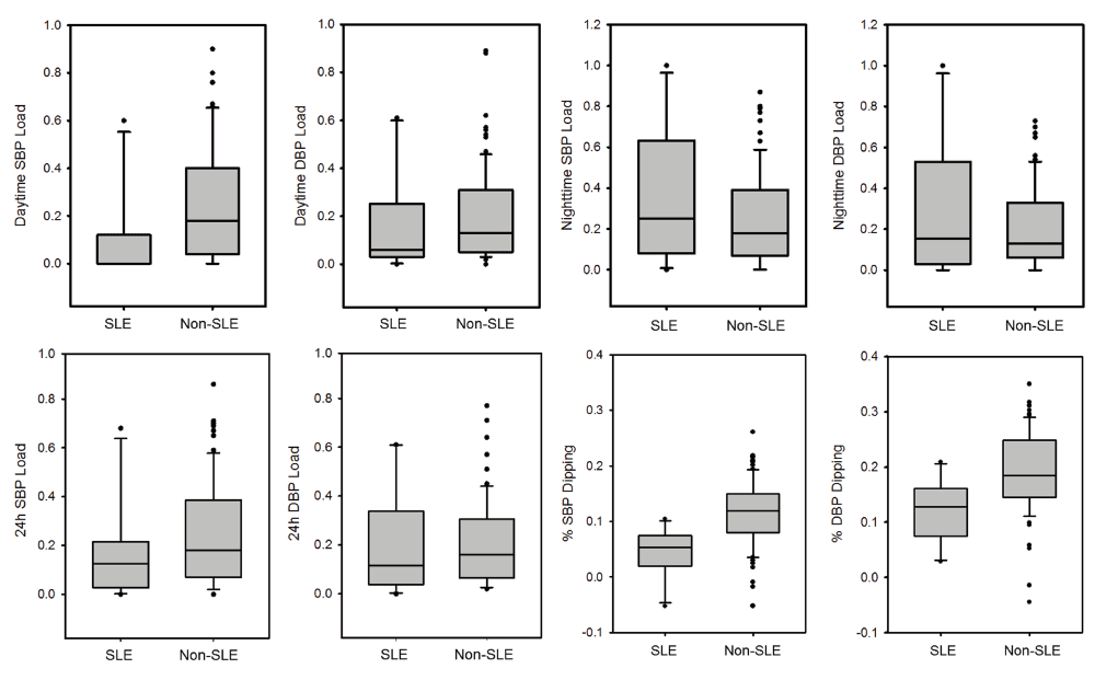
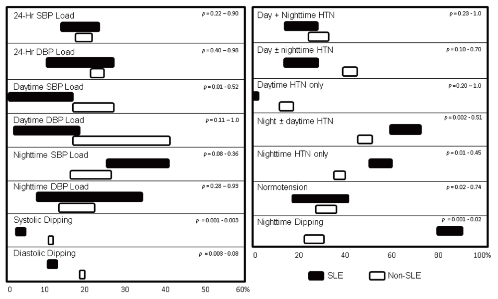
Comments on this article Comments (0)