Ajani MA, Salami AA, Awolude OA and Oluwasola AO. Pattern of triple negative epithelial ovarian cancer in indigenous African women [version 1; peer review: 2 approved]. F1000Research 2016, 5:2415 (https://doi.org/10.12688/f1000research.9632.1)
NOTE: If applicable, it is important to ensure the information in square brackets after the title is included in all citations of this article.
1Department of Pathology, University College Hospital, Ibadan, Nigeria 2Department of Obstetrics and Gynaecology, University College Hospital, Ibadan, Nigeria
OPEN PEER REVIEW
REVIEWER STATUS
Abstract
Background: Triple negative epithelial ovarian cancer (TNEOC) refers to ovarian carcinomas that do not express estrogen receptor (ER), progesterone receptor (PR) and human epidermal growth factor receptor- type 2 (HER-2/neu). The aim of this study is to determine the pattern of triple negative epithelial ovarian cancer in indigenous African women.
Methods: We performed a retrospective review of ER, PR and HER-2/neu expression in 90 Nigerian patients with histologically diagnosed epithelial ovarian cancer. Lack of expression of ER, PR and HER2/neu antigens was used to determine carcinomas that are among the TNEOC. We also compared the clinicopathological parameters (age, International Federation of Gynaecology and Obstetrics (FIGO) stage, grade and histological subtype) in patients with TNEOC and non- TNEOC .
Results: Thirty-eight (42.2%) of the 90 tumours diagnosed as EOC were negative for ER, PR and HER2/neu expression. There was no significant association between TNEOC with other parameters such as age, FIGO stage and histological grade. Sixteen (66.7%) of the 24 mucinous carcinomas were triple negative, while only 21 (33.3%) of the 63 serous carcinomas were triple-negative and one (50%) of the two endometrioid carcinomas was triple negative. There was a significant association between triple-negative tumours and histological subtypes of EOC (p = 0.034).
Conclusions: A subtype of epithelial ovarian cancer that is negative for ER, PR and HER-2/neu has been discovered in indigenous African women. TNEOC expression is high and is comparable to the triple negative breast cancer subtype seen in people of African ancestry. Future study of TNEOC in a large sample size should be considered.
Epithelial ovarian cancer (EOC) remains one of the leading causes of death in gynaecological malignancies in developed countries1–4. The initial symptoms of ovarian cancer are often ambiguous, therefore it goes undiagnosed until after the disease is far advanced and has spread throughout the abdomen or to distant organs5,6.
Steroid hormone receptors expression in epithelial ovarian cancers have been proposed to have therapeutic and prognostic relevance, as is the case in breast cancers7. The determination of tumour characteristics such as age, International Federation of Gynaecology and Obstetrics (FIGO) stage, grade and histological subtypes has been associated with clinical behaviour and impact on treatment and prognosis but have been found to be limited8. Among the biological parameters proposed as possible prognostic factors in ovarian cancer, estrogen receptor (ER), progesterone receptor (PR) and human epidermal growth factor receptor- type 2 (HER-2/neu) have been tested as potential biomarkers that guide individualized treatment of the cancer5,6,9. Epithelial ovarian carcinoma results from repeated ovulations, where the cumulative effects of each minor trauma on the ovarian epithelium can lead to malignant transformation10. PR has been observed to predict better prognosis because of its protection against ovarian carcinoma development11,12. On the other hand, overexpression of ER has been found to be associated with poor prognosis due to its contribution to initiation and/or promotion of ovarian carcinogenesis10,13. The HER-2/neu has been shown to be over-expressed in approximately 20–30% of EOC with associated poor prognosis14–16.
Triple negative epithelial ovarian cancer (TNEOC) cases have been found to be more aggressive and display a worse prognosis than non-TNEOC cases17. This was similarly observed in the studies of triple negative breast cancer18,19.
This study was designed to determine the pattern of TNEOC among indigenous African women and correlate it with clinicopathological parameters.
Methods
Patient selection
We performed a retrospective review of ER, PR and HER-2/neu expression in 90 patients with histologically diagnosed epithelial ovarian cancer seen at the University College Hospital, Ibadan, Nigeria between January 2006 and December 2012. Non-epithelial primary ovarian cancers and metastatic cancers in the ovary were not included in this study. The demographic data and clinical history of these cases were obtained from the case notes, surgical daybooks, surgical pathology request forms, post-mortem records and Cancer Registry data. Formalin-fixed paraffin-embedded tissue blocks of histologically diagnosed solid EOC between January 2006 and December 2012 were retrieved and used for the study. The microscopic grading (three-grade system) of Shimizu and Silverberg was used, which assesses architectural pattern, nuclear pleomorphism and mitotic activity20. All histological classification of the EOC was based on the 2013 World Health Organisation (WHO) classification of ovarian tumours21. The FIGO staging of the cases used for this study was extracted from the case notes of the patients.
Ethics
The ethical clearance for this study was obtained from the Joint University of Ibadan/University College Hospital Ethical Review Committee (approval number UI/EC/13/0050) according to the Declaration of Helsinki.
Immunohistochemistry
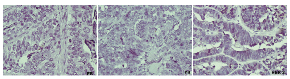
The immunostaining procedure for HER-2/neu was carried out in accordance with the previously published article22. For the immunostaining procedure, three sections each for ER, PR and HER-2/neu at 5µm were cut from each of the paraffin-embedded tissue blocks after deparaffinization in xylene (two aliquots for five minutes each with the xylene covering the slide entirely). The sections were then rehydrated in graded alcohol concentrations (two aliquots each of 100% and 95% each and a single aliquot of 70%) in 250ml couplin jars. The antibodies used were monoclonal mouse anti-human ERα (Dako USA; clone ID5) and monoclonal mouse anti-human PR (Dako USA; clone PgR636) which identify the ER and PR nuclear protein antigens. The primary antibody used for HER-2/neu antigen was polyclonal rabbit anti-human C-erbB-2 (MBO/TEG, Dako USA, 1:800). The tissue sections were immersed in EDTA buffer (pH 9.0) for ER, citrate buffer (pH 6.0) for PR and in 1M Tris buffer (pH 9.0) for HER-2/neu. These slides were then incubated at room temperature for 20 minutes with primary monoclonal antibodies against ER (Dako USA, clone 1D5; 1:50), PR (Dako USA, clone PgR636; 1:50) and polyclonal rabbit anti-human C-erbB-2 (MBO/TEG, Dako USA, 1:800) followed by incubation in biotin-labelled secondary antibodies, polyclonal goat anti-mouse antibody for both ER and PR, (Dako USA, REF: K0675, LOT: 10081219) and polyclonal goat anti-rabbit antibody for HER-2/neu, (kitR, K5001, Dako Denmark) for 20 minutes and streptavidin-peroxidase complex (Dako USA, REF: K0675, LOT: 10084687) for another twenty minutes. The antigen-antibody complex was precipitated with di-aminobenzidine (DAB) for light microscopy with DAB substrate and DAB chromogen in the ratio of 1ml to 1 drop respectively. This was thereafter counterstained in Mayer’s haematoxylin (Dako USA). Dehydration of the sections was performed in ascending grades of alcohol and cleared in xylene. The slides were coversliped with DPX mountant. Known cases of breast cancer with positive reactions for ER, PR and HER-2/neu were used as a positive control. Negative controls were cases of tumour sections that were pretreated in Tris but without primary antibody immunostaining. All slides were reviewed independently by the three of the authors and cases with discordant scores were re-evaluated to have a consensus score. Grading of nuclear ER and PR staining was performed using an immunoreactive H-scoring system {none= 0 (negative); 1–25%=1+ (weak); 26–50%=2+ (moderate); >50%=3+(strong)}11. HER-2/neu membrane staining was graded in according to the Hercep Test protocol system as 0, 1+, 2+ or 3+. Samples scored as 0 or 1+ were considered negative for HER-2/neu overexpression, 2+ was weakly positive and 3+ was strongly positive22,23. Photomicrographs of the specimens were taken using Olympus digital camera, DP 21 at 400X magnification (Figure 1).
Figure 1. Photomicrographs showing ER (left), PR (middle) and HER-2/neu (right) negative expression (TNEOC) (immunostaining, 400X).
Statistical analysis
The data obtained were subjected to statistical analysis using Statistical Package for Social Sciences (SPSS) version 20. Statistical analysis was used to evaluate statistical associations between TNEOC and clinicopathological parameters i.e. age, FIGO stage grade, and histological subtypes. Continuous variables were compared using the student’s T test and categorical variables were compared using the chi-square test, with the level of significance set at p <0.05.
Results
RAW DATA FOR PATTERN OF TRIPLE NEGATIVE EPITHELIAL OVARIAN CANCERS IN INDIGENOUS BLACK WOMEN
S/NO
AGE/years
BIOPSY SIDE
HISTOLOGICAL DIAGNOSIS
FIGO Stage
Histological Grading
Estrogen receptor (ER)
Progesterone receptor (PR)
HER-2/neu
1
55
Right
SEROUS CARCINOMA
II
II
negative
negative
negative
2
31
Right
MUCINOUS CARCINOMA
II
I
negative
negative
negative
3
72
Bilateral
SEROUS CARCINOMA
II
II
negative
weakly positive
weakly positive
4
50
Right
SEROUS CARCINOMA
II
I
weakly positive
strongly positive
strongly positive
5
59
Right
SEROUS CARCINOMA
II
I
strongly positive
negative
negative
6
60
Right
SEROUS CARCINOMA
II
II
negative
negative
strongly positive
7
44
Left
MUCINOUS CARCINOMA
I
II
negative
negative
negative
8
36
Right
SEROUS CARCINOMA
III
III
negative
negative
negative
9
52
Bilateral
SEROUS CARCINOMA
II
III
weakly positive
strongly positive
weakly positive
10
75
Bilateral
SEROUS CARCINOMA
III
II
negative
negative
negative
11
65
Left
SEROUS CARCINOMA
IV
III
weakly positive
weakly positive
strongly positive
12
48
Left
MUCINOUS CARCINOMA
II
I
negative
negative
weakly positive
13
54
Right
SEROUS CARCINOMA
II
III
weakly positive
negative
negative
14
37
Right
MALIGNANT BRENNER TUMOUR
I
I
strongly positive
negative
strongly positive
15
61
Bilateral
SEROUS CARCINOMA
II
II
strongly positive
weakly positive
weakly positive
16
43
Right
SEROUS CARCINOMA
II
I
negative
weakly positive
negative
17
41
Right
SEROUS CARCINOMA
IV
III
negative
negative
negative
18
49
Right
MUCINOUS CARCINOMA
III
II
negative
negative
negative
19
40
Left
MUCINOUS CARCINOMA
III
II
negative
weakly positive
negative
20
50
Right
SEROUS CARCINOMA
IV
III
negative
negative
strongly positive
21
26
Left
SEROUS CARCINOMA
II
III
negative
negative
negative
22
45
Bilateral
ENDOMETRIOID CARCINOMA
I
II
negative
negative
negative
23
30
Right
SEROUS CARCINOMA
IV
III
negative
negative
negative
24
47
Left
SEROUS CARCINOMA
III
III
negative
weakly positive
negative
25
38
Left
SEROUS CARCINOMA
II
II
weakly positive
strongly positive
negative
26
48
Left
SEROUS CARCINOMA
III
II
negative
strongly positive
strongly positive
27
45
Left
SEROUS CARCINOMA
II
II
weakly positive
weakly positive
negative
28
70
Right
SEROUS CARCINOMA
II
III
weakly positive
negative
negative
29
35
Bilateral
MUCINOUS CARCINOMA
III
III
weakly positive
negative
negative
30
53
Right
MUCINOUS CARCINOMA
IV
II
negative
negative
weakly positive
31
42
Bilateral
SEROUS CARCINOMA
III
I
negative
negative
negative
32
75
Bilateral
MUCINOUS CARCINOMA
III
I
negative
negative
negative
33
55
Bilateral
SEROUS CARCINOMA
IV
I
negative
negative
negative
34
50
Bilateral
SEROUS CARCINOMA
IV
III
strongly positive
negative
negative
35
65
Bilateral
SEROUS CARCINOMA
III
II
negative
negative
negative
36
49
Right
SEROUS CARCINOMA
III
III
strongly positive
srong
strongly positive
37
50
Right
SEROUS CARCINOMA
IV
III
negative
weakly positive
strongly positive
38
45
Left
SEROUS CARCINOMA
III
III
weakly positive
strongly positive
strongly positive
39
52
Left
SEROUS CARCINOMA
II
II
weakly positive
negative
negative
40
56
Right
SEROUS CARCINOMA
II
III
negative
negative
negative
41
46
Bilateral
MUCINOUS CARCINOMA
II
II
negative
strongly positive
negative
42
16
Bilateral
MUCINOUS CARCINOMA
II
II
negative
negative
negative
43
55
Left
MUCINOUS CARCINOMA
II
I
negative
negative
negative
44
59
Bilateral
SEROUS CARCINOMA
III
I
negative
negative
negative
45
69
Bilateral
SEROUS CARCINOMA
III
III
weakly positive
weakly positive
weakly positive
46
47
Bilateral
SEROUS CARCINOMA
III
II
negative
negative
negative
47
82
Right
MUCINOUS CARCINOMA
II
III
negative
negative
negative
48
44
Left
MUCINOUS CARCINOMA
II
I
negative
negative
negative
49
44
Right
ENDOMETRIOID CARCINOMA
II
II
negative
strongly positive
weakly positive
50
60
Left
SEROUS CARCINOMA
IV
II
negative
negative
strongly positive
51
32
Right
MUCINOUS CARCINOMA
I
I
negative
negative
negative
52
59
Bilateral
MUCINOUS CARCINOMA
II
II
negative
negative
negative
53
68
Right
SEROUS CARCINOMA
III
III
weakly positive
weakly positive
weakly positive
54
57
Left
SEROUS CARCINOMA
III
III
negative
negative
weakly positive
55
36
Left
MUCINOUS CARCINOMA
I
I
negative
negative
negative
56
49
Right
SEROUS CARCINOMA
I
I
negative
negative
strongly positive
57
39
Left
MUCINOUS CARCINOMA
II
II
negative
negative
negative
58
65
Right
SEROUS CARCINOMA
III
I
negative
negative
negative
59
64
Left
SEROUS CARCINOMA
IV
III
negative
weakly positive
negative
60
75
Bilateral
MUCINOUS CARCINOMA
II
I
weakly positive
negative
strongly positive
61
47
Bilateral
MUCINOUS CARCINOMA
II
II
negative
negative
negative
62
51
Left
SEROUS CARCINOMA
IV
III
negative
negative
negative
63
50
Right
MUCINOUS CARCINOMA
III
III
negative
negative
negative
64
28
Right
SEROUS CARCINOMA
III
III
negative
negative
weakly positive
65
53
Right
SEROUS CARCINOMA
IV
III
negative
negative
negative
66
46
Left
SEROUS CARCINOMA
III
II
weakly positive
weakly positive
negative
67
69
Left
SEROUS CARCINOMA
IV
III
negative
weakly positive
negative
68
40
Right
SEROUS CARCINOMA
III
III
negative
weakly positive
negative
69
75
Right
SEROUS CARCINOMA
III
II
negative
negative
strongly positive
70
52
Right
SEROUS CARCINOMA
III
II
negative
negative
strongly positive
71
47
Bilateral
SEROUS CARCINOMA
IV
III
negative
weakly positive
weakly positive
72
69
Bilateral
MUCINOUS CARCINOMA
I
I
negative
weakly positive
weakly positive
73
56
Bilateral
SEROUS CARCINOMA
II
II
negative
negative
negative
74
66
Right
SEROUS CARCINOMA
II
III
negative
weakly positive
negative
75
40
Left
SEROUS CARCINOMA
II
III
negative
negative
negative
76
60
Right
SEROUS CARCINOMA
II
III
negative
negative
negative
77
54
Bilateral
SEROUS CARCINOMA
III
III
weakly positive
strongly positive
negative
78
54
Right
SEROUS CARCINOMA
IV
II
negative
negative
weakly positive
79
58
Right
MUCINOUS CARCINOMA
II
II
negative
negative
negative
80
53
Bilateral
SEROUS CARCINOMA
IV
II
negative
negative
strongly positive
81
64
Bilateral
SEROUS CARCINOMA
IV
III
strongly positive
strongly positive
strongly positive
82
57
Right
SEROUS CARCINOMA
III
III
negative
negative
negative
83
54
Left
MUCINOUS CARCINOMA
II
I
negative
negative
strongly positive
84
42
Right
SEROUS CARCINOMA
III
III
negative
strongly positive
weakly positive
85
35
Left
SEROUS CARCINOMA
II
II
negative
negative
negative
86
60
Bilateral
SEROUS CARCINOMA
II
II
negative
negative
weakly positive
87
62
Bilateral
SEROUS CARCINOMA
IV
III
negative
strongly positive
strongly positive
88
67
Bilateral
SEROUS CARCINOMA
III
II
negative
negative
negative
89
56
Bilateral
MUCINOUS CARCINOMA
II
II
negative
negative
negative
Dataset 1.Raw data for ‘Pattern of triple negative epithelial ovarian cancer in indigenous African women’.
Thirty-eight (42.2%) of the 90 epithelial ovarian cancers (EOC) were negative for ER, PR and HER-2/neu expression (Figure 1). There was no significant association between triple-negative EOC and age (p = 0.218), FIGO stage (p = 0.425) and histological grade (p= 0.269). There were more TNEOC cases seen in patients older than 40 years than those below 40 years of age. Of 38 cases of TNEOC, 21 (55.3%) were found in the early stage (FIGO stage I and II) of epithelial ovarian cancer and 17 (44.7%) were at the advanced stage.
However, sixteen (66.7%) of the 24 mucinous carcinomas were triple-negative, while only 21 (33.3%) of the 63 serous carcinomas were triple-negative and one (50%) of the two endometrioid carcinomas was triple-negative (Table 1). There was therefore a significant association between triple-negative tumours and histological subtypes of EOC (p = 0.034).
Table 1. Correlation between triple negative epithelial ovarian cancer and clinicopathological parameters.
TNEOC
Non-TNEOC
Total
P value
Age
<40
9
4
13
>40
29
48
77
0.218
FIGO Stage
I-II
21
22
43
III-IV
17
30
47
0.425
Histological grade
I
10
9
19
II
16
18
34
III
12
25
37
Histological subtypes
Serous Carcinoma
21
42
63
Mucinous Carcinoma
16
8
24
Endometrioid Carcinoma
1
1
2
Malignant Brenner tumour
0
1
1
0.034
Total
38
52
90
Discussion
A subgroup of epithelial ovarian cancer that is negative for ER, PR and HER-2/neu expression has been identified among indigenous African women. This subgroup is known as triple negative epithelial ovarian cancer (TNEOC). According to ER, PR, and HER-2/neu expressions, a breast cancer subtype known as triple negative breast cancer (TNBC) has been identified24.
In our study, triple negative tumours accounted for 42.2% of EOC. This value contrasts with the results of other studies17,25 and compares with the results of previous study26. A significant percentage (66.7%) of mucinous carcinoma were negative for ER, PR and HER-2/neu and this was statistically significant (p=0.034). This finding contrasts what was found from previous studies where there was no significant association between the TNEOC and histological subtypes17,25,26. No significant association was also found between the TNEOC and histological grade unlike what was observed by Liu et al.17 and de Toledo et al.26 where TNEOC was significantly correlated with histological grade. There was no significant association between TNEOC and age and FIGO stage compared to the findings of other studies17,25,26.
Our findings were comparable with what was found by Huo et al. in the population differences in breast cancer where triple negativity was predominant (27%)19. In view of the fact that triple-negative breast cancers are more often seen in black Africans and African-Americans and are associated with a poorer prognosis than non-triple-negative breast cancers, further studies of TNEOC in different environments are required.
Conclusions
A subtype of epithelial ovarian cancer that is negative for ER, PR and HER-2/neu has been discovered in Nigeria. Its (TNEOC) expression is high and is comparable to the triple negative breast cancer subtype seen in people of African ancestry. Future study of TNEOC in a large sample size should be considered.
Data availability
F1000Research: Dataset 1. Raw data for ‘Pattern of triple negative epithelial ovarian cancer in indigenous African women’, 10.5256/f1000research.9632.d13677727
Author contributions
MAA conceived and designed the study. MAA, AAS and AOO carried out the research. MAA and AOO prepared the first draft of the manuscript. All authors contributed to the experimental design and preparation of the manuscript. All authors were involved in the revision of the draft manuscript and have agreed to the final content.
Competing interests
No competing interests were disclosed.
Grant information
The author(s) declared that no grants were involved in supporting this work.
Acknowledgements
Special thanks to Mr S Ajagboye and Mr SP Otegbade of the Department of Pathology, University of Ibadan and University College Hospital respectively for the technical assistance they rendered with the slides used for this study and Mr Abayomi Odetunde of Institute for Advanced Medical Research and Training, College of Medicine, University of Ibadan for carrying out the immunohistochemical staining on the cases.
Faculty Opinions recommended
References
1.
Hunn J, Rodriguez GC:
Ovarian cancer: etiology, risk factors, and epidemiology.
Clin Obstet Gynaecol.
2012; 55(1): 3–23. PubMed Abstract
| Publisher Full Text
4.
Swamy GG, Satyanarayana N:
Clinicopathological analysis of ovarian tumors--a study on five years samples.
Nepal Med Coll J.
2010; 12(4): 221–223. PubMed Abstract
5.
Dinh P, Harnett P, Piccart-Gebhart MJ, et al.:
New therapies for ovarian cancer: cytotoxics and molecularly targeted agents.
Crit Rev Oncol Hematol.
2008; 67(2): 103–12. PubMed Abstract
| Publisher Full Text
6.
Yap TA, Carden CP, Kaye SB:
Beyond chemotherapy: targeted therapies in ovarian cancer.
Nat Rev Cancer.
2009; 14(3): 167–81. PubMed Abstract
| Publisher Full Text
7.
Scambia G, Ferrandina G, D’Agostino G, et al.:
Oestrogen and progesterone receptors in ovarian carcinoma.
Endocrine Rel Cancer.
1998; 5: 293–301. Publisher Full Text
8.
de Toledo MC, Barreta A:
The Role of Steroid Receptors and HER2 in Ovarian Cancer.
J Carcinog Mutagen.
2014; 5: 1. Publisher Full Text
9.
Tuefferd M, Couturier J, Penault-Llorca F, et al.:
HER2 status in ovarian carcinomas: a multicenter GINECO study of 320 patients.
PLoS One.
2007; 2(11): e1138. PubMed Abstract
| Publisher Full Text
| Free Full Text
11.
Ayadi L, Chaabouni S, Khabir A, et al.:
Correlation between immunohistochemical biomarkers expression and prognosis of ovarian carcinomas in Tunisian patients.
World J Oncol.
2010; 1(3): 118–128. Publisher Full Text
12.
Lee P, Rosen DG, Zhu C, et al.:
Expression of progesterone receptor is a favorable prognostic marker in ovarian cancer.
Gynaecol Oncol.
2005; 96(3): 671–677. PubMed Abstract
| Publisher Full Text
13.
Zhao D, Zhang F, Zhang W, et al.:
Prognostic role of hormone receptors in ovarian cancer: a systematic review and meta-analysis.
Int J Gynecol Cancer.
2013; 23(1): 25–33. PubMed Abstract
| Publisher Full Text
14.
Hellström I, Goodman G, Pullman J, et al.:
Overexpression of HER-2 in ovarian carcinomas.
Cancer Res.
2001; 61(6): 2420–2423. PubMed Abstract
15.
Cirisano FD, Karlan BY:
The role of the HER-2/neu oncogene in gynecologic cancers.
J Soc Gynecol Investig.
1996; 3(3): 99–105. PubMed Abstract
16.
Yu D, Wolf JK, Scanlon M, et al.:
Enhanced c-erbB-2/neu expression in human ovarian cancer cells correlates with more severe malignancy that can be suppressed by E1A.
Cancer Res.
1993; 53(4): 891–898. PubMed Abstract
17.
Liu N, Wang X, Sheng X:
The clinicopathological characteristics of ‘triple-negative’ epithelial ovarian cancer.
J Clin Pathol.
2010; 63(3): 240–243. PubMed Abstract
| Publisher Full Text
19.
Huo D, Ikpatt F, Khramtsov A, et al.:
Population differences in breast cancer: survey in indigenous african women reveals over-representation of triple-negative breast cancer.
J Clin Oncol.
2009; 27(27): 4515–4521. PubMed Abstract
| Publisher Full Text
| Free Full Text
20.
Shimizu Y, Kamoi S, Amada S, et al.:
Toward the development of a universal grading system for ovarian epithelial carcinoma. I. Prognostic significance of histopathologic features--problems involved in the architectural grading system.
Gynecol Oncol.
1998; 70(1): 2–12. PubMed Abstract
| Publisher Full Text
21.
Tassovoli FA, Devilee P:
Pathology and Genetics. Tumours of the Breast and Female Genital Organs. Lyon, IARC Press. 2003; 114. Reference Source
22.
Ajani MA, Salami A, Awolude OA, et al.:
The expression status of human epidermal growth factor receptor 2 in epithelial ovarian cancer in Ibadan, Nigeria.
South Afr J Gynaecol Oncol.
2016; 8(1): 9–13. Publisher Full Text
23.
Wolff AC, Hammond ME, Schwartz JN, et al.:
American Society of Clinical Oncology/College of American Pathologists guideline recommendations for human epidermal growth factor receptor 2 testing in breast cancer.
J Clin Oncol.
2007; 25(1): 118–145. PubMed Abstract
| Publisher Full Text
24.
Reis-Filho JS, Tutt AN:
Triple negative tumours: a critical review.
Histopathology.
2008; 52(1): 108–118. PubMed Abstract
| Publisher Full Text
25.
Demir L, Yigit S, Sadullahoglu C, et al.:
Hormone Receptor, HER2/NEU and EGFR expression in ovarian carcinoma--is here a prognostic phenotype?
Asian Pac J Cancer Prev.
2014; 15(22): 9739–9745. PubMed Abstract
| Publisher Full Text
26.
de Toledo MC, Sariana LO, Salluma LF, et al.:
Analysis of the contribution of immunologically-detectable HER2, steroid receptors and of the “triple-negative” tumor status to disease-free and overall survival of women with epithelial ovarian cancer.
Acta Histochemica.
2014; 116(3): 440–447. PubMed Abstract
| Publisher Full Text
27.
Ajani MA, Salami AA, Awolude OA, et al.:
Dataset 1 in: Pattern of triple negative epithelial ovarian cancer in indigenous African women.
F1000Research.
2016. Data Source
1
Department of Pathology, University College Hospital, Ibadan, Nigeria 2
Department of Obstetrics and Gynaecology, University College Hospital, Ibadan, Nigeria
Ajani MA, Salami AA, Awolude OA and Oluwasola AO. Pattern of triple negative epithelial ovarian cancer in indigenous African women [version 1; peer review: 2 approved]. F1000Research 2016, 5:2415 (https://doi.org/10.12688/f1000research.9632.1)
NOTE: If applicable, it is important to ensure the information in square brackets after the title is included in all citations of this article.
track
receive updates on this article
Track an article to receive email alerts on any updates to this article.
Share
Open Peer Review
Current Reviewer Status:
?
Key to Reviewer Statuses
VIEWHIDE
ApprovedThe paper is scientifically sound in its current form and only minor, if any, improvements are suggested
Approved with reservations
A number of small changes, sometimes more significant revisions are required to address specific details and improve the papers academic merit.
Not approvedFundamental flaws in the paper seriously undermine the findings and conclusions
Ahmed SA. Reviewer Report For: Pattern of triple negative epithelial ovarian cancer in indigenous African women [version 1; peer review: 2 approved]. F1000Research 2016, 5:2415 (https://doi.org/10.5256/f1000research.10379.r16697)
This is an interesting manuscript, well conceived and has addressed the objective of the research.
The methodology is simple and reproducible.
Result was clearly outlined and discussion
... Continue reading
This is an interesting manuscript, well conceived and has addressed the objective of the research.
The methodology is simple and reproducible.
Result was clearly outlined and discussion was adequate.
Conclusion is apt.
This research finding will add to the few literature on TNEOC in the population studied. It will serve as a baseline for future reference and interestingly the research has prognostic and therapeutic implications.
Competing Interests: No competing interests were disclosed.
I confirm that I have read this submission and believe that I have an appropriate level of expertise to confirm that it is of an acceptable scientific standard.
Ahmed SA. Reviewer Report For: Pattern of triple negative epithelial ovarian cancer in indigenous African women [version 1; peer review: 2 approved]. F1000Research 2016, 5:2415 (https://doi.org/10.5256/f1000research.10379.r16697)
I have gone through the whole paper and these are my comments:
It is a well conceived and conducted research paper with the aims and objectives and rationale for the study well spelled out.
... Continue reading
I have gone through the whole paper and these are my comments:
It is a well conceived and conducted research paper with the aims and objectives and rationale for the study well spelled out.
It addresses the peculiarities of the African race which is fundamental to adequate care.
The authors are very clear and consistent in the message that they aimed to convey and have passed this through.
All parts of the research work have fulfilled the guidelines for sound scientific work.
The discussion addresses crucial details.
The conclusion is a brilliant and excellent summation of the author's scientific work.
Competing Interests: No competing interests were disclosed.
I confirm that I have read this submission and believe that I have an appropriate level of expertise to confirm that it is of an acceptable scientific standard.
Alongside their report, reviewers assign a status to the article:
Approved - the paper is scientifically sound in its current form and only minor, if any, improvements are suggested
Approved with reservations -
A number of small changes, sometimes more significant revisions are required to address specific details and improve the papers academic merit.
Not approved - fundamental flaws in the paper seriously undermine the findings and conclusions
Spreadsheet data files may not format correctly if your computer is using different default delimiters (symbols used to separate values into separate cells) - a spreadsheet created in one region is sometimes misinterpreted by computers in other regions. You can change the regional settings on your computer so that the spreadsheet can be interpreted correctly.
How to fix it
Save downloaded CSV file
Open spreadsheet program (e.g. Excel)
Click the ‘Data’ tab at the top
Click the ‘From text’ icon (top left)
Browse for downloaded CSV file, click ‘Import’
Ensure ‘Delimited’ radio button is selected, click ‘Next’
Check one of the appropriate delimiter checkboxes (you can visualize the formatting by looking at the data preview below these options)
Ajani MA, Salami AA, Awolude OA and Oluwasola AO. Dataset 1 in: Pattern of triple negative epithelial ovarian cancer in indigenous African women. F1000Research 2016, 5:2415 (https://doi.org/10.5256/f1000research.9632.d136777)
Adjust parameters to alter display
View on desktop for interactive features
Includes Interactive Elements
View on desktop for interactive features
Competing Interests Policy
Provide sufficient details of any financial or non-financial competing interests to enable users to assess whether your comments might lead a reasonable person to question your impartiality. Consider the following examples, but note that this is not an exhaustive list:
Examples of 'Non-Financial Competing Interests'
Within the past 4 years, you have held joint grants, published or collaborated with any of the authors of the selected paper.
You have a close personal relationship (e.g. parent, spouse, sibling, or domestic partner) with any of the authors.
You are a close professional associate of any of the authors (e.g. scientific mentor, recent student).
You work at the same institute as any of the authors.
You hope/expect to benefit (e.g. favour or employment) as a result of your submission.
You are an Editor for the journal in which the article is published.
Examples of 'Financial Competing Interests'
You expect to receive, or in the past 4 years have received, any of the following from any commercial organisation that may gain financially from your submission: a salary, fees, funding, reimbursements.
You expect to receive, or in the past 4 years have received, shared grant support or other funding with any of the authors.
You hold, or are currently applying for, any patents or significant stocks/shares relating to the subject matter of the paper you are commenting on.
Stay Updated
Sign up for content alerts and receive a weekly or monthly email with all newly published articles
Comments on this article Comments (0)