Keywords
Progressive contact lens, myopia, hyperopic defocus, peripheral retina
This article is included in the Eye Health gateway.
Progressive contact lens, myopia, hyperopic defocus, peripheral retina
The most common type of eye refractive error is called myopia, which is considered a global health problem1. With the beginning of 21st century, Atchison et al.2 2006 and Mutti et al.3 2007 observed that myopic eyes have more hyperopic peripheral refraction than emmetropes in the horizontal visual field. Studies conducted by Smith and colleagues in monkeys have shown that not only the fovea, but also the peripheral retina, is capable of regulating the emmetropiszation process4–6. This indicates that the peripheral retina is important in determining ocular development and refractive error.
Studies have shown that conventional correction of myopia using spectacles lenses may increase hyperopic defocus in the periphery7,8. Hyperopic defocus worsens with a higher degree of myopia and eccentricity9. In 2009, Tabernero et al.8 suggested that by changing the peripheral optics of corrective devices, relative hyperopic defocus in myopic eyes could be inverted into peripheral relative myopia. This could be a possible strategy to counterbalance the unknown stimulus that triggers the eye elongation and subsequent progression of myopia.
Specially designed spectacle lenses10 and contact lenses11 have employed the change in peripheral optics of the optical design. Some commercially available progressive contact lenses (PCL) (dominant-design) intended for presbyopic patients might render a similar effect. The peripheral add power area, which was primarily intended to increase spherical aberration and depth of focus in presbyopic patients, has been shown to induce significant changes in the peripheral refractive error profile of the eye. Lopes-Ferreira et al.12 found that a +3.00 D add dominant design Proclear progressive contact lens in 20 emmetropic and 28 myopic eyes inverted the hyperopic defocus to myopic defocus in the periphery. In another study by Rosén et al.13, they were able to induce approximately 0.50 D of myopic defocus 30° using a +2.00 D lens in 1 myopic and three emmetropic patients. These studies were done in adults, and it is unclear if the hyperopic defocus could be inverted to myopic defocus in myopic children. This knowledge is important because myopia progression occurs mainly in children. The aim of this study was to compare the changes in relative peripheral refractive error produced by using two different designs of commercially available progressive soft contact lenses in myopic schoolchildren.
Twenty-seven myopic schoolchildren (24 females, 3 males) aged between 13 and 15 years were recruited in this study. The purpose and procedure of the study were explained to all participants and their parents. Then, written informed consent was obtained before enrolment into the study. The study was conducted at the University Kebangsaan Malaysia (UKM) Optometry Clinic and Vision Science Lab. This research was approved by the Ethics Committee of the Universiti Kebangsaan Malaysia (UKM 1.5.3.5/244/NN-144-2013) and followed the tenets of the Declaration of Helsinki in using human subjects.
The inclusion criteria for this study were having visual acuity of 6/9 or better in both eyes, having normal ocular condition with a spherical component refractive error range between -3.00 and -6.00 D, astigmatism not more than -1.00 D and anisometropia of less than 1.50 D between both eyes. Children with manifest strabismus, amblyopia, any ocular conditions associated with myopia, a history of bifocal or progressive spectacle wear, orthokeratology contact lens wear, or those currently wearing soft contact lenses were excluded from participation in this study.
A comprehensive ocular examination, which included fundus evaluation, anterior segment assessment, and axial length calculation, was conducted by an experienced optometrist to select the candidates. The spherical equivalent refractive error (M) for each subject was determined using non-cycloplegic objective and subjective refraction. An ultrasound A-scan (Tomey AL-2000) was used to measure axial length using a handheld probe. The final outcome was calculated as the mean of 5 measurements.
Central and peripheral refraction were measured using an open-view autorefractometer (Grand-Seiko WR-5100K,Grand Seiko Co., Ltd., Hiroshima, Japan). The examination room illumination was dimmed (mean of three measurements: 9.91 ± 1.73 lux, measured using a Topcon Luxmeter) in order to obtain a pupil size sufficiently large enough to measure peripheral retina without using dilatation drops. The measurement was obtained initially without contact lenses (WL), then re-measured again using Multistage progressive contact lenses (Multistage PCL, from SEED Co. Japan) and Proclear progressive contact lenses (Proclear PCL, from CooperVision) in random order. Subjects were masked to the type of each lens, while the practitioner was unmasked. The subjects were instructed to fixate on targets (green light laser) located at 4 metres arranged horizontally in the positions corresponding to eccentricities from 35° temporal to 35° nasal, in 5° steps. The straight ahead viewing technique was used in this study, in which the subjects rotated their eyes to view a series of fixation targets. Five refraction measurements were taken at each target fixation for the right eye only, while the left eye was occluded. For statistical analysis, the sphero-cylindrical refractive error measurements were converted into vector components of refraction M, J0, J45 using the equations recommended by Thibos et al.14 M, J0 and J45 according to Fourier analysis,
M = sph + (cyl/2), J0 = (-cyl/2) cos (2 α), J45 = (-cyl/2) sin (2 α),
where sph, cyl and (α) represent sphere, cylinder and axis, respectively. Relative peripheral refractive error (RPRE) was calculated as the difference between eccentric peripheral refraction and central refraction. A one-way repeated measures ANOVA with Bonferroni’s post-hoc test was conducted to determine the changes in RPRE values for the mean spherical equivalent M, J0 and J45 components between the groups.
All subjects were fitted with Multistage PCL and Proclear PCL to their right eyes in random order on the same days. Lens powers fully corrected the central refractive error. The Multistage PCL used in the study was a biweekly soft contact lens made of 42% Group IV (ionic high water content) and 58% water content, with diameter of 14.2 mm and a base curve of 8.6 mm. The B-Design used in this study is spherical distance power at the centre zone (2.5 mm), a junction zone (2.5 mm to 3.5 mm) and a near zone (3.5 mm to 8.0 mm) with a maximum addition power of +1.50 D in the periphery.
The Proclear progressive D® design contact lens was a monthly disposable lens made from omafilcon A, with a water content of 62% and an overall diameter of 14.4 mm, with a base curve of 8.7 mm. The lens design has a 2.3-mm inner distance central spherical area, surrounded by an annular aspheric zone where the addition power increases gradually to reach its maximum power of +1.50D at 5 mm. There is a second spherical zone with a maximum addition power of +1.50D from 5 mm to 8 mm diameter. Table 1 illustrates the parameters of contact lenses used in this study.
Analysis was performed using SPSS statistical software version 20 (SPSS Inc., IL, USA). Only data from the right eye was analysed. A Shapiro–Wilk test was used to evaluate the normality of the data distribution. A paired t-test was used for paired comparisons of RPRE within each group at the different eccentricities with respect to the centre. When normality could not be assumed, the Wilcoxon signed-ranks test was used. The differences were considered statistically significant when the p value was lower than 0.05. Then, repeated measures analysis of variance (ANOVA) was performed to compare the RPRE between the different groups at the different eccentricities.
*Values are expressed in diopters (D). N is nasal visual field; T is temporal visual field; C is centre; p represents the value of statistical significance according to Paired t-test or Wilcoxon Signed Ranks Test. Bold indicates statistically significant power difference from central point (95% confid
A total of 27 myopic schoolchildren with a mean age of 14.18 ± 0.88 years (range: 13 years to 15 years) participated in this study. The mean central spherical equivalent refractive error was found to be -4.39 ± 0.95 D (range: -3.12D to -5.93D) without correction, with a mean axial length of 24.72 ± 0.92 mm (range: 23.51 mm to 26.39 mm). Table 2 presents the mean values of refractive error and standard deviations of eyes without contact lenses (WL), with Multistage PCL and Proclear PCL.
Table 3 shows the RPRE and standard deviations (SD) for mean spherical equivalent values (M), horizontal astigmatism component (J0) and oblique astigmatism component (J45) in WL conditions, with Multistage PCL and Proclear PCL. A paired t-test showed that without contact lenses, there was a significant hyperopic defocus at and beyond 30° in the nasal visual field (N30° p= 0.001, N35° p< 0.001) and at and beyond 25° in the temporal visual field (T25° p= 0.018, T30° p= 0.001, and T35°< 0.001). When Multistage PCL was used, the peripheral defocus was only present at 35° in the nasal (p= 0.009) and temporal visual fields (p= 0.026). However, with Proclear PCL there was significant hyperopic defocus at and beyond 30° nasally (N30° p= 0.004, N35° p< 0.001) and at 25° temporally (T25° p= 0.031, T30° p= 0.004, and T35° p= 0.001). Multistage PCL shows a significant myopic defocus at nasal and temporal 35° for the J0 component, while for the J45 component, there was a significant hyperopic defocus at 20° but a significant myopic defocus at 15° nasal and 20° temporal.
With the Multistage PCL, the peripheral hyperopic defocus was decreased and only present at eccentricity of 35° nasally and temporally. However, with Proclear PCL, the hyperopic shift was still present at and beyond 30° in the nasal visual field and 25° in the temporal visual field. The hyperopic defocus was much smaller for Multistage PCL (+0.67 ±1.23 D at 35° nasal and +0.52 ±1.14 D at 35° temporal) as compared to Proclear PCL (+1.13 ±1.31 D at 35° nasal and +0.81 ±1.10 D at 35° temporal).
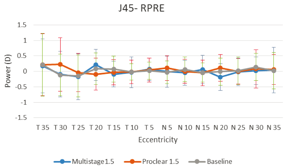
Figure 1, Figure 2 and Figure 3 illustrate the RPRE without contact lenses (baseline), with Multistage PCL and Proclear PCL for spherical equivalent value M, horizontal astigmatic component J0 and oblique astigmatic component J45, respectively. The hyperopic defocus is obvious at nasal and temporal visual fields with spherical equivalent value M. The J0 and J45, however, showed few changes at the peripheral field.


Progressive contact lens design
(a) Proclear progressive contact lens from Coopervision (b) Multistage progressive contact lens from Seed.
A one-way repeated measures ANOVA was conducted to determine the changes in RPRE values for the mean spherical equivalent M, J0 and J45 components between the groups. The results of the ANOVA indicated a significant difference in mean spherical equivalent between groups with a Greenhouse–Geisser correction (F(7.218, 43.794) = 4.285, p= 0.032). A post-hoc test using Bonferroni’s correction indicated a statistically significant difference in mean spherical equivalent RPRE (M) between the baseline and Multistage PCL (p=0.015), while Proclear PCL showed no statistically significant different in comparison to the baseline (p=0.830). The results showed no statistically significant difference between without contact lenses and all contact lenses used in this study for J0 and J45F(1.772, 52.926) = 0.871, p= 0.425, and F(0.440, 67.258) = 0.172, p=0.844, respectively. Therefore, it can be concluded that wearing a Multistage PCL can reduce hyperopic defocus in the retinal periphery.
With the extensive range of powers, materials and designs of soft contact lenses, they have become one of the most popular modes of myopia correction widely used by young adults. The present study compared the effect of RPRE along the horizontal visual field between two different designs of progressive contact lens (Multistage PCL and Proclear PCL). Although both progressive contact lenses in this study are simultaneous vision lenses, and had the same addition power (+1.50 D), the results showed a greater reduction in hyperopic defocus with Multistage PCL in comparison with Proclear PCL. The Multistage PCL had a decreased mean hyperopic defocus along the horizontal visual field up to 30° nasally and temporally, which indicated possible control of myopia progression for prolonged wear. However, the Proclear PCL showed significant hyperopic defocus from 30°, and 25° and beyond at the nasal and temporal visual fields, respectively.
The reason for the difference in hyperopic defocus at the periphery between both PCLs could be due to the difference in lens design. The Proclear PCL has a distance centre design, where the centre zone is 2.3 mm in diameter, and the added power increases progressively in a wide annular aspheric zone (from 2.3 to 5 mm/1.35 width), and ends in a spherical near zone (from 5 to 8.5 mm/1.75 width) where the full addition power of +1.50D exists. However, with the Multistage PCL, the design is different in diameters and power progression. The centre distance zone is 2.5 mm in diameter, surrounded by a narrow aspheric multifocal zone “junction zone” (from 2.5 to 3.5 mm/0.5 mm width), followed by a large spherical near zone (from 3.5 to 8.0 mm/2.25 width). With the dim illumination used in this study, the subject’s pupil size was approximately 4 to 5 mm. Hence, children were unable to view from the spherical near zone in Proclear PCL, where the near zone in this lens starts at 5 mm in diameter until 8 mm in diameter. However, with Multistage PCL the pupil size was sufficient to view the junction zone (2.5 mm to 3.5 mm), and part of the near spherical zone where the maximum addition power exists.
Phillips and Anstice15 used dual-focus soft contact lenses on children aged 11 to 14 years old. The lens had a central distance correction zone followed by a concentric treatment zone with +2.00 D of peripheral retinal defocus. They reported a 36% reduction of myopia progression (-0.44 D versus -0.69 D) over 10 months of treatment as compared to a single vision contact lens. However, Sankaridurg et al.11 2011, found a reduction of myopia progression of 34% (-0.57 D versus -0.87 D) over one year of using multifocal contact lenses with a distance centre zone. The design had a progression increase of +2.00 D additional power compared to the control group. In 2013, Walline controlled myopia progression by 51% over 2 years of treatment by using Proclear PCL with +2.00 D additional power. However, the axial length elongation was slowed down by approximately 29% over this 2-year period16. The authors could not explain why the myopia progression was slowed almost twice as much as the axial elongation. The reasons for not matching the myopia progression with the axial elongation in the Walline study might have been due to the fact that subjects were not randomly allocated to treatment groups, had a high drop-out rate (32.5%) with uncollected reasons for subjects’ withdrawals and there were 5 years of difference in data collection between the treatment group (June 2007 to May 2009) and the control group (September 2003 to Oct 2004).
Although neither PCL used in this study is made for myopia control, and they are commercially used for presbyopic older patients, the results of the present study illustrate no significant effect of relative peripheral hyperopic defocus with Proclear PCL +1.50D addition power. However, in 2013, Lopes-Ferreira et al. reported that a minimum addition of +2.00 D Proclear PCL D-design was necessary to induce a significant effect on peripheral refractive error, which explains why no statistical difference was found with Proclear PCL +1.50 D in the present study17.
The mean central refractive error was -1.08 ±0.29D and -1.11 ±0.36D with Multistage PCL and Proclear PCL, respectively. Since the refractive error was fully corrected with contact lenses, the measurement of central refractive error was expected to be zero. This could be due to the infrared light beam used to measure the refractive error in the open-view Grand-Seiko WR-5100K autorefractometer. The size of the infrared light beam is about 2.3 mm in diameter, which is similar in size to the central zone of PCLs used in this study. Therefore, a small decentration of the lens (<0.5 mm) could have made the instrument read part of the addition power zone. However, by using the same procedure to measure all points of peripheral refraction with the same light beam, the relative peripheral refractive error would give the same myopic shift of readings, and therefore, the measurements were still valid and reliable along the 70° of the horizontal visual field.
It was possible to decrease the peripheral retinal hyperopic defocus by using soft progressive contact lenses with a distance centre design. This study suggested that PCL designed with a narrow junction zone and wider spherical near zone had a greater effect on the pattern of peripheral refractive error, which may show better control of myopia in comparison to PCLs designed with a progressive increase of addition power.
F1000Research: Raw data for ‘Peripheral refraction with different designs of progressive soft contact lenses in myopes’, 2016, 10.5256/f1000research.9971.d14367718
| Views | Downloads | |
|---|---|---|
| F1000Research | - | - |
|
PubMed Central
Data from PMC are received and updated monthly.
|
- | - |
Click here to access the data.
Spreadsheet data files may not format correctly if your computer is using different default delimiters (symbols used to separate values into separate cells) - a spreadsheet created in one region is sometimes misinterpreted by computers in other regions. You can change the regional settings on your computer so that the spreadsheet can be interpreted correctly.
Provide sufficient details of any financial or non-financial competing interests to enable users to assess whether your comments might lead a reasonable person to question your impartiality. Consider the following examples, but note that this is not an exhaustive list:
Sign up for content alerts and receive a weekly or monthly email with all newly published articles
Already registered? Sign in
The email address should be the one you originally registered with F1000.
You registered with F1000 via Google, so we cannot reset your password.
To sign in, please click here.
If you still need help with your Google account password, please click here.
You registered with F1000 via Facebook, so we cannot reset your password.
To sign in, please click here.
If you still need help with your Facebook account password, please click here.
If your email address is registered with us, we will email you instructions to reset your password.
If you think you should have received this email but it has not arrived, please check your spam filters and/or contact for further assistance.
Comments on this article Comments (0)