Keywords
nerve sheath tumor, adrenal, lung, metastasis
nerve sheath tumor, adrenal, lung, metastasis
We have addressed the reviewer concerns and added to the Abstract and the Introduction. We agree with changing the title from "collision tumor" to “cancer to cancer" metastasis given the ambiguous definitions.
See the authors' detailed response to the review by Ahmed Abu-Zaid
See the authors' detailed response to the review by Shweta Gera
Soft tissue sarcomas of the extremity are a rare disease process, comprising less than 1% of all malignancies1. The majority of soft-tissue sarcomas occur in the limb or limb girdle or within the abdomen, with 40% being found in the lower extremities. These sarcomas are a histologically heterogeneous group of tumors (over 50 tumor subtypes have been identified) with a predilection for hematogenous spread. Distant metastatic disease is found in approximately 20–30% of patients1–3, with pulmonary lesions accounting for 75% of these cases1,4,5. Because of the relatively low incidence of extra-pulmonary metastasis, the current guidelines from the National Comprehensive Cancer Network (NCCN) recommends to only “consider” abdominal/pelvic CT imaging for certain subgroups of sarcomas (myxoid/round cell liposarcoma, epithelial sarcoma, angiosarcoma, leiomyosarcoma)6. A similar recommendation is made by the European Society of Medical Oncology (ESMO)/European Sarcoma Network Working Group7. No specific recommendation is made for any additional sarcoma subtype regardless of size of the tumor, despite this being a known prognostic factor.
Malignant peripheral nerve sheath tumors (MPNST) are highly malignant sarcomas which originate from peripheral nerves or from cells associated with the nerve sheath, such as Schwann cells, perineural cells, or fibroblasts. MPNSTs compromise 5–10% of all soft tissue sarcomas with 22–50% of the tumors associated with a diagnosis of Neurofibromatosis-1 (NF1)8–10. We report a case of a malignant peripheral nerve sheath tumor of the thigh, without evidence of concomitant pulmonary metastases, which was found to have metastasized to the adrenal gland. Thus, we pose the question of should the staging work up for large sarcomas be expanded to include abdominal imaging, even if the CT chest is unrevealing?
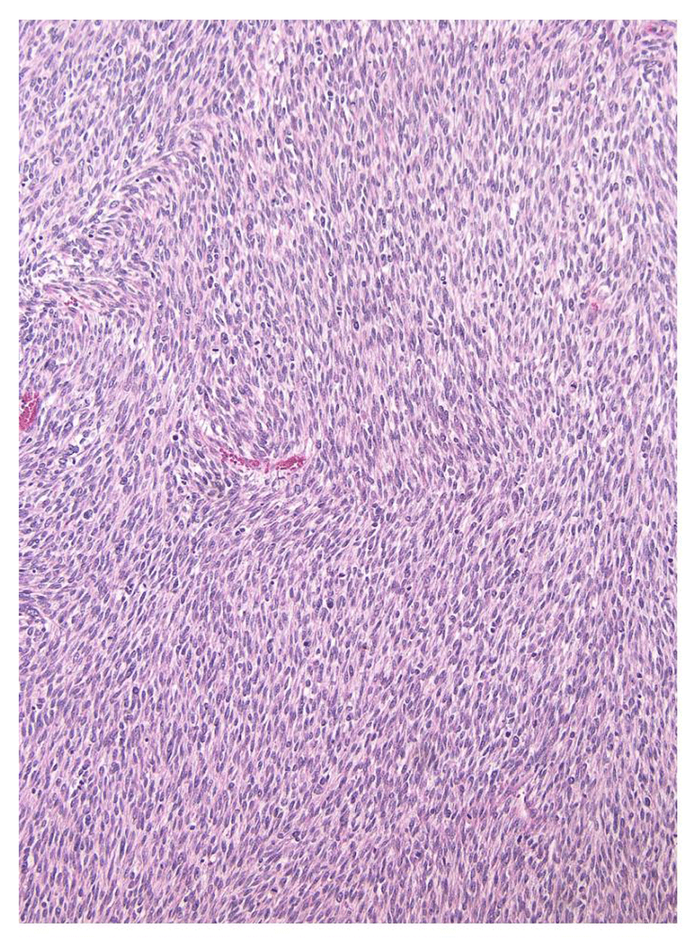
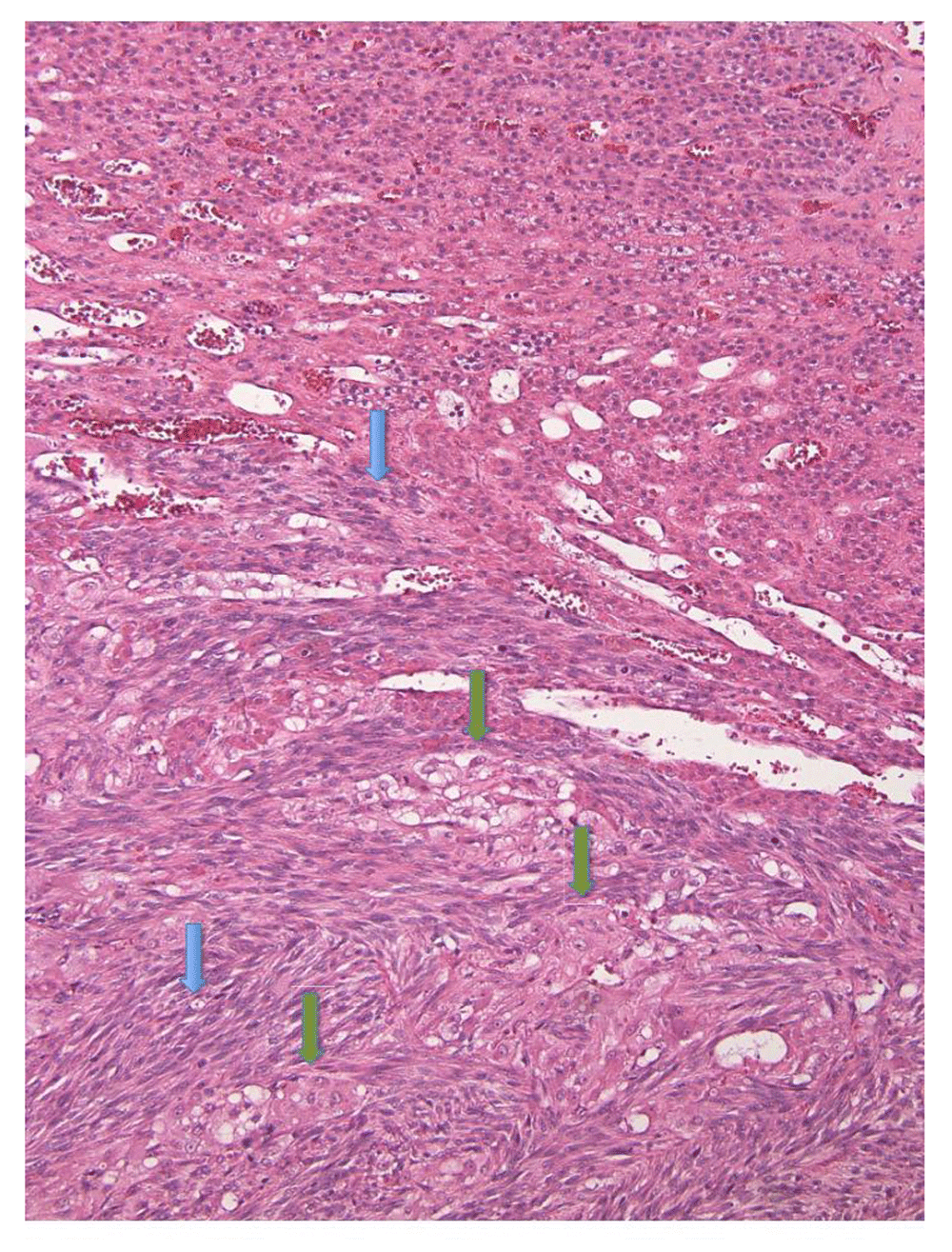
A 40 year old woman withNF1 presented for a hemorrhagic right thigh mass which had been enlarging over the past 3 months. The mass had previously been stable in size for a few years and was thought to be consistent with a neurofibroma. On presentation her exam was notable for a very large soft tissue tumor of the right posterior upper leg (>15cm). The distal portion of the lesion revealed skin breakdown and active bleeding with exposed muscle and tumor. Laboratory analyses revealed a white blood cell count of 23.0k/µL and hemoglobin of 10.7 g/dL with an unremarkable basic metabolic panel. Computerized tomogram (CT) of the extremities revealed a large heterogeneously enhancing mass within the posterior compartment of the right thigh, measuring 13.4 × 13.4 × 24.8 cm. In addition, an incidental 2 × 2 cm left adrenal gland mass was noted. Of note, a CT scan of the thorax, completed for staging purposes, did not reveal any suspicious pulmonary nodules or masses. After an initial excisional biopsy of the thigh mass, she underwent a radical resection of the right thigh mass. Pathology confirmed a high grade spindle cell sarcoma, negative for S100 protein and SOX10 (variable expression in MPNST), Desmin (rhabdomyosarcoma differentiation), CD31 and AE1:AE3 (vascular sarcomas, myoepitheliomas), HMB45, MelanA (epitheloid MPNST). Given the clinical history of NF-1 and the lack of immunoreactivity for all performed markers for this tumor was most compatible with an undifferentiated MPNST (Figure 1) AMRI of the abdomen, to further evaluate the adrenal mass, revealed 2 small left adrenal gland lesions in the medial and lateral limbs. Given the known association of neurofibromatosis with pheochromocytoma, a biochemical workup was pursued and confirmed this diagnosis. The patient underwent a laparoscopic adrenalectomy, with gross pathological exams revealing two tumor nodules and with a histological exam revealing an intermixed “cancer to cancer metastasis” involving pheochromocytoma and sarcoma, consistent with MPNST(Figure 2). The patient refused radiation therapy and did not follow up with oncology.

The spindle-shaped nuclei have clumped chromatin. These features are compatible with a malignant peripheral nerve sheath tumor.

One population is composed of hypercellular malignant spindle cells with hyperchromatic nuclei (blue arrow) that are infiltrating the adjacent adrenal tissue. This is morphologically compatible with malignant peripheral nerve sheath tumor. The other population is composed of the nests of polygonal cells with abundant eosinophilic cytoplasm (green arrow), compatible with pheochromocytoma.
The above case describes a patient with a MPNST of the thigh with pathologically confirmed metastases to the adrenal gland, yet without evidence of pulmonary metastases. The adrenal mass was found incidentally upon imaging of the thigh mass, but based on current NCCN guidelines6, a screening CT of the abdomen/pelvis (A/P) would not be indicated and thus could have potentially missed the presence of metastatic disease.
To the best of our knowledge there have only been 2 case series that comment on the indication for A/P imaging in sarcoma patient and they reached contradictory conclusions.. In the first case series, King, et al. evaluated 124 adult patients with sarcoma who underwent CT chest(C)/A/P imaging at their institution for staging and surveillance. Twenty (16%) of the patients had evidence of A/P metastasis, 7 on the initial scan and 13 on the surveillance11. Of note, six of the 20 patients (5% of the cohort) were found to have isolated A/P metastases without the development of pulmonary metastases during the study period. MPNST, specifically, made up 6% of the sarcomas evaluated and while no A/P metastases were found on screening, 2 patients had evidence on surveillance scans. Based on the finding that a wide variety of sarcoma subtypes were found to have extra-pulmonary disease, the authors conclude that A/P imaging should be included in the evaluation of all sarcoma subtypes. In contrast, Thompson et al. reviewed 140 patients of all ages who had a diagnosis of a malignant neoplasm of the upper or lower extremity and underwent screening and/or surveillance with a CT C/A/P12. Of those patients, 14 (10%) had evidence of abdominal/pelvic metastasis, with only 4 (2.9%) with evidence of isolated A/P disease. Additionally, of the 10 patients who developed metastases to both the chest and abdomen/pelvis, none developed evidence of disease in the abdomen/pelvis prior to the chest. Of note, though, there were only 2 MPNSTs in the entire cohort and neither one had evidence of abdominopelvic disease on imaging. Based on their results, the authors offer up the contrary opinion from King and do not support routine abdomen/pelvis imaging.
Our case adds a patient to the literature with a peripheral MPNST who was found to have an isolated adrenal metastasis without evidence of concomitant pulmonary disease. One striking feature of this case is the very large size of the primary tumor(>20 cm). Factors known to be associated with the development of metastases are tumor grade, tumor size, tumor depth, and certain histopathologies. Specifically, tumors > 5cm have been found to be associated with an increased risk of metastatic recurrence13. Currently, though, guidelines only recommend CT of the chest for all patients and to consider CT A/P in patients with myxoid/round cell liposarcoma, epithelial sarcoma, angiosarcoma and leiomyosarcoma. Based on our case and the literature, we also would suggest adding screening CT A/P for large (>5 cm), deep tumors of any histology.
Soft tissue sarcomas are a heterogeneous group of tumors with a variety of prognoses. Deciding on a unified set of guidelines will be challenging, but given the clinical significance of finding metastatic disease, adding additional parameters (size and depth) to a more complete screening process would seem prudent.
Written informed consent was obtained by the patient for publication of their clinical details. There are no potentially identifying images included in this paper.
| Views | Downloads | |
|---|---|---|
| F1000Research | - | - |
|
PubMed Central
Data from PMC are received and updated monthly.
|
- | - |
Competing Interests: No competing interests were disclosed.
Reviewer Expertise: Pathology, oncology
Competing Interests: No competing interests were disclosed.
Reviewer Expertise: Medical oncology, surgical oncology
Is the background of the case’s history and progression described in sufficient detail?
Partly
Are enough details provided of any physical examination and diagnostic tests, treatment given and outcomes?
Partly
Is sufficient discussion included of the importance of the findings and their relevance to future understanding of disease processes, diagnosis or treatment?
Partly
Is the case presented with sufficient detail to be useful for other practitioners?
Partly
Competing Interests: No competing interests were disclosed.
Reviewer Expertise: Pathology, oncology
Is the background of the case’s history and progression described in sufficient detail?
Yes
Are enough details provided of any physical examination and diagnostic tests, treatment given and outcomes?
Yes
Is sufficient discussion included of the importance of the findings and their relevance to future understanding of disease processes, diagnosis or treatment?
Partly
Is the case presented with sufficient detail to be useful for other practitioners?
Partly
Competing Interests: No competing interests were disclosed.
Reviewer Expertise: Medical Oncology, Surgical Oncology
Alongside their report, reviewers assign a status to the article:
| Invited Reviewers | ||
|---|---|---|
| 1 | 2 | |
|
Version 2 (revision) 09 Apr 18 |
read | read |
|
Version 1 07 Nov 17 |
read | read |
Provide sufficient details of any financial or non-financial competing interests to enable users to assess whether your comments might lead a reasonable person to question your impartiality. Consider the following examples, but note that this is not an exhaustive list:
Sign up for content alerts and receive a weekly or monthly email with all newly published articles
Already registered? Sign in
The email address should be the one you originally registered with F1000.
You registered with F1000 via Google, so we cannot reset your password.
To sign in, please click here.
If you still need help with your Google account password, please click here.
You registered with F1000 via Facebook, so we cannot reset your password.
To sign in, please click here.
If you still need help with your Facebook account password, please click here.
If your email address is registered with us, we will email you instructions to reset your password.
If you think you should have received this email but it has not arrived, please check your spam filters and/or contact for further assistance.
Comments on this article Comments (0)