Keywords
infective endocarditis, prosthetic valve endocarditis, Aerococcus urinae
infective endocarditis, prosthetic valve endocarditis, Aerococcus urinae
We added new references to the introduction about favorable outcome to IE caused by Aerococcus.
We added also new references to the introduction about diagnosing IE based on the Dukes criteria or their modifications.
We also discussed the rapid & sudden deterioration of the patient. The appropriate arrangements of treatment were prepared but the patient deceased prior to surgery.
We also included a reference confirming that surgery isn’t indicated most of these cases.
Discussion about MALDI-TOF MS remained, as we wish our readers to know that improved methods of isolation are important and could help with management
Abstract and introduction re-written to reflect changes as per recommendations.
See the authors' detailed response to the review by Jens J Christensen
See the authors' detailed response to the review by Magnus Rasmussen
IE is a serious and potentially life-threatening condition. Expedite recognition, diagnosis, and treatment is critical. The diagnosis of IE is based on Dukes criteria or its modifications1. Risk factors for IE include advanced age (> 60 years), male gender, history of intravenous drug use, poor dentition, structural or valvular heart disease and presence of prosthesis. Here, we describe a rare case of IE secondary to Aerococcus urinae, a gram-positive, catalase-negative coccus that grows in clusters. Aerococcus urinae is a rare organism and since its isolations in 1967, has been increasingly recognized as a causative pathogen of urinary tract infections and rarely IE, as can be seen in this case. In the past, reported cases showed poor outcome; however recent Swedish epidemiological study reported the favorable outcome2.
A 75-year-old Caucasian man presented to his local hospital with malaise, fever, and nausea for five days. He had a bio prosthetic aortic valve replacement for mixed aortic valve disease 12 years ago; further significant past medical history included placement of a permanent pacemaker for complete heart block, right total hip replacement, hypertension and benign prostatic hyperplasia (BPH). The patient had no history of smoking, alcohol consumption or illicit drug use. The patient had no recent surgeries or dental work, and the review of systems was unremarkable. The physical exam revealed vital parameters of HR 97 bpm regular, BP 134/87, the temperature of 101.5°F, respiratory rate of 18 per minute and oxygen saturation of 96% on room air. On precordial auscultation, a systolic and a diastolic murmur were heard in the aortic area, mild bi-basal crackles, but no jugular venous distention or peripheral edema. The rest of the physical exam was unremarkable. The labs showed a normal white cell count (WCC) of 9.9 × 10⁶/L, elevated C-reactive protein to 214.9 and a hemoglobin of 11.2 g/dl), the other labs were unremarkable. His mid-stream urine showed WCC < 20; red cell count (RCC) of 20–50 and it grew mixed organisms, all considered part of the normal flora. Chest X-ray, CT scan of the brain, thorax, abdomen, and pelvis did not show any source of infection.
The patient was empirically commenced on IV piperacillin-tazobactam and vancomycin. Blood cultures collected at the time of admission grew Aerococcus urinae in both bottles. A repeat set of blood cultures corresponding to a spike of fever in the following 24 hours also grew Aerococcus urinae in both bottles; all cultures were sensitive to ampicillin (MIC 0.064 mg/L) and gentamicin (MIC 2 mg/L).
A trans-thoracic echocardiogram showed mild aortic regurgitation and mitral regurgitation with no clear vegetation, however, trans-esophageal echocardiogram (TOE) showed normal left ventricular function with moderate aortic regurgitation due to large mobile vegetation on the bio-prosthetic aortic valve. There was no peri-valvular abscess or features of the paravalvular abscess noted (See Image 1a and 1b). Pacemaker lead and right-sided valves were not involved.
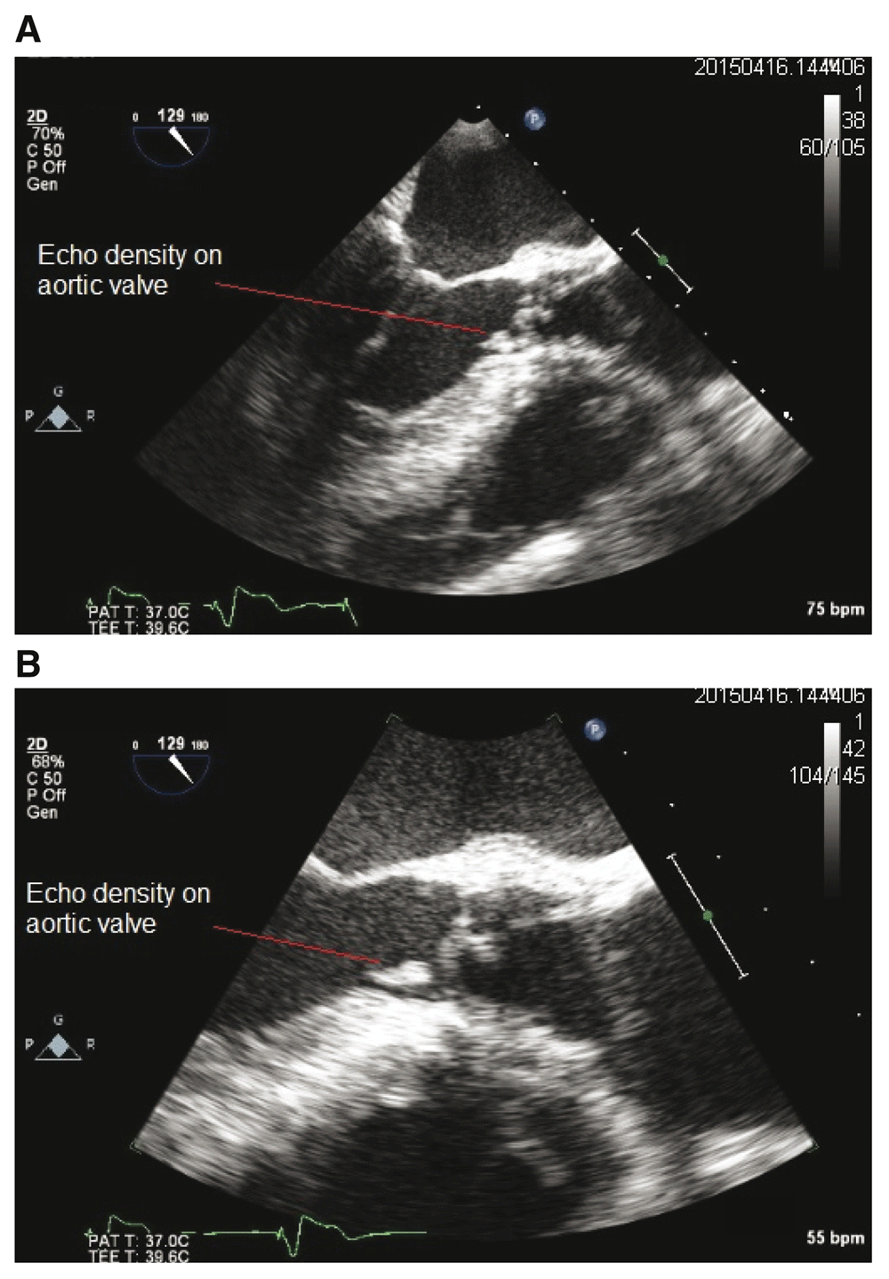
1A: Transesophageal echocardiogram (TEE), mid-esophageal view showing mobile echo density on the prosthetic aortic valve. 1B: Transesophageal echocardiogram (TEE), mid-esophageal view enlarged to show mobile echo density on the prosthetic aortic valve.
Clinical presentation, echocardiographic findings, and positive blood cultures fulfilled Duke’s criteria for IE. The patient was managed as prosthetic aortic valve endocarditis from Aerococcus urinae with IV amoxicillin 2 grams every 4 hours, and gentamicin 1 mg/kg twice daily as per hospital guidelines for IE. IV antibiotic therapy for six weeks in total with possible surgery for prosthetic valve replacement was planned.
Despite prompt initiation of appropriate antibiotic treatment and intensive clinical monitoring, the patient failed to improve this hospitalization and developed sudden pulmonary edema and worsening aortic regurgitation on repeat transthoracic echo and unfortunately died due to rapid deterioration before surgery. As per family’s wishes, an autopsy was not performed.
Aerococcus urinae is a gram-positive, catalase-negative coccus which grows in clusters. It is mostly associated with urinary tract infections in elderly men, especially in the setting of structural abnormalities, e.g. BPH, urethral strictures and nephrolithiasis. It has been associated with culture-negative infective endocarditis3. It is reported to be sensitive to penicillins/cephalosporins and resistant to sulfonamides and aminoglycosides4. By now, more than 40 cases of IE caused by Aerococcus urinae have been reported5 likely due to improvements in diagnostics.
Despite the fact that Aerococcus urinae is rare organism causing infective endocarditis, most cases respond well to antibiotic therapy and surgery is often not needed2. The indications for surgical intervention for PVE include severe prosthetic dysfunction, severe heart failure, large vegetation, and abscess or peri-valvular involvement6.
This case highlights the importance of source control by expediting prosthesis removal in the presence of overt symptoms of worsening cardiac failure and worsening prosthesis dysfunction (regurgitation in this case), as medical therapy alone may not be sufficient to effectively treat Aerococcus urinae IE despite appropriate sensitivities. Early identification is crucial and can be life-saving. The main problem is current diagnostic testing for microorganisms – whereas partial 16S rRNA gene sequencing analysis would be the most time-efficient method, it’s rarely done, as the expertise is limited and costs are high. Recently, there is good evidence for the use of MALDI-TOF7,8 due to increased detection rates, even in direct comparison to 16s sequencing.
In conclusion, Aerococcus urinae has been increasingly identified as the cause of infective endocarditis due to advancement in isolation methods. Therefore establishing a concise and broadly acknowledged protocol for diagnosis up to patient management is critical.
Written informed consent for publication of their clinical details was obtained from the patient. Permission was also granted from a next of kin for publication of the manuscript.
| Views | Downloads | |
|---|---|---|
| F1000Research | - | - |
|
PubMed Central
Data from PMC are received and updated monthly.
|
- | - |
Provide sufficient details of any financial or non-financial competing interests to enable users to assess whether your comments might lead a reasonable person to question your impartiality. Consider the following examples, but note that this is not an exhaustive list:
Sign up for content alerts and receive a weekly or monthly email with all newly published articles
Already registered? Sign in
The email address should be the one you originally registered with F1000.
You registered with F1000 via Google, so we cannot reset your password.
To sign in, please click here.
If you still need help with your Google account password, please click here.
You registered with F1000 via Facebook, so we cannot reset your password.
To sign in, please click here.
If you still need help with your Facebook account password, please click here.
If your email address is registered with us, we will email you instructions to reset your password.
If you think you should have received this email but it has not arrived, please check your spam filters and/or contact for further assistance.
Comments on this article Comments (0)