Keywords
primary, lung, choriocarcinoma, brain, metastasis
primary, lung, choriocarcinoma, brain, metastasis
Primary lung choriocarcinoma is an extremely rare entity1. Choriocarcinoma is the malignant proliferation of the syncytial cells of trophoblastic origin following gestational events, such as a term pregnancy, molar pregnancy or an abortion. We herein report one such rare case of multiple hemorrhagic metastases to the brain from primary lung choriocarcinoma in a 22 year old young woman. We also review the literature regarding primary lung choriocarcinoma and discuss recent advancements in the management of this disease.
A 22 year old woman presented to our emergency department with a history of a recent onset progressive headache for 15 days, followed by decreased level of consciousness and multiple episodes of vomiting for the last 5 days. The patient had a history of normal vaginal delivery one month past. The patient had no history of fever, chills or any rigor associated with these symptoms, and there was no history of abnormal discharge or bleeding from the vagina. There was no other significant past medical and surgical illnesses or any relevant family history. On presentation, the patient was slightly drowsy with a Glasgow Coma Scale of E4V1M3, with bilateral pupils equal and reacting. She had bilateral sixth nerve palsies (left >> right; Figure 1). Neck rigidity was absent. There was no pallor or any lymphadenopathy. Remaining systemic examination was normal. Pelvic and genital examination from a gynecologist did not reveal any abnormal findings.
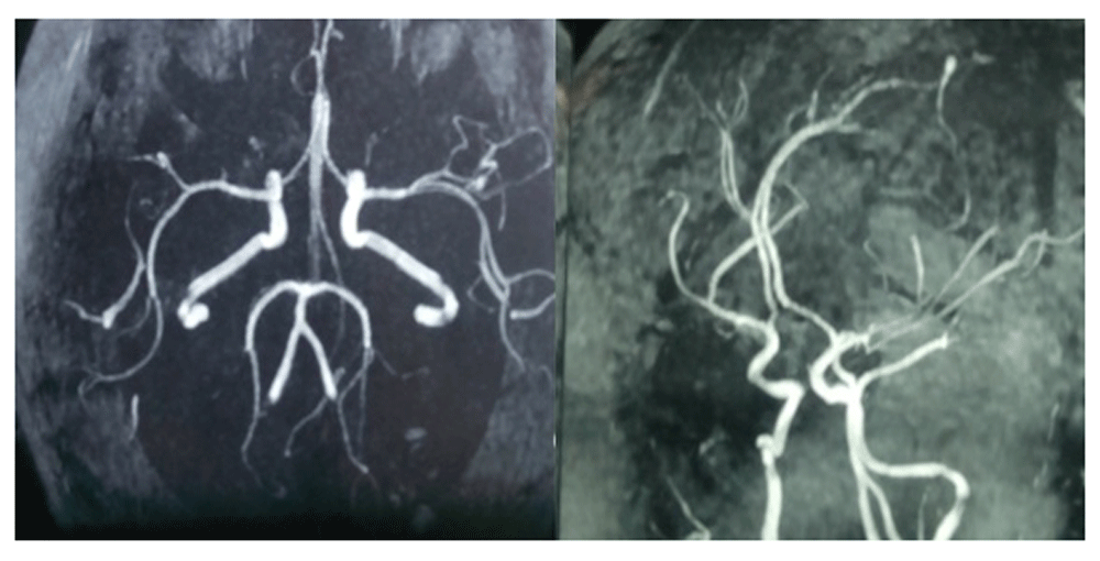
CT and MRI images of the head revealed multiple hemorrhagic lesions both in the supra and the infra-tentorial compartment with evidence of effacement of the forth ventricle and evolving hydrocephalus (Figure 2 and Figure 3). There was no vascular blush seen within the brain in the MR angiography (Figure 4). Routine chest X-ray revealed the presence of a right lung mass (Figure 5). Urine for pregnancy test was also positive. However, an ultrasound of the abdomen and pelvis was normal. Therefore, choriocarcinoma was suspected and serum B-human chorionic gonadotropin (HCG) levels were assessed, >2,20,000 mIU/ml (normal range: <1 mIU/ml). The patient’s hemoglobin was 14.5 gm% (normal range: 12.1–15.1 gm%) and a platelet count of 2,15,000 (normal range: 1,50,000–4,00,000). Peripheral smear for cytology was normal. Her immune status was normal.

Consequently, a differential diagnosis of multiple hemorrhagic metastases to the brain from the primary lung choriocarcinoma was made. The patient’s husband was informed about the disease condition and the immediate need for the removal of the posterior fossa lesion in order to prevent tonsillar herniation. The patient was in a poor medical condition, so could not decide on her treatment plan.
The patient immediately underwent sub-occipital craniactomy and excision of the well capsulated hemorrhagic lesion from the left cerebellar hemisphere (Figure 6). The patient made an uneventful recovery from the surgery and wound sutures were removed on the seventh day.
Histopathological study of the excised lesion showed diffuse cohesive sheets of trimorphic malignant trophoblasts, consisting of intermediate trophoblasts and cytotrophoblast, and rimmed with syncytiotrophoblast with the presence of a central hemorrhage and necrosis (Figure 7). The cells showed striking cytological atypia, high mitotic activity and absence of villi consistent to choriocarcinoma.
The CT chest of the patient following her surgery revealed a vascular right apical lesion (Figure 8).
A final diagnosis of multiple hemorrhagic lesions in the brain from primary lung choriocarcinoma was eventually made. The patient was referred to the National Cancer Centre for chemotherapy. The patient was started on the EMA-CO regime (Etoposide, Methotrexate and Actinomycin by drip over 2 days, followed by Cyclophosphamide and Oncovin the following week). The patient’s B-HCG decreased sharply after the first session of chemotherapy (serum B-HCG dropped to 1,50,000 mIU/ml). The patient was given three cycles of chemotherapy and has been on regular follow up at the cancer centre.
Primary lung choriocarcinoma is a very rare entity, with < 50 cases reported currently2. This case report discusses an even rarer phenomenon of multiple hemorrhagic metastasis in the brain from primary lung choriocarcinoma.
There are various theories behind the etiology of primary lung choriocarcinoma. The foremost being embolism of trophoblastic cells during abortion, or even normal delivery, to the lung vasculature, thereby causing the cells to proliferate therein3. This may have occurred in the present case study. Other theories discuss the probable role of primordial germ cells and the genesis of metaplasia4. Choriocarcinoma can have either a gestational or non-gestational origin5,6.
Sometimes large cell anaplastic carcinoma, mediastinal germ cell tumor, bronchogenic carcinoma show ectopic HCG secretion, but this elevation is mild7,8. A high B-HCG level, as in our case, suggests a trophoblastic origin1.
The pathogenesis behind multiple hemorrhagic lesions in the brain is the tendency of such malignant trophoblastic cells to invade the vessels, and sometimes even leads to distal aneurysms9. Radiation has a poor response to such entity10. Therefore, the preferred therapy for gestational trophoblastic neoplasm is the EMA-CO regimen, similar to what was prescribed to our patient11.The prognosis of the condition is poor, with previous reports of a 5 year survival of <5%. However, recent advancements in chemoradiation therapy has helped to increase the overall 5 year survival rate up to 50%4,12. A multimodal approach is also required, constituting of neo-adjuvant chemotherapy followed by excision of the lung lesion9.
Primary lung choriocarcinoma metastasis should be recognized as a differential diagnosis in hemorrhagic lesions of the brain, especially in patients of a child bearing age. Early diagnosis and rapid initiation of therapy is the cornerstone for a better outcome in such patients.
Written informed consent for the publication of the clinical case study and accompanying images was taken from the patient.
| Views | Downloads | |
|---|---|---|
| F1000Research | - | - |
|
PubMed Central
Data from PMC are received and updated monthly.
|
- | - |
Is the background of the case’s history and progression described in sufficient detail?
Yes
Are enough details provided of any physical examination and diagnostic tests, treatment given and outcomes?
Partly
Is sufficient discussion included of the importance of the findings and their relevance to future understanding of disease processes, diagnosis or treatment?
Partly
Is the case presented with sufficient detail to be useful for other practitioners?
Yes
References
1. Kobayashi T, Kida Y, Yoshida J, Shibuya N, et al.: Brain metastasis of choriocarcinoma.Surg Neurol. 1982; 17 (6): 395-403 PubMed AbstractCompeting Interests: No competing interests were disclosed.
Reviewer Expertise: Neurology
Is the background of the case’s history and progression described in sufficient detail?
Partly
Are enough details provided of any physical examination and diagnostic tests, treatment given and outcomes?
Yes
Is sufficient discussion included of the importance of the findings and their relevance to future understanding of disease processes, diagnosis or treatment?
Partly
Is the case presented with sufficient detail to be useful for other practitioners?
Yes
Competing Interests: No competing interests were disclosed.
Reviewer Expertise: Radiation
Alongside their report, reviewers assign a status to the article:
| Invited Reviewers | ||
|---|---|---|
| 1 | 2 | |
|
Version 1 23 May 17 |
read | read |
Provide sufficient details of any financial or non-financial competing interests to enable users to assess whether your comments might lead a reasonable person to question your impartiality. Consider the following examples, but note that this is not an exhaustive list:
Sign up for content alerts and receive a weekly or monthly email with all newly published articles
Already registered? Sign in
The email address should be the one you originally registered with F1000.
You registered with F1000 via Google, so we cannot reset your password.
To sign in, please click here.
If you still need help with your Google account password, please click here.
You registered with F1000 via Facebook, so we cannot reset your password.
To sign in, please click here.
If you still need help with your Facebook account password, please click here.
If your email address is registered with us, we will email you instructions to reset your password.
If you think you should have received this email but it has not arrived, please check your spam filters and/or contact for further assistance.
Comments on this article Comments (0)