Keywords
aerobic exercise, telomere length, high fat-diet
aerobic exercise, telomere length, high fat-diet
Obesity is a global problem that is associated with high mortality and morbidity. Several studies has shown that obesity increases the risk of cardiovascular disease, renal impairment, diabetes, and even certain cancers1–3. Meanwhile, the World Health Organization (WHO) showed that in the last 4 decades obesity rates have increased 10 times worldwide3. This phenomenon is worrisome because there is an increasing number of people who have a high risk of developing various diseases associated with obesity.
Some studies show that there is a strong association between the incidence of obesity with the level of physical activity and high fat diet intake. Individuals with low physical activity levels are known to have a higher risk of developing obesity4,5. Physical activity is also known to have a role on lowering blood glucose levels, improving homeostasis in people with diabetes mellitus, decreasing production of oxidative stress, lowering triglyceride levels in the body, increasing endogenous antioxidants, and can also maintain telomere length, thus reducing cardiovascular metabolic disease risk6–8.
The role of physical activity in decreasing the risk of cardiovascular metabolic mortality and morbidity is thought to be mediated by maintaining the length of telomeres9. A study performed by Goglin et al. demonstrates the association between telomere shortening and increased 5-year mortality in patients with acute coronary syndromes. This study also showed that a decrease in telomere shortening rate would be followed by a decrease in mortality rate10.
The rate of shortening of the telomere can be suppressed through a healthy lifestyle such as healthy diet and physical activity9,11. Certain types of food have been shown to have a correlation with telomere length. A Mediterranean-rich diet of olive oil (38% of total energy as fat) has been observed to maintain telomere length12. In contrast, a high western type diet of sugar and red meat is associated with shortening of the telomere12.
The aim of this study was to explore the effect of aerobic physical exercise on telomere length under high fat-diet conditions to provide information for further research.
Twelve male Sprague-Dawley rats (Rattus norvegicus), weighing 250–450g, aged 12–13 months, were obtained from Central Animal Facility (Bogor Agricultural University) and divided into two groups: control and trained high fat diet. They were acclimatized for 1 week in a controlled room temperature of 24+1°C, with a 12-hour light/dark cycle, and access to food pellets and filtered water ad libitum to adapt to the new environment. They were housed in plastic cages (50×34×25 cm), two animals in each cage. All protocols used in this experiment received approval from the Ethical Animal Care and Use Committee of Faculty of Medicine Universitas Indonesia with approval number 164/UN2.F1/ETIK/2017.
Before the beginning of the study all rats were acclimatized with high fat-diet for 10 weeks, consisting of 19% fat, 24% protein, and 47.77% carbohydrate. After 10 weeks of acclimatization, the rats were evenly and randomly assigned using a random number into two groups (n=6 per group): (1) the control group (without aerobic exercise) and (2) the treatment group (with aerobic physical exercise). The treatment group received aerobic exercise for 8 weeks. During 8 weeks of intervention, both treatment and control groups were still given a high-fat diet.
Aerobic exercise was conducted using an animal treadmill, with a speed of 20 m/min for 20 minutes, 5 days/week, every morning around 6 am until 8 am. The intervention was carried out at the Biochemistry and Molecular Laboratory, Faculty of Medicine, Universitas Indonesia. All aerobic exercise protocols were supervised by experienced researchers.
Blood was collected from both groups after an overnight fasting. All animals were anesthetized intraperitoneally with a ketamine-xylazine (KX) solution before blood was taken. Approximately 1 ml of whole blood was taken from the sinus orbitalis on week 0, week 4 and week 8. Genomic DNA in leukocytes was extracted from peripheral blood. DNA isolation were then performed using DNA isolation kit (GeneAll® ExgeneTM Clinic SV mini). Relative telomere length from isolated DNA were measured on a real-time PCR detection system using a Quantitative PCR method. Kit used for qPCR were pipettes (and tips), optical PCR plates and caps, and master mix. The type of taq used was AmpliTaq Gold DNA polymerase. The model number/name of the PCR machine was Applied Biosystems 7300. The cycling conditions used were 10 min at 95°C, followed by 40 cycles of 95°C for 15 sec, 60°C for 1 min, followed by a dissociation (or melt) curve. The primer sequences were as follows:
Telo F: CGGTTTGTTTGGGTTTGGGTTTGGGTTTGGG TTTGGGTT
Telo R: GGCTTGCCTTACCCTTACCCTTACCC TTACCCTTACCCT
36B4 F: ACTGGTCTAGGACCCGAGAAG
36B4 R: TCAATGGTGCCTCTGGAGATT
The primers were obtained from rodent (GenScript®). Relative telomere length was calculated using the formula of 2-ΔΔCt13.
The characteristics of the animals are shown in Table 1. All the animals were in good condition throughout the length of study. There was no significant difference in age, body weight, Lee index and telomere length between control and treatment groups.
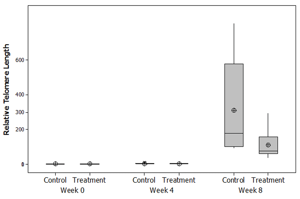
There was an increase in relative telomere length at weeks 4 and 8 compared to week 0 in both groups. At week 4, the relative telomere length of the control group (2.231) did not differ much with the treatment group (1.802) when compared to week 0 of control group. At week 8, there was a progressive increase of relative telomere length in both groups compared to week 0 and week 4. Relative telomere length increase in week 8 of the control group was much higher (178.62) compared to week 8 treatment group (74.86) (Figure 1; Table 2).

Median (minimum-maximum).
One important structures located at the ends of the linear chromosomes is the telomere. In human cells, they are composed of TTAGGG repeats and a number of proteins. Their function is to protect the integrity and stability of the DNA14.
Many studies showed that telomere length is influenced by a number of factors9,12,15. Sedentary lifestyle, high blood glucose levels, and increased percentage of body fat have a negative influence on telomere length. The underlying mechanism have been suggested as being mediated through oxidative stress and inflammation16.
Exercise as a lifestyle intervention has been associated with longer leukocytes telomere length15. A study by Cherkas on 2401 subject showed that telomere length has a direct relation with increased level of physical activity17. An observation of physical activity and telomere length by Du et al. on 7813 adult women concluded that moderate and vigorous intensity activity increased telomere length compared with least active women18. Ludlow et al. studied the effect of physical activity on telomere length in three different groups: sedentary, moderate and overtraining. The result showed a positive effect on telomere length in the moderate group19.
To date, to our knowledge, very few studies have investigated the relative effect of a specific diet on telomere length. Cassidy et al. found that total fat intake was only inversely associated with leukocytes telomere length and higher polyunsaturated fatty acids (PUFA) intake, specifically linoleic acid intake, was inversely associated with leukocytes telomere length11. Li et al. found that there was no difference in telomere length between consumption of fish oil-rich diet and soy oil-rich diet20. Kiecolt-Glaser et al. compared telomere length between subjects with n-6 PUFA and n-3 PUFA supplementation. They found that telomere length is longer in subject with omega 3 or n-3 PUFA supplementation (high in fish oil) compared to n-6 PUFA supplementation21.
Results from our current study showed a lengthening of relative telomere length in both groups in week 4 and week 8. This was in contrast with studies showing telomeres usually shortened with age9., but there are also studies which indicate that in vivo, telomere may shorten or elongate, and leukocyte telomere length may fluctuates within months22,23.
In general, preserved telomere length and lengthening of telomere are considered as something good because it is thought to play an important role in extending the biological age of cells12,21,24.
Nevertheless, studies have also shown that telomere lengthening can be an initial response that arises after exposure to low doses of various carcinogenic chemicals in vitro and in experimental animals23. Zhang et al. concluded from their study that subject with longer telomere length had a higher risk of getting lung cancer, and this was especially true for men25.
The positive associations between high fat-diet conditions and telomere length is difficult to explain because a high fat-diet is associated with increased risk of various diseases. Telomeres generally shorten with age, thus, the discovery of telomer elongation in the provision of high-fat diet can be regarded as something that deviates from normal condition. Therefore, the positive associations between high fat-diet conditions and telomere length observed in this study are notable.
Telomere will shorten at each cell division. Telomeres that elongate excessively in both groups in this study may indicate a prolonged period before apoptosis, and this could indicates a change from normal cell function. Currently, the implications of excessive telomere lengthening are still unknown. Our result shows that aerobic exercise can act as a barrier to progressive changes that occur in the relative telomere length caused by a high-fat diet condition. Modulation of oxidative stress in the body is one possible mechanism that may explain how aerobic exercise resist relative telomere changes. Aerobic exercise upregulate genes that encode various antioxidant enzymes. Several studies shows that regular physical exercise increase the body's endogenous antioxidant activity and thus increase body’s resistance to oxidation events15,26.
Our study showed that exposure to a high fat-diet plays an important role to the emergence of altered telomere length, and aerobic exercise could reduce the progression of the alteration in length. Our results support the hypothesis that leukocyte telomere length is associated with daily dietary intake and physical activities. Further investigation is still needed to explore the mechanism and implications of telomere length changes found in this study.
Dataset 1: Raw data including the relative telomere length for control and treatment groups at week 0, 4 and 8, and 2-ΔΔCt calculations. DOI, 10.5256/f1000research.15127.d21168127.
This research was funded by Hibah Publikasi Internasional Terindeks untuk Tugas Akhir (PITTA) 2017.
The funders had no role in study design, data collection and analysis, decision to publish, or preparation of the manuscript.
| Views | Downloads | |
|---|---|---|
| F1000Research | - | - |
|
PubMed Central
Data from PMC are received and updated monthly.
|
- | - |
Is the work clearly and accurately presented and does it cite the current literature?
Partly
Is the study design appropriate and is the work technically sound?
Partly
Are sufficient details of methods and analysis provided to allow replication by others?
Partly
If applicable, is the statistical analysis and its interpretation appropriate?
No
Are all the source data underlying the results available to ensure full reproducibility?
Partly
Are the conclusions drawn adequately supported by the results?
Partly
References
1. McEachern MJ, Blackburn EH: Runaway telomere elongation caused by telomerase RNA gene mutations.Nature. 1995; 376 (6539): 403-9 PubMed Abstract | Publisher Full TextCompeting Interests: No competing interests were disclosed.
Is the work clearly and accurately presented and does it cite the current literature?
Partly
Is the study design appropriate and is the work technically sound?
Yes
Are sufficient details of methods and analysis provided to allow replication by others?
Yes
If applicable, is the statistical analysis and its interpretation appropriate?
Yes
Are all the source data underlying the results available to ensure full reproducibility?
Partly
Are the conclusions drawn adequately supported by the results?
Partly
References
1. Lesmana R, Iwasaki T, Iizuka Y, Amano I, et al.: The change in thyroid hormone signaling by altered training intensity in male rat skeletal muscle.Endocr J. 2016; 63 (8): 727-38 PubMed Abstract | Publisher Full TextCompeting Interests: No competing interests were disclosed.
Alongside their report, reviewers assign a status to the article:
| Invited Reviewers | ||
|---|---|---|
| 1 | 2 | |
|
Version 1 27 Jul 18 |
read | read |
Click here to access the data.
Spreadsheet data files may not format correctly if your computer is using different default delimiters (symbols used to separate values into separate cells) - a spreadsheet created in one region is sometimes misinterpreted by computers in other regions. You can change the regional settings on your computer so that the spreadsheet can be interpreted correctly.
Provide sufficient details of any financial or non-financial competing interests to enable users to assess whether your comments might lead a reasonable person to question your impartiality. Consider the following examples, but note that this is not an exhaustive list:
Sign up for content alerts and receive a weekly or monthly email with all newly published articles
Already registered? Sign in
The email address should be the one you originally registered with F1000.
You registered with F1000 via Google, so we cannot reset your password.
To sign in, please click here.
If you still need help with your Google account password, please click here.
You registered with F1000 via Facebook, so we cannot reset your password.
To sign in, please click here.
If you still need help with your Facebook account password, please click here.
If your email address is registered with us, we will email you instructions to reset your password.
If you think you should have received this email but it has not arrived, please check your spam filters and/or contact for further assistance.
Comments on this article Comments (0)