Keywords
fibroblast, chlorhexidine, essential oils, essential oils with sodium fluoride, mouthwash.
This article is included in the Advances in Fibroblast Research collection.
fibroblast, chlorhexidine, essential oils, essential oils with sodium fluoride, mouthwash.
Fibroblasts are the common cell type in connective tissue and plays a major role in normal function and in pathologic changes of oral tissues1,2. Various antiseptic agents are available in the form of mouthwashes, among them are essential oils (EOs), EOs with sodium fluoride (EOF) and chlorhexidine (CHX). The antibacterial action of these agents had been widely examined1,3,4.
In fibroblasts, EOs can induce depolarization of the mitochondrial membranes by decreasing the membrane potential, affecting ionic Ca++ cycling5,6. They change the permeability of membranes resulting in liberation of cytochrome, leading to apoptosis7. Fluoride inhibits protein synthesis and thus it influences certain signalling pathways included in apoptosis such as the P53 pathway8,9.
CHX inhibits protein and DNA synthesis, resulting in cell apoptosis3,10. Moreover, CHX can damage mitochondrial membranes resulting in leakage of apoptosis-inducing proteins11,12.
Many previous studies have compared the effect of both CHX and EOs mouthwashes on cultured fibroblasts3,5. However, to the best of our knowledge, the combined effect of EOs and EOF hasn’t been studied yet. In this study, we investigated the effect of different doses and different exposure periods of mouthwashes containing EOs, EOF and CHX on a fibroblast cell line.
This study was performed on BHK-21 cell line in (Vacsera Co., Egypt). It was cultured according to a previous protocol3. Briefly, BHK-21 cells were cultured in Eagle's Minimum Essential Medium (ATCC, Manassas, VA, USA). They were sub-cultured into 96-well plates (ATCC, USA).
Commercial mouthwashes (3 total) were diluted to 15% and 25% using distilled water. Mouthwashes used were composed as followed:
- EO mouthwash: containing thymol (0.0060%), eucalyptol (0.09%), mentol (0.042%) and methyl-salicylate (0.064%) in a 26.9% hydroalcoholic vehicle;
- EOF mouthwash: containing EOs as before and 0.2% sodium fluoride;
- CHX mouthwash: containing 0.12% chlorhexidine hydrochloric acid.
Cells were incubated with mouthwashes for 0, 1, 2, 3, 5, 10 minutes in the 96-well plates at 37oC.
After cell culturing, RNA extraction and quantitative real time polymerase chain reaction (PCR) was performed as follows1:
1. Total RNA was isolated using the RNeasy Micro Kit (Qiagen, Germany) and RNA concentration was measured spectrophotometrically using Nanodrop ND-1000.
2. Reverse transcription was performed on 600 ng of total RNA using oligo dT primers and MMLV Reverse Transcriptase in a final volume of 20 mL (Invitrogen, CA, USA) for 5 minutes at 65oC, followed by one hour at 37oC.
3. Samples were subsequently heated for 15 minutes at 70oC to terminate the reverse transcription reaction.
4. Real-time quantitative PCR was performed on the cDNA samples using an Applied Biosystem Real-Time Detection System. Annexin V was the target gene for apoptosis. Primer sequences for Annexin V: sense primer, 5' CAGTCTAGGTGCAGCTGCCG 3' and antisense primer, 5' GGTGAAGCAGGACCAGTGT3'. The following primer sequences were used for GAPDH (housekeeping gene): sense primer, 5' ATG GCC TTC CGT GTT CCT AC 3' and antisense primer, 5' GCC TGC TTC ACC ACC TTC TT 3'
5. Real-time PCR was conducted by amplifying the cDNA with iQ SYBR Green Universal Master Mix (Applied Biosystems, CA, USA).
6. Melting curve analysis of amplification products was performed at the end of the PCR reaction to confirm that a single PCR product was detected. For every PCR reaction, GAPDH was used as the internal control.
7. A relative quantification method 2−ΔΔCT method was used13. The relative quantitation value of target was normalized to the internal control (GAPDH).
Microscopic examination of fibroblast cell line after application of different mouthwashes at different concentrations and for different time durations is shown in Figure 1.
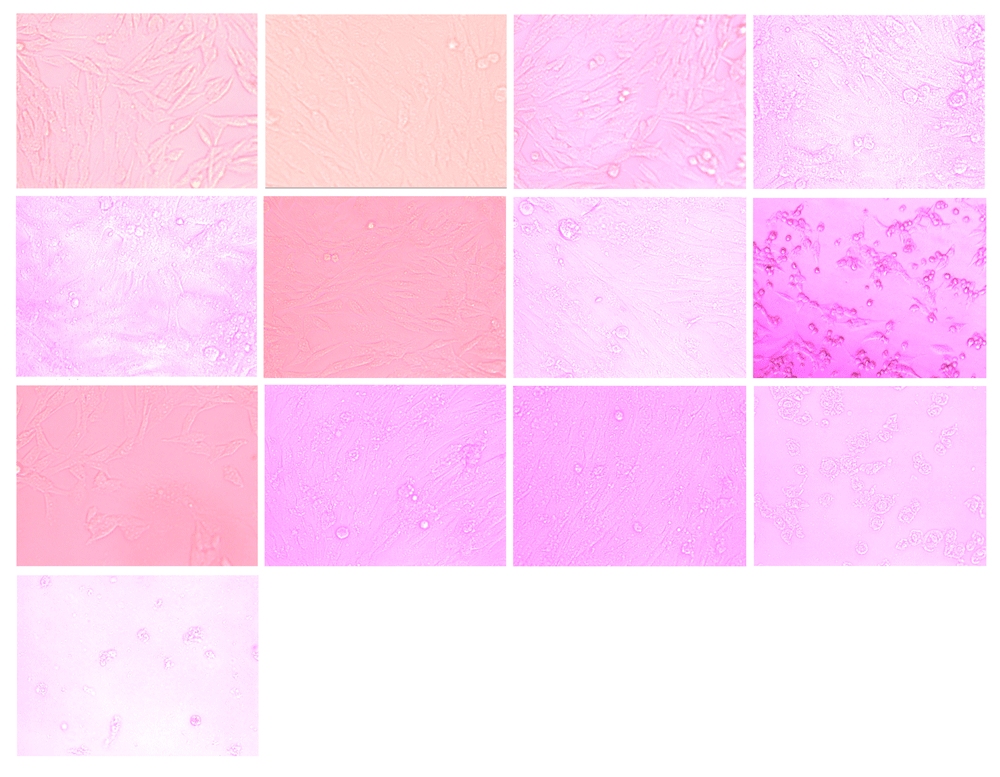
From L-R (A) Untreated fibroblast cell line demonstrated confluent growth of elongated cells. Some cells appeared bipolar and some were multipolar. (B–E) Essential oil mouthwash: (B) 15% for 1 minute showed many viable spindle shaped fibroblasts; (C) 15% for 10 minutes showed decreased spindle shaped fibroblasts; (D) 25% for 1 minute showed few viable spindle shaped fibroblasts; (E) 25% for 10 minutes showed obvious cell free areas. (F–I) Sodium fluoride mouthwash: (F) 15% for 1 minute showed many viable spindle shaped fibroblasts and few apoptotic cells; (G) 15% for 10 minutes showed destroyed fibroblasts; (H) 25% for 1 minute showed large cell free areas; (I) 25% for 10 minutes showed more obvious large cell free areas. (J-M) Chlorhexidine mouthwash: (J) 15% for 1 and (K) 10 minutes many viable fibroblasts were detected; (L) 25% for 1 minute showed massive reduction in cell viability, many dead or destroyed fibroblasts surrounded by large cell free areas; (M) 25% for 10 minutes, only few remnants of dead fibroblasts were obvious.
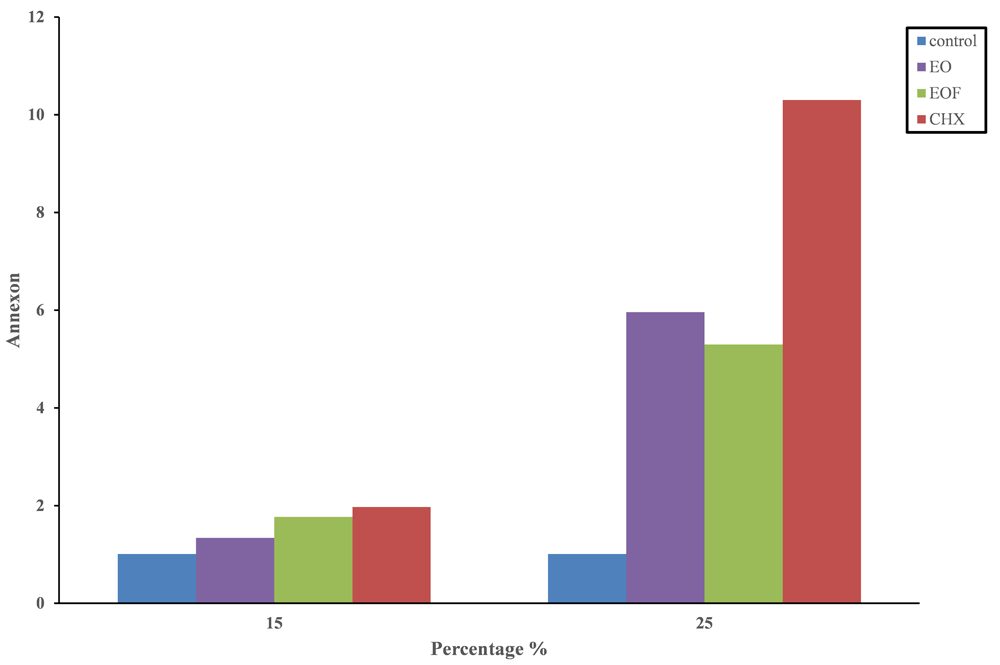
In all mouthwashes 25% concentration showed a statistically significant increase in apoptosis compared with 15% and the untreated control (Table 1 and Figure 2).
| Mouthwash | Untreated | 15% mouthwash dilution | 25% mouthwash dilution | P-value** |
|---|---|---|---|---|
| EO | 1.01±-0.2a | 1.34±0.470a | 5.97±1.90b | 0.0000 |
| EOF | 1.01±-0.2a | 1.77±0.710a | 5.30±1.93b | 0.0000 |
| CHX | 1.01±-0.2a | 1.97±0.52a | 10.3±2.61c | 0.0000 |
EO, essential oil; EOF, sodium fluoride; CHX, chlorhexidine (n=3).
In the EO mouthwash, values for apoptosis continued to significantly increase after 2, 3, 5 and 10 minutes for the 25% concentration (Table 2 and Figure 3).
| Time | |||||||
|---|---|---|---|---|---|---|---|
| Mouthwash | 0 | 1 | 2 | 3 | 5 | 10 | P-value** |
| 15% | 1.01±1.5a | 1.15±0.18a | 1.17±0.21a | 1.22±0.46a | 1.5±0.502a | 1.74±0.65a | 0.6448 |
| 20% | 1.01±1.5a | 4.66±1.28b | 5.26±1.58b | 5.99±1.64b | 6.57±1.65c | 6.78±2.3c | 0.0001 |

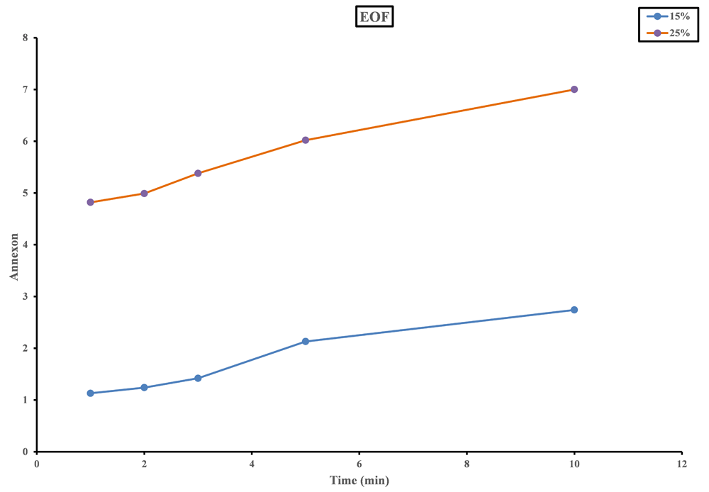
In the EOF mouthwash, at 25% concentration, a significant increase in apoptosis values was detected after 5 and 10 minutes (Table 3 and Figure 4).
| Time | |||||||
|---|---|---|---|---|---|---|---|
| Mouthwash | 0 | 1 | 2 | 3 | 5 | 10 | P-value** |
| 15% | 1.01±1.5a | 1.19±0.15a | 1.24±0.18a | 1.42±0.34a | 2.13±0.31a | 2.74±0.25a | 0.0019 |
| 20% | 1.01±1.5a | 4.82±2.06a | 4.99±0.95a | 5.38±1.36a | 6.02±2.7b | 7±1.96b | 0.0008 |
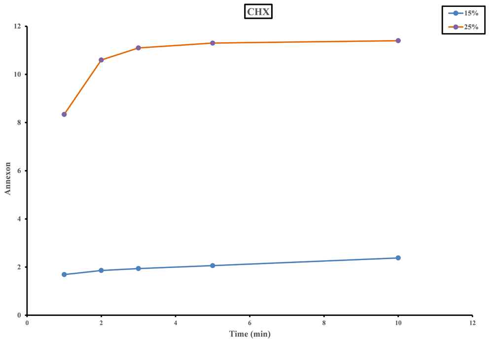
In the CHX mouthwash, at 25% concentration, a significant increase in apoptosis values started after 1 minute and continued to increase by time (Table 4 and Figure 5).
| Time | |||||||
|---|---|---|---|---|---|---|---|
| Mouthwash | 0 | 1 | 2 | 3 | 5 | 10 | P-value** |
| 15% | 1.01±1.5a | 1.86±0.71a | 1.87±0.36a | 1.94±0.69a | 2.06±0.25a | 2.38±0.45a | 0.1606 |
| 20% | 1.01±1.5a | 8.34±1.9c | 10.6±3.4b | 11.1±1.96d | 11.3±0.83b | 11.4±1.5b | 0.0000 |
The effectiveness of CHX and EOs mouthwashes in controlling the formation of plaque and gingivitis has been demonstrated14. However, there are concerns that these products are harmful to oral cells15–17.
CHX is toxic, even in low concentrations, for different cell types including fibroblasts in culture18,19. Topical application of CHX can result in its penetration through the epithelial barrier leading to tissue damage20. In our study, and increase in concentration for EOs, EOF and CHX mouthwashes resulted in an increase in apoptosis. This was also observed by Faria et al.1 who reported that CHX induced apoptosis of cultured fibroblasts in a concentration dependent manner. Values for apoptosis at 15% and 25% concentrations of EOF were slightly higher than EOs alone in our study, demonstrating the ability of fluoride to enhance apoptosis induction, as previously described9.
A significant increase in apoptosis induction was seen at 15% concentration CHX and at 25% concentration EOs and EOF. This suggests that CHX is more effective than EOs and EOF in apoptosis induction. This result was in agreement with Tsourounakis et al.5 who reported that there was a significant reduction in cell survival that occurred at concentrations of 15% CHX and 25% EOs21.
The increase in duration of application of EOs, EOF and CHX didn’t significantly increase apoptosis at the low concentration of 15% in the present study. However, at 25% CHX, increase in the duration of treatment significantly enhanced apoptosis, which was significant obvious after 1 minute. This means that CHX is more efficient than EOs and EOF in apoptosis induction at lower concentrations. This was in accordance with Flemingson et al.3 who stated that the apoptotic effect of CHX on fibroblasts occurs early, after 1 minute exposure, and added that CHX had the maximum cytotoxicity followed by EOs.
CHX mouthwash is the most cytotoxic to fibroblasts compared to EOs and EOF containing mouthwashes. Adding fluoride to EOs in low concentrations didn’t worsen the adverse effects of Eos as shown by the combination mouthwash containing EOs and fluoride.
F1000Research: Dataset 1. File containing: Part A, Raw numerical data of RNA concentration at each concentration and duration of application of the mouthwashes; part B, raw phase contrast photomicrograph of fibroblast cell line after application of mouthwashes at different concentrations and durations., 10.5256/f1000research.16337.d22068422.
| Views | Downloads | |
|---|---|---|
| F1000Research | - | - |
|
PubMed Central
Data from PMC are received and updated monthly.
|
- | - |
Click here to access the data.
Spreadsheet data files may not format correctly if your computer is using different default delimiters (symbols used to separate values into separate cells) - a spreadsheet created in one region is sometimes misinterpreted by computers in other regions. You can change the regional settings on your computer so that the spreadsheet can be interpreted correctly.
Provide sufficient details of any financial or non-financial competing interests to enable users to assess whether your comments might lead a reasonable person to question your impartiality. Consider the following examples, but note that this is not an exhaustive list:
Sign up for content alerts and receive a weekly or monthly email with all newly published articles
Already registered? Sign in
The email address should be the one you originally registered with F1000.
You registered with F1000 via Google, so we cannot reset your password.
To sign in, please click here.
If you still need help with your Google account password, please click here.
You registered with F1000 via Facebook, so we cannot reset your password.
To sign in, please click here.
If you still need help with your Facebook account password, please click here.
If your email address is registered with us, we will email you instructions to reset your password.
If you think you should have received this email but it has not arrived, please check your spam filters and/or contact for further assistance.
Comments on this article Comments (0)