Keywords
Toxoplasma gondii, Toxoplasmosis,Seroprevalence,IgG, IgM, Pregnant women
Toxoplasma gondii, Toxoplasmosis,Seroprevalence,IgG, IgM, Pregnant women
Toxoplasmosis is a widely distributed zoonotic illness caused by Toxoplasma gondii, an obligate intracellular parasite1,2. Globally, the distribution of this disease is extremely variable even inside the countries3,4. In all host species, including humans, Toxoplasmosis is generally acquired either vertically from mother to fetus (congenital infection), or through ingestion of oocysts in contaminated food or water5. Rarely, T. gondii can transmit through organ transplantation and the transfusion of infected blood6,7. Following ingestion, the intestinal epithelium is the primary portal of entrance for T. gondii; next, it spreads to other tissues, where it can cause more severe pathogenesis8,9. If toxoplasmosis is acquired during pregnancy, severe infection may develop, especially in immunocompromised individuals, such as those with defects in T-cell-mediated immunity10. In patients with AIDS, toxoplasmosis may leads to severe, life-threatening disease11. For example, cerebral focal lesions are caused by cerebral toxoplasmosis (CT) in HIV-infected patients12.
The signs and symptoms of this illness are markedly divergent and range from asymptomatic to serious infection13. This variation depends on several factors includes inoculums size, virulence of the strain of toxoplasma, the individual’s genetic background and the status of the immune system of the infected individual14. In addition, since the organism has an affinity for muscular and neural tissues as well as the other visceral organs, many hosts harboring latent tissue cysts following toxoplasmosis15.
Fetuses may acquire toxopasmosis during delivery or through the placenta during pregnancy16. Early infection of the fetus may cause severe damage, or either pre- or post-natal death17. The clinical manifestation of congenital toxoplasmosis generally depends on the gestational stage, and can includes seizures, mental retardation, severe neurological defects, chorioretinitis, epilepsy and blindness10,16,18.
Approximately 90% of pregnant women infected with T. gondii are asymptomatic, and recover spontaneously19,20. Only a small percentage of pregnant women show the clinical symptoms of disease19,21. In pregnant women, the clinical signs are no more severe than in non-pregnant women, and typically an influenza-like illness is seen after an incubation period of 5 to 18 days19,22,23. Early diagnosis and treatment of mothers during pregnancy prevents fetal infection and minimizes the probability of complications24,25.
Laboratory diagnosis of toxoplasmosis is usually performed by serological detection of T. gondii-specific IgG and IgM antibodies26. Worldwide, the screening of T. gondii infection in pregnant women is preferably performed during the first trimester and subsequently every month or trimester in seronegative women, as applied in many countries27.
Our study was undertaken to determine the prevalence and geographic distribution of toxoplasmosis. In addition, it sought to evaluate the role of animal contact in disease development among pregnant women through the serological detection of toxoplasma IgM and IgG antibodies, as well as to estimate the seropositivity of these antibodies among different age groups. It also attempted to identify the percentage of toxoplasma IgM seropositivity (indicative of acute infection) among different pregnancy trimesters.
This was a descriptive cross-sectional hospital-based study carried out in the District Head quarter Hospital (Mansehra, Hazara, Pakistan) and Ayub Medical Complex Hospital (Abbottabad, Khyber Pakhtunkhwa, Pakistan) over a period of 4 months (April to July 2015).
Our study included pregnant women of different trimesters, ages and ethnic groups who visited our study areas hospitals; the only eligibility criteria were pregnancy and visiting the hospitals in our study area. Patients were recruited by the researchers face-to-face. During this study duration, a total of 500 pregnant women (convenience sample) fulfilled the inclusion criteria. Out of the total of participants, 204 were recruited from Abbottabad and 296 from Mansehra district.
A total of 5 ml venous blood was collected from each participant using a sterile syringe and transferred to a blood container without anticoagulant, allowed to clot at room temperature for 15 minutes, then centrifuged at 3000 rpm for 10 minutes to obtain serum, which was transferred into a 1.5-ml microcentrifuge tube and stored at −80°C for further analysis. In this study, every sample was screened and confirmed for toxoplasmosis through the serological tests.
All sera samples were screened for T. gondii IgG and IgM antibodies using Rapid Diagnostic immunochromatographic test (Tox IgG/IgM Rapid Test Dip strip, CTK BIOTECH, San Diego, USA) according to manufacture instructions. In order to avoid false-positive results due to the incomplete specificity of the screening test, every positive sample was further subject to confirmation step by ELISA. Each positive individual also answered a questionnaire concerning their age, trimester and whether they had been in recent contact with animals (Supplementary File 1).
Following screening, the positive samples (n=150) were further confirmed to toxoplasmosis using IgM and IgG ELISA kit (Monobind, San Diego, USA) according to the manufacturer’s protocol. The samples positive for T. gondii IgG titers indicated chronic infection and those with high IgM titers indicates recent or acute infection. All ELISA tests were performed in triplicate.
Our study was approved by Hazara University. Further approval was provided by the Ayub Medical Complex Hospital. From every participant, written informed consent was obtained for conduction of the study. In addition, all the performed steps in this study were completely in accordance with Helsinki Declaration and the rules defined by the World Medical Association, including samples collection and processing.
The obtained results were analyzed by Graph Pad Prism 5 (Graph Pad Software, La Jolla, CA, USA). A χ2 test was involved to test the statistical differences in seropositivity and negativity of anti-toxoplasma antibodies among the participants of different study areas and gestational periods or those had/had no prior history of animal contact, at 95% level of significance. Moreevore, ANOVA was tested the statistical difference of these antibodies among the participants of every age group. Difference was considered statistically significant when P <0.05.
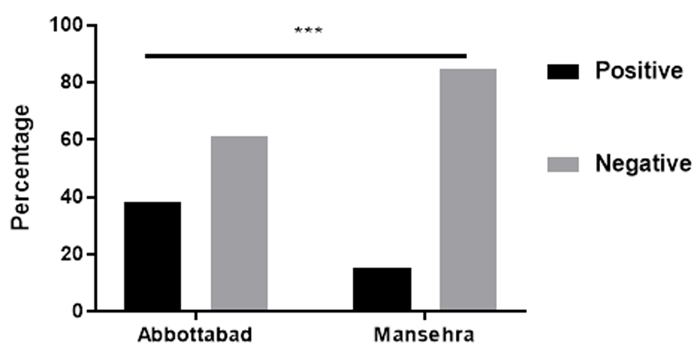
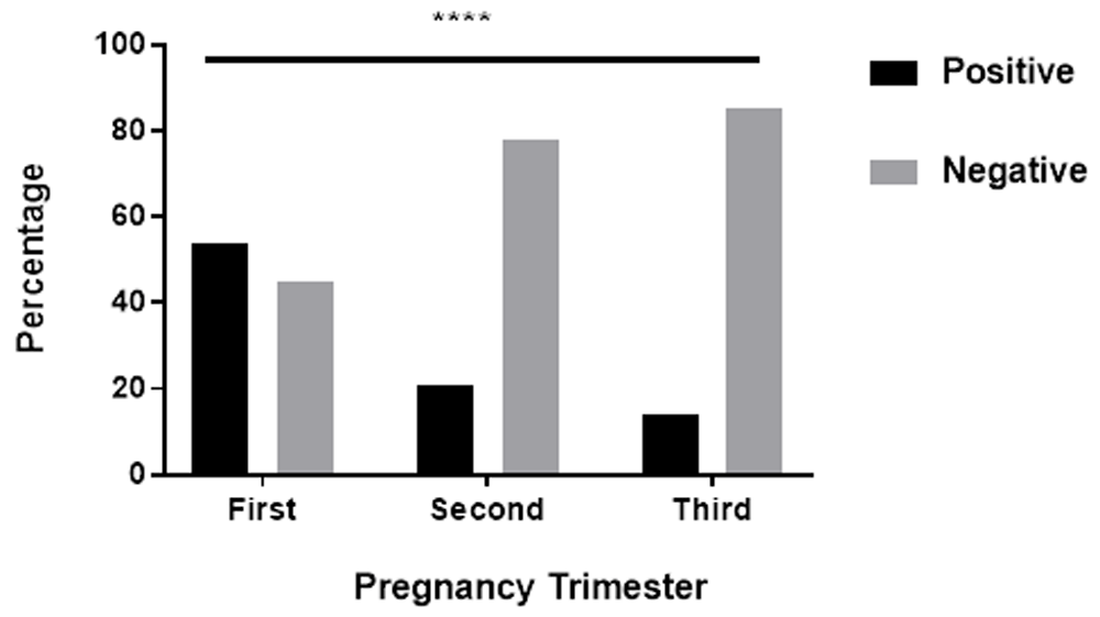
Out of 500 women, using ELISA the overall seroprevalence of toxoplasmosis was 24.8% (124/500). Statistically significant differences were observed between the seroprevalence of disease in Abbottabad and Mansehra district (Figure 1). In addition, the prevalence of toxoplasma antibodies among pregnant women revealed out of the total of 500 participants, only 8% had a serological marker of acute toxoplasmosis (Figure 2).

Out of the total of participants in every district, 38.7% (79/204) had the serologic marker of toxoplasmosis in Abbottabad district and 15% (45/296) in Mansehra. ***P = 0.0002.
Among the positive cases (n=124), the seropositivity of toxoplasma antibodies was shown to be statistically significant different among different age groups (Table 1). There was also a statistically significant difference in the seropositivity of toxoplasma IgM (indicating acute infection) between different gestational trimesters, the highest level of IgM seropositivity was observed in first trimester (54.34%) (Figure 3).
| Age, years | Positive cases | IgG | IgM | IgG and IgM |
|---|---|---|---|---|
| 17–24 | 46 | 43.5% (20/46) | 32.6% (15/46) | 23.9% (11/46) |
| 25–32 | 54 | 40.7% (22/54) | 35.2% (19/54) | 24.1% (13/54) |
| 33–40 | 24 | 50% (12/24) | 25% (6/24) | 25% (6/24) |
| P value | 0.003 | |||

Among the total of positive cases in every trimester, the seropositivity of IgM revealed statistically significant difference. Out of 46, 51, and 27 toxoplamosis infected cases in first, second and third trimesters, respectively, 54.34% (25/46) were seropositive to IgM (acute infection) in first trimester, 21.56% (11/51) seropositive to IgM in second trimester, and 14.81% (4/27) seropositive to IgM in third trimester. ****P = 0.0001.
Our study findings revealed the previous animal contact is associated with toxoplasmosis (Table 2). In addition, the occurrence of Toxoplasmosis is also influenced by the species of animal to a statistically significant level. As we displayed in Table 2, out of 40 women with previous history of cow/buffalo contact, 40% were seropositive for toxoplasma IgM. As well as, out of 30 women with previous history of dog contact, 16.66% had serologic marker of acute toxoplasmosis.
Toxoplasmosis in pregnancy can predispose the fetus to serious complications28. The fetus can be severely damaged when the infection is acquired during pregnancy29. Therefore, testing the serum of pregnant women for toxoplasma IgG and IgM is important to avoid intrauterine infection and complications. The current study was conducted on 500 blood samples collected from pregnant women in Mansehra and Abbottabad district of Pakistan, and examined for T. gondii IgM (acute infection) and IgG (chronic infection) antibodies. Out of the total of 500 pregnant women, 24.8% (124 women) had serologic marker of toxoplasmosis. Among the 124 positive cases, 54 were seropositive for toxoplasma IgG antibody, 40 cases for Toxo-IgM and 30 cases for both IgM and IgG antibody. In addition, out of 500 participants, 8% had a serologic marker of acute toxoplasmosis. In 2007, Obeed reported the prevalence of IgG (chronic infection) and IgM (acute infection) antibodies were 36% and 26.6%, respectively, which are greater than those seen in our study results30. In addition, the seroprevalence of toxoplasmosis in Saudi Arabia was reported as 21.8%31. In pregnant women from South Korea, a low prevalence rate was observed (0.79%)32, with rates of 20% reported in Finland33 and 24% in Prague34. These findings indicate the prevalence of toxoplasmosis is markedly difference in different countries.
Moreover, our study revealed that the geographic distribution of toxoplasmosis is significantly different among the study areas. Out of the 296 participants analyzed from Mansehra and 204 from Abbottabad, the overall prevalence of toxoplasmosis was 15% and 38.7%, respectively. The higher prevalence in Abbottabad when compared with Mansehra may because Abbottabad is an area where agricultural practices are common, and domestic animals like cats and goats were generally kept in or near the homes. Thus, contact with these animals may be the main risk factor of the disease. In addition, low educational and socioeconomic level may have contributed.
In our study, a high percentage of IgM seropositivity was reported in the 1st trimester, which indicated a high prevalence of acute toxoplasmosis or recent infection in this trimester compared with the others. Furthermore, as reported in this study, there is a mild difference in the seropositivity of toxoplasma antibodies among age groups, which requires further study to assess whether, is there any significant association exists between toxoplasmosis and age.
Usually T. gondii does not cause clinical illness in the majority of animal species35. Human often acquire this infection from animals by ingestion of improperly cooked or raw animal meat, or via consumption of contaminated food and water with animals waste36. However, there is a need for detailed knowledge about the risk factors of toxoplasmosis. Previously, it was reported that there are some of risk factors are associated with toxoplasmosis, such as owning cats37. Our study found that a considerable percentage of acute toxoplasmosis-infected women had a previous history of close contact with animals; for example, 15% had the history of contact with cats, 16.66% with dogs, 35% with goats and 40% with cows/buffalos. These results indicate that contact with domestic animals carry a risk for this disease. Similar results are also reported by many studies38,39.
In this study, a high prevalence of toxoplasmosis was revealed. Lack of awareness together with contact with domestic animals are potential risk factors in Mansehra and Abbottabad district of Pakistan. Moreover, in the first and second trimester of pregnancy, the prevalence of acute toxoplasmosis seems to be higher compare with third. Thus it is necessary to test every pregnant women for toxoplasmosis and distinguish the type of infection. In addition, urgent treatment and medicine is essential to decrease the risk of intra-uterine infection and congenital toxoplasmosis. Additionally, there is a need to conduct public health education to create greater awareness about the disease, its transmission, symptoms, and prevention. In addition, screening of T. gondii infection and maternal care should be considered as the main stratagem to reduce the risks of congenital toxoplasmosis.
Dataset 1. Raw data associated with this study. The results of immunochromatographic and ELISA screening, and history of animal contact are included. DOI: https://doi.org/10.5256/f1000research.15344.d22433540.
The authors acknowledge the study participants and staff of District Head Quarter Hospital and Ayub Medical Complex Hospital.
| Views | Downloads | |
|---|---|---|
| F1000Research | - | - |
|
PubMed Central
Data from PMC are received and updated monthly.
|
- | - |
Is the work clearly and accurately presented and does it cite the current literature?
Partly
Is the study design appropriate and is the work technically sound?
Partly
Are sufficient details of methods and analysis provided to allow replication by others?
Yes
If applicable, is the statistical analysis and its interpretation appropriate?
Partly
Are all the source data underlying the results available to ensure full reproducibility?
Yes
Are the conclusions drawn adequately supported by the results?
Partly
References
1. Luft BJ, Remington JS: AIDS commentary. Toxoplasmic encephalitis.J Infect Dis. 1988; 157 (1): 1-6 PubMed AbstractCompeting Interests: No competing interests were disclosed.
Is the work clearly and accurately presented and does it cite the current literature?
Yes
Is the study design appropriate and is the work technically sound?
Yes
Are sufficient details of methods and analysis provided to allow replication by others?
Yes
If applicable, is the statistical analysis and its interpretation appropriate?
Yes
Are all the source data underlying the results available to ensure full reproducibility?
Yes
Are the conclusions drawn adequately supported by the results?
Yes
Competing Interests: No competing interests were disclosed.
Alongside their report, reviewers assign a status to the article:
| Invited Reviewers | ||
|---|---|---|
| 1 | 2 | |
|
Version 3 (revision) 18 Jun 19 |
read | read |
|
Version 2 (revision) 25 Mar 19 |
read | read |
|
Version 1 20 Nov 18 |
read | read |
Click here to access the data.
Spreadsheet data files may not format correctly if your computer is using different default delimiters (symbols used to separate values into separate cells) - a spreadsheet created in one region is sometimes misinterpreted by computers in other regions. You can change the regional settings on your computer so that the spreadsheet can be interpreted correctly.
Provide sufficient details of any financial or non-financial competing interests to enable users to assess whether your comments might lead a reasonable person to question your impartiality. Consider the following examples, but note that this is not an exhaustive list:
Sign up for content alerts and receive a weekly or monthly email with all newly published articles
Already registered? Sign in
The email address should be the one you originally registered with F1000.
You registered with F1000 via Google, so we cannot reset your password.
To sign in, please click here.
If you still need help with your Google account password, please click here.
You registered with F1000 via Facebook, so we cannot reset your password.
To sign in, please click here.
If you still need help with your Facebook account password, please click here.
If your email address is registered with us, we will email you instructions to reset your password.
If you think you should have received this email but it has not arrived, please check your spam filters and/or contact for further assistance.
Comments on this article Comments (0)