Keywords
Polyarteritis nodosa, Henoch-Schonlein purpura, vasculitis
Polyarteritis nodosa, Henoch-Schonlein purpura, vasculitis
Polyarteritis nodosa (PAN) is a systemic vasculitis that mostly involves medium sized arteries, and sometimes involves small arteries1. The prevalence of PAN is estimated to be 2 to 33 million individuals worldwide2,3. The annual incidence in some areas of Europe estimate 4.4 to 9.7 per million population4. The diagnosis is most commonly made in middle-aged or older adults, and increases with age, and its peak is in the sixth decade of life2. Polyarteritis nodosa can mimic the clinical manifestations of Henoch-Schonlein purpura (HSP). It is difficult to differentiate between PAN and HSP at an early stage. If PAN is not diagnosed and treated at an early stage it has a high morbidity5. Considering that PAN is a rare disease and requires a high clinical suspicion for diagnosis, here, we report a case of PAN and the reasoning behind its diagnosis in our patient.
The patient was a 65 year old woman from the south of Iran that came to our hospital due to abdominal pain and skin lesion on right upper and right lower extremities, which were was mostly on the distal of extremities since 2 weeks preadmission. Other complaints of the patient were diarrhea, vomiting, chills, fever and anorexia. In the past medical history, the patient had diabetes, hypertension and Bell's palsy (treated with 40mg prednisolone daily).
On examination of the skin, the patient had palpable plaque in the erythematous and purpuric context with vesicular and bulla lesion on right upper and right lower extremities that mostly extended to the distal part (Figure 1). An abdominal examination revealed mild tenderness in the epigaster. The extremities were warm and end pulses were normal. Neurologic exam of the right lower extremity revealed decreased motor function (muscle power 4/5).
Laboratory tests: HCV, HBV, HIV, ANA (antinuclear antibodies), crayoglobulin, anti-double-stranded DNA (dsDNA) antibodies, complement (C3 and C4), perinuclear antineutrophil cytoplasmic antibodies (P-ANCA and C-ANCA), all were normal. Urine analysis, amylase and lipase levels were normal. ESR was 40mm/h (normal range, <20mm/h), occult blood one pluses positive, and hemoglobin was 11/9 g/L (normal range, 13–16g/l).
Skin biopsy: Mild hyperkeratosis, slight spongiosis with intact basal layer. The dermis showed moderate to severe perivascular PMN infiltration with vessel wall degeneration and extravasation of RBCs. A diagnosis of a vasculitis leukocytoclastic variant (immunofluorescence is not available at our center).
Evaluation of patient anemia and GI tract were done via endoscopy and colonoscopy.
Endoscopy: Patchy erythematous lesions were observed.
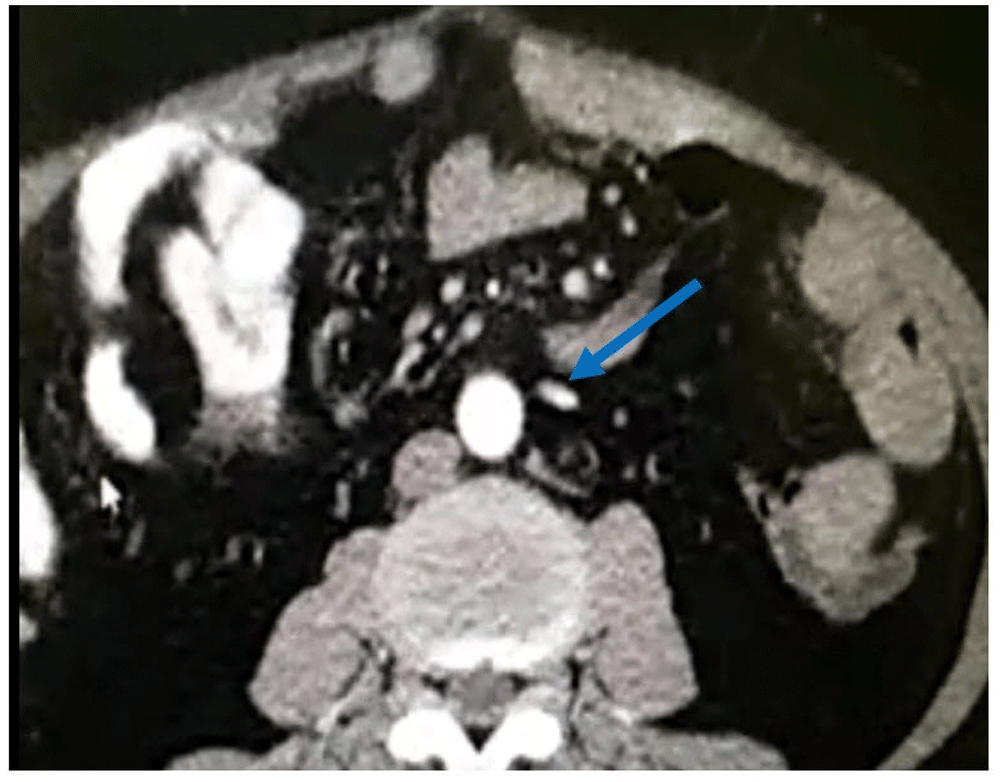
Abdominopelvic CT scan (Figure 2): A 130mm of segment of terminal ileum had diffuse wall thickening (3–8mm) associated with mesenteric fat. Narrow enhancement of inferior mesenteric artery with patchy filling defect, poor enhancement of terminal branches. Therefore, suspicions were: 1)vasculitis, 2)mesenteric ischemia.

Narrow enhancement of the inferior mesenteric artery can be observed (blue arrow).
Colonoscopy: Diffuse mucosal erythema and erosions were seen in the rectum until 6cm of anal verge. Hemorrhoid without active bleeding in anus, few erythema and ophtus ulcer in cecum. Terminal ileum was not intubated. A diagnosis of a rectal erosion maybe due to vasculitis.
Electromyogram test and nerve conduction velocity: Upper extremities reported bilateral mild carpal tunnel syndrome, and in right lower extremities mononeuritis multiplex could not be ruled out.
Echocardiography: No evidence of any other disorder.
Final diagnosis: Vasculitis (PAN or complicated HSP)
Unlike other vasculitis, such as microscopic polyarthritis or Wegener’s, PAN is not associated with ANCA6. The organs most affected in PAN are the skin, renal and GI tract. Cardiac involvement can manifest itself with hypertension, or even ischemic heart disease7. In the skin, PAN may manifest by erythematous nodules, livedo reticularis, ulcer, bullous or vesicular eruption and purpura6,8,9. Gastrointestinal symptoms that may be seen include abdominal pain, nausea, vomiting, melena, and bloody or non-bloody diarrhea10. One of the most common manifestations of patients with PAN is mononeuropathy multiplex that typically involves both motor and sensory deficits in up 70% of patients6,11. Most cases of PAN are idiopathic, although hepatitis B virus infection, hepatitis C virus infection, and hairy cell leukemia are important in the pathogenesis of some cases3,4,12,13. PAN can mimic the clinical manifestations of HSP. It is difficult to differentiate between PAN and HSP at an early stage5. The biopsy pattern helps to differentiate between PAN and HSP; in tissue studies of HSP leukocytoclastic vasculitis in post capillary venules together with IgA deposition is observed14. As already mentioned, PAN is most commonly seen in middle-aged or older adults3, while HSP is a childhood disease that occurs between the ages of 3 and 15 years15. Neurologic manifestation in HSP is rare. Single reports and case series document neurologic manifestations including headaches, intracerebral hemorrhage, focal neurologic deficits, ataxia, seizures, and central and peripheral neuropathy in children with HSP16. In the present case, using clinical manifestations and laboratory tests, we excluded other differential diagnosis apart from PAN. Considering that PAN and HSP have narrowing clinical manifestation, we differentiated between the two diseases by age and neuropathy. However, although the diagnosis of the present patient is PAN, for a better diagnosis, immunofluorescence of the biopsy is needed, which is not available in our center. Overall, diagnosis and treatment of PAN is important, and PAN should be considered in a patient with skin lesions and neurological impairment.
Written informed consent was obtained from the patient for the publication of the patient’s clinical details and accompanying images.
| Views | Downloads | |
|---|---|---|
| F1000Research | - | - |
|
PubMed Central
Data from PMC are received and updated monthly.
|
- | - |
Is the background of the case’s history and progression described in sufficient detail?
Partly
Are enough details provided of any physical examination and diagnostic tests, treatment given and outcomes?
Partly
Is sufficient discussion included of the importance of the findings and their relevance to future understanding of disease processes, diagnosis or treatment?
No
Is the case presented with sufficient detail to be useful for other practitioners?
Partly
Competing Interests: No competing interests were disclosed.
Is the background of the case’s history and progression described in sufficient detail?
Partly
Are enough details provided of any physical examination and diagnostic tests, treatment given and outcomes?
Partly
Is sufficient discussion included of the importance of the findings and their relevance to future understanding of disease processes, diagnosis or treatment?
Partly
Is the case presented with sufficient detail to be useful for other practitioners?
Yes
References
1. De Virgilio A, Greco A, Magliulo G, Gallo A, et al.: Polyarteritis nodosa: A contemporary overview.Autoimmun Rev. 2016; 15 (6): 564-70 PubMed Abstract | Publisher Full TextCompeting Interests: No competing interests were disclosed.
Alongside their report, reviewers assign a status to the article:
| Invited Reviewers | ||
|---|---|---|
| 1 | 2 | |
|
Version 2 (revision) 16 Apr 18 |
read | read |
|
Version 1 12 Jan 18 |
read | read |
Provide sufficient details of any financial or non-financial competing interests to enable users to assess whether your comments might lead a reasonable person to question your impartiality. Consider the following examples, but note that this is not an exhaustive list:
Sign up for content alerts and receive a weekly or monthly email with all newly published articles
Already registered? Sign in
The email address should be the one you originally registered with F1000.
You registered with F1000 via Google, so we cannot reset your password.
To sign in, please click here.
If you still need help with your Google account password, please click here.
You registered with F1000 via Facebook, so we cannot reset your password.
To sign in, please click here.
If you still need help with your Facebook account password, please click here.
If your email address is registered with us, we will email you instructions to reset your password.
If you think you should have received this email but it has not arrived, please check your spam filters and/or contact for further assistance.
Comments on this article Comments (0)