Keywords
High frequency jet ventilation, Liver ablation, Stereotactic surgery
High frequency jet ventilation, Liver ablation, Stereotactic surgery
Thermal ablation of primary and secondary liver tumours is a potentially curative treatment, and an alternative for patients not eligible for surgical resection due to severe comorbidity or underlying liver disease. Its efficacy has been proven for tumours smaller than 30mm in diameter, especially in treatment of hepatocellular carcinomas1. Adequate imaging of the tumour and precise guidance of the ablation device are crucial for accurate local ablative treatment. Accurate targeting is essential for an effective treatment, reducing the risk for local recurrence and need of retreatment1,2.
Recent developments in image guidance systems, with robotic and computer-assisted navigation, may help correct needle placement and improved ablation efficacy. Needle navigation and placement is based on pre-interventional imaging. Early phantom and clinical experiences with navigation systems suggest good procedural accuracy, reduced procedure time and reduced patient radiation exposure compared to freehand techniques3.
The high frequency jet-ventilation technique (HFJV) was developed in the seventies by Klain and Smith, and mostly applied in the field of ear-nose-and-throat (ENT) surgery. It does not rely on conventional tidal volumes but uses a high frequency forced gas move4. The potential advantage of using HFJV in abdominal surgery is to minimise the amplitude of respiration-related upper-abdominal organs compared to conventional tidal volume lung ventilation (TV)5,6,7.
The aim of this clinical methodological trial was to measure the liver movements during open surgery under general anaesthesia and compare HFJV with conventional ventilation.
Five consecutive patients who were scheduled for elective, open liver ablations were included in the clinical protocol.
General anaesthesia was induced and maintained by total intravenous technique (TIVA) with target controlled infusion (TCI - Alaris, PK CareFusion, Sarl, Switzerland) of propofol 2–6µg/ml according to Marsh pharmacokinetic model (Propofol Sandoz®, Sandoz, Copenhagen, Denmark) and remifentanil 2-10ng/ml according to Minto pharmacokinetic model (Ultiva®, GlaxoSmithKline,Solna, Sweden) with muscle relaxation achieved by rocuronium 0,6 mg/kg during induction of anaesthesia, followed by incremental doses of 0,15mg/kg during surgery (Rocuronium, Fresenius Kabi, Uppsala, Sweden).
Endotracheal intubation with a conventional endotracheal (ET) tube was performed at the induction of anaesthesia, followed by the initiation of conventional lung ventilation with pressure control/volume guarantee ventilation (PCV/VG - Aisys Carestation, GE Healthcare, Helsinki, Finland) as well as a lung-protective regime to achieve normo-ventilatory status. Tidal volumes have been calculated after the reduced body weight with 6-7ml/kg target and fixed 5cmH20 positive end-expiratory pressure (PEEP). Laparotomy was performed with a right subcostal incision. A HFJV cannula (LaserJet Catheter, Acutronic Medical Systems AG, Hirzel, Switzerland) was inserted endotracheally with the tip at the end of the ET-tube. HFJV (Monsoon HFJV ventilator, Acutronic Medical Systems, AG, 8816 Hirzel, Switzerland) was then initiated and continued during the liver ablation procedure. HFJV driving pressure (DP) was adjusted downwards, beginning at 1.8 bar, until satisfactory operation-field conditions were reached in accordance with the operating surgeon's assessment.
In the first phase, after the induction of anaesthesia, end-tidal carbon dioxide tension (EtCO2) was continuously monitored through the use of classical side-stream capnography towards the normocapnic state. During the HFJV phase, sequential measurements were taken with 10 minute intervals (Integrated Monsoon ventilator etCO2 module). After the termination of the last tumour ablation and the completion of liver movement measurement, the conventional lung ventilation was restored. Lastly, the etCO2 measurement was repeated following the same method as the one used at the start of the procedure. Cut-off values for discontinuation of HFJV was either etCO2 rise over 10 kPa, or oxygen de-saturation under 90%. With etCO2 exceeding 8 kPa, the DP down-regulation has been stopped and instead increased by 0.1 bar increments every 5 minutes until the target etCO2 was reached.
Patients were selected at the regional liver multidisciplinary team conference and were regarded as unresectable due to multiple metastases involving too many liver segments, but numbered less than twenty and none larger than thirty millimeters in diameter8. Multiple ablations were then performed using an intraoperative ultrasound and a stereotactic targeting device, CAS-one (Cascination AG, Bern, Switzerland) where a previously acquired computed tomography scan was merged with previous scans in cases of vanished lesions, and a 3D model of the liver reconstructed by MeVIS medical solutions AG (Bremen, Germany) was used as a surgical map with optical navigation of ablation antennae, as previously described8. For tracking of liver movement a rigid marker shield with a set of retroreflective marker spheres was placed on the liver surface in the vicinity of the lower border of segment 4b and tracked with an optical position measurement system (Polaris Vicra, NDI, Canada) incorporated into the CAS-One system which was positioned in the vicinity of the operative field thus providing a constant line of sight. A 4D position of the marker shield was measured for approximately 2–3 minutes for HFJV and conventional ventilation.
At each time point t, the magnitude of liver displacement d, was calculated as an Euclidean distance between translational component of the marker shield’s 3D position, and previously estimated centroid of the translational motion , i.e. an average translational position of the marker, as listed in the equation below:
d t = ||⬚ c − ||
All displacement errors d were described quantitatively using mean (µ) and standard deviation (σ) as well as a maximum error value. Statistically significant differences were tested with the two-tailed, nonparametric, unpaired t-test, where p < 0.05 was defined as statistically significant.
Patient demographics, medical status and extent of surgery is presented in Table 1.
| Patient ID | Sex | Age (years) | ASA | Weight (kg) | Height (cm) | BMI | No lesions |
|---|---|---|---|---|---|---|---|
| 1 | m | 74 | 3 | 78 | 178 | 25 | 4 |
| 2 | m | 58 | 4 | 99 | 178 | 31 | 8 |
| 3 | m | 76 | 3 | 82 | 181 | 25 | 2 |
| 4 | m | 82 | 3 | 77 | 185 | 23 | 9 |
| 5 | f | 48 | 3 | 73 | 163 | 27 | 30 |
Ventilator settings and readings are shown in in Table 2. The following parameters have been registered: end tidal CO2 concentrations before and after HFJV phase, respiratory pressures on conventional tidal volume ventilation before and after HFJV, peak inspiratory pressure and mean airway before and after HFJV, mean airway pressure, dynamic lung compliance both before and after HFJV phase as well as tidal volumes on conventional lung ventilation, at liver displacement measurement point. HFJV ventilator settings: respiratory frequency and target driving pressure as well as the measured respiratory parameters: peak inspiratory pressure and mean airway pressure as well as maximum end tidal carbon dioxide tension on HFJV.
In one case (patient number 2) an increase in DP was needed, because the etCO2 rose to 8.3 kPa and the optimal value was set on to 1.5 bar where in other four cases it was set to 1.1-1.2 bar.
The mean Euclidean liver displacement was 0.80 (0.10 SD) mm and 2.90 (1.03 SD) mm for HFJV and TV respectively with maximum displacement going as far as 12 mm on standard ventilation (p=0.0001). Data shown in Figure 1.
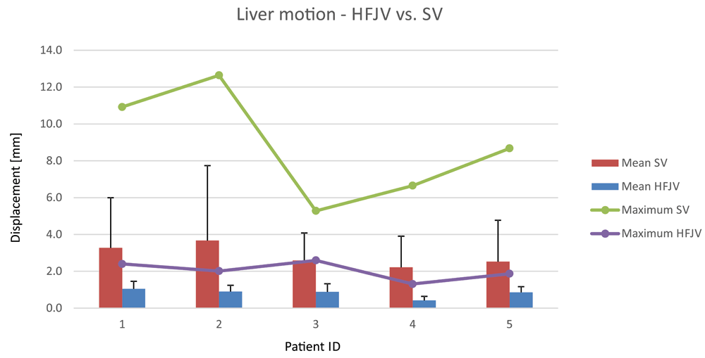
Displacement of measured point on liver surface during High-Frequency Jet Ventilation (HFJV) and standard ventilation (TV) Error bars mark standard deviation.
End tidal CO2 (etCO2)registered before and after High frequency jet ventilation (HFJV) phase (etCO2 - pre jet and post jet), Respiratory pressures on conventional tidal volume ventilation before and after HFJV phase, expressed in cm.H2O: peak inspiratory pressure (PeakP) pre jet, mean airway pressure (MaP) pre jet, dynamic lung compliance both before and after HFJV phase (Compliance pre and post jet) as well as tidal volumes on conventional lung ventilation, at liver displacement measurement point (TV post jet). HFJV ventilator settings: respiratory frequency (FQ on jet) and target driving pressure (T-DP on jet) as well as the measured respiratory parameters: peak inspiratory pressure (PIP) and mean airway pressure (MaP) and maximum end tidal carbon dioxide tension during HFJV phase (Max etCO2 on jet).
One of the most important challenges the anaesthesiologist faces perioperatively is the maintaining of the patient’s homeostasis and the facilitating of the course of surgery. In certain clinical situations, such as in stereotactic ablative procedures, it can be difficult to establish since there is a demand for keeping respiratory organ displacement to a minimum.
The recent investigation provides evidence for the claim that respiration induced liver motion during intervention can be reduced by more than two thirds when using HFJV instead of TV. This is the only study measuring this effect dynamically. This can have a decisive bearing on the risk of local recurrence rates and risk for collateral damage after image guided stereotactic treatment of liver tumours. The benefits in terms of radiation dose and respiratory organ shifting, when using HFJV in interventional radiology has previously been reported from several groups5,9,10, but these were all conducted in the setting of a CT-guided ablation with non-dynamic measurements of target organ displacement.
Stereotactic navigation is often performed on rigid registration between the intraoperative target organ with the images obtained before the surgery. With this setting, soft tissue deformation and patient motion will affect the navigation system and can cause significant inaccuracy11. The minimization of deformation-induced errors can be done in several ways. From experimental research point of view, the position of the moving target can be measured by implanted navigation aids or by using electromagnetic tracking devices11,12. Implantation of invasive needles is however not prudent in a clinical setting due to high risk of haemorrhage, tumour seeding, and long-term risks with leaving foreign bodies in situ.
Another approach is the mathematical modelling of mechanical tissue properties and organ motion in order to predict the target location based on a statistical model derived from preoperative 4D CT. This approach is frequently used in intensity modulated radiation therapies (IMRT)13. The relationship between the respiratory cycle and the movement of a target is however complex to predict and not possible in real time due to highly intensive computational requirements and obvious risks of differences in outcome during the acquisition of preoperative images and a situation with artificial respiration and an open or laparoscopically affected abdomen.
Therefore, respiratory gating methods that reliably reproduce a known breathing stage (temporarily disconnecting endotracheal tube in anaesthetized patients) seem to be a more reliable approach13,14. An overall internal target movement of 1.41 ± 0.75 mm was reported. However, periods of apnea are usually limited to 1–2 minutes depending on the health condition of the patient. HFJV overcomes these restrictions.
Use of HFJV outside the ENT and Thorax suites have been the subject of several, but rather anecdotal reports. In minimally invasive oncological procedures HFJV have been used in percutaneous, laparoscopic as well as in open approaches3,5,9,15. In cardiology it has been beneficial in catheter ablations16. In urology it can be helpful to minimize the numbers of shocks needed during ESWL-treatment16,17.
The present study is small and though the liver displacement data is solid, further studies on the physiological effects of HFJV is needed to elucidate the limitations. Carbon dioxide control is one of the important aspects of perioperative management. In the treatment protocol established during the study, it remained even more challenging because of the “less is better” strategy, favouring relatively low respiratory driving pressures.
Introducing HFJV in the management of computer-assisted abdominal surgery to a wider extent remains promising. The wider use of this method is, however, limited by the equipment availability and staff experience. Nevertheless, in the scale of a highly specialized centre, the acceptable skill level can easily be achieved, and the overall cost of the equipment as well as materials and utilities remains reasonable. HFJV is a promising lung ventilation modality for patients undergoing stereotactic surgical procedures in general anaesthesia when reduction of target organ displacement is crucial.
All procedures performed were in accordance with the ethical standards of the institution at which the studies were conducted. Since this was a retrospective analysis of the clinical material collected before, the written consent has been obtained only from two patients still alive at the time when the decision of data analysis and publication have been made. Other three patients have already died.
Dataset 1: Demographic data and ventilation readings. metodological_study_1.xls 10.5256/f1000research.14873.d20721218
Dataset 2: Liver positioning data. 14-09-01-open-liver-all.xlsx 10.5256/f1000research.14873.d20721319
| Views | Downloads | |
|---|---|---|
| F1000Research | - | - |
|
PubMed Central
Data from PMC are received and updated monthly.
|
- | - |
Click here to access the data.
Spreadsheet data files may not format correctly if your computer is using different default delimiters (symbols used to separate values into separate cells) - a spreadsheet created in one region is sometimes misinterpreted by computers in other regions. You can change the regional settings on your computer so that the spreadsheet can be interpreted correctly.
Provide sufficient details of any financial or non-financial competing interests to enable users to assess whether your comments might lead a reasonable person to question your impartiality. Consider the following examples, but note that this is not an exhaustive list:
Sign up for content alerts and receive a weekly or monthly email with all newly published articles
Already registered? Sign in
The email address should be the one you originally registered with F1000.
You registered with F1000 via Google, so we cannot reset your password.
To sign in, please click here.
If you still need help with your Google account password, please click here.
You registered with F1000 via Facebook, so we cannot reset your password.
To sign in, please click here.
If you still need help with your Facebook account password, please click here.
If your email address is registered with us, we will email you instructions to reset your password.
If you think you should have received this email but it has not arrived, please check your spam filters and/or contact for further assistance.
Comments on this article Comments (0)