Keywords
Occlusal veneer preparation designs, Planar preparation, Circumferential Preparation, Intracoroanal extension preparation, Fatigue resistance, 3D Finite Element Analysis.
Occlusal veneer preparation designs, Planar preparation, Circumferential Preparation, Intracoroanal extension preparation, Fatigue resistance, 3D Finite Element Analysis.
Minimally invasive dentistry has recently become an area of interest. Maximum sound tooth structure conservation is crucial for tooth restorations and durability1. This concept can be applied successfully with occlusal veneers (thin onlay/overlay with non-retentive design), which are posterior extra coronal Restorations needing less preparation guided by interocclusal space and teeth anatomy2.
The evaluation of various preparation models showed that the amount of tooth structure removed in the posterior teeth for onlay and partial coverage preparation could be reduced by as much as 40% compared to full coverage preparation2.
In the standard onlay configurations, sharply defined angles and cusp edges magnify stresses on ceramic restorations, increasing the incidence of their fracture3, Moreover, the precise reestablishment of the breached sound proximal contact is challenging. Finally, milling machine errors resulting from milling tool tip diameter may represent a limitation while milling the restoration, since the milling tool tip diameter smaller than diameter of prepared cusp tip will result in incomplete seating of the restoration whereas a diameter larger than the prepared cusp tip will result in poor restoration adapt.
The emergence of strong, etchable and machinable ceramics such as lithium disilicate has expanded the uses of bondable restorations4. Silanization and cementation using adhesive bonding to the available tooth tissue improved the mechanical properties of definitive ceramic restorations compared to non-adhesive cementation5.
Recently, new classes of hybrid ceramics, as zirconia-reinforced lithium silicate (ZLS) ceramics (e.g. Celtra Duo, Vita Suprinity) have been developed. While elevated translucency and elevated strength are mutually exclusive for most kinds of monolithic ceramics, zirconia-reinforced lithium silicate has flexural strength values similar to lithium disilicate6.
The samples are compelled to fail, in load- to -failure experiment under displacement control of loading device. This offers valuable data under harsh conditions but of negligible importance to clinical sustenance. Dental materials’ fatigue behaviour is defined by an exact fatigue limit, above that limit the material quickly fails and below it long term survival of the material is expected4.
Gradual resistance degradation of the ceramic was recorded at low ongoing or cyclic loads, particularly in humid settings1. Fatigue is therefore an essential factor that limits the longevity of dental restorations and is a requirement for reliable in vitro testing1.
The fatigue manner of dental materials is characterized by a well-established fatigue limit, above that limit the material fails rapidly and below it there is long-standing survival4.
The use of 3D finite element analysis (FEA) to evaluate the behaviour of materials and designs has been expanding. FEA analyses how distinct materials and restoration configurations interact in a non-invasive, time-saving manner, as well as overcoming ethical dilemmas of clinical investigations7.
The purpose of this research was to examine the impact of two modified occlusal veneer preparations on the fatigue resistance and stress distribution of bonded occlusal veneers compared to conventional preparation design.
This study was approved by the Research Ethics Committee of the Faculty of Dentistry, Cairo University. Approval number: 1587.
Sample size estimation. To compare the fatigue resistance of machine milled ceramic occlusal veneers with new preparation designs versus conventional design, one-way analysis of variance (ANOVA) was done. Based on a previous study by Stappert et al.8, the outcome variable is normally distributed and an expected effect size of approximately 0.44 is estimated, therefore a total sample size of 54 (18 in each group) would be sufficient to detect large effect size (f=0.44) with power 80% and 5% significance level. (G*Power program).
Sample fabrication
Teeth collection, periodontal ligament simulation and occlusal veneer preparation
A total of 54 human mandibular molars, obtained from the Outpatient Clinic of the Department of Oral Surgery, Faculty of Dentistry, Cairo University, which were free of any caries, defects and cracks were chosen. Teeth mean dimensions of the anatomical crown bucco-lingual width (distance between the maximum convexities on buccal and lingual surfaces) (11±1mm), mesio-distal widths of (10±2 mm) at the level of the cementoenamel junction (CEJ). An ultrasonic scaler was used to remove calculus deposits, debris and soft tissue remnants. The teeth were stored until use in distilled water.
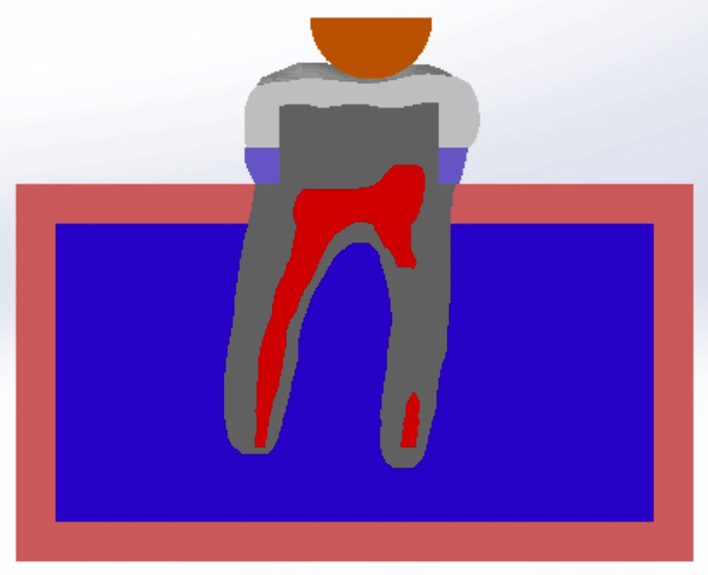
Root surfaces were plunged into blue inlay melted wax (Bego crown and bridge wax) 2.0 mm below the CEJ, resulting in a 0.2- to 0.3-mm thick wax layer9. Teeth were then mounted in Epoxy resin (CMB, Egypt) using a plastic ring (2.5 cm in diameter and 2 cm in length) and a custom-made paralleling device was used to allow precise vertical centralization of the long axis of each tooth parallel to the long axis of the plastic ring.
The wax was separated from the root surface and the resin cylinder “alveolus” after resin polymerization. Silicone elastomeric material (Elite Light Body Zhermack GmbH Deutschland) was then injected in the resin cylinders, the tooth was repositioned in the epoxy cylinder and the surplus elastomeric material was removed with a scalpel blade.
Randomization and grouping. The mounted teeth were randomly distributed into three equal groups of 18 teeth each according to the occlusal veneer preparation design (Table 1). Randomisation was performed using the random.org random number generator.
Tooth preparation. A special milling machine (AF30 Nouvag, Switzerland) was used for tooth preparation. The assembly incorporates a conventional-speed straight hand-piece perpendicular to the surveyor platform. A customized base was constructed to accurately fit the epoxy resin block, which was held stable in place on the magnetic table of the milling machine. Then the researcher operates the machine to prepare all the teeth in a standardized manner (Figure 1).
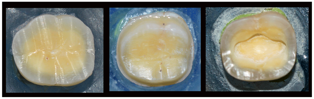
Group I (left), Group II (middle) and Group III (right).
Construction of occlusal veneers. CAD/CAM (CEREC AC, Sirona, Bensheim, Germany) was used for the fabrication of all samples in this research. Each prepared tooth was scanned using the CEREC Omnicam and designing was carried out using CEREC Premium 4.4 software. Cusp height thickness was adjusted at 1.5 mm in all groups while thickness of the restorations at the central groups was adjusted as follows: Group I and Group II at 1 mm and Group III at 2.5 mm. Milling of the Occlusal veneers was done from Zirconium Lithium Silicate (ZLS) blocks (VITA Suprinity, VITA Zahnfabrik H. Rauter GmbH & Co.KG) in 4-axis milling machine CEREC MC XL). This process was repeated 18 times to end up with 18 milled VITA Suprinity occlusal veneers for each of the three groups. Finally, occlusal veneers were fully crystallized and glazed in Programat P510 furnace (Ivoclar Vivadent AG. Principality of Liechtenstein) (Figure 2).
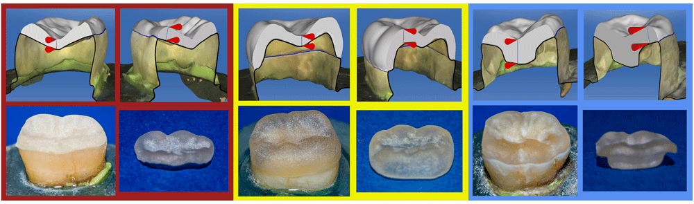
Group I, red; Group II (yellow), Group III (blue).
Cementation of Occlusal veneers. For surface treatment of occlusal veneers, etching of the intaglio surfaces of the veneers was conducted using Bisco Porcelain Etch (BISCO Dental Products, Illinois, U.S.A.) for 20 seconds, then rinsed with water and dried with air, then silanized using Bisco Silane (BISCO Dental Products, Illinois, U.S.A.) for 60 seconds then air dried for 5 seconds.
For surface treatment of the prepared natural tooth, the prepared surfaces of the tooth were acid-etched using 35% phosphoric acid (ScotchBond Universal Etchant, 3M ESPE AG. ESPE Platz Seefeld. Germany) then rinsed for 10 seconds. Two successive coats of Adper Single Bond 2 Adhesive (3M ESPE AG. ESPE Platz Seefeld. Germany) were applied with a micro-brush and gently air-dried for 5 seconds, then light polymerized for 10 seconds.
Rely X Ultimate (3M ESPE AG. Seefeld, Germany) Dual cure, adhesive resin cement was used to cement 54 Occlusal Veneers. The occlusal veneer was seated on the corresponding tooth and held in place with light pressure. A gel state was achieved by tack-curing excess with light curing (Elipar DeepCure-S LED Curing Light) (3M ESPE AG. Seefeld, Germany) for approximately 2 seconds and the excess cement was removed with an explorer, then a glycerin-based gel (K-Y Jelly; Johnson & Johnson, USA) was applied at the margins of the occlusal veneer to prevent the formation of the oxygen inhibiting layer. During cementation, a custom-made loading device was used to apply a steady load of 3 kg parallel to each tooth’s long axis10 for 5 minutes to allow cement to self-cure as recommended by the manufacturer11,12. All surfaces (axial and occlusal) were light cured for 20 seconds. (Figure 3)
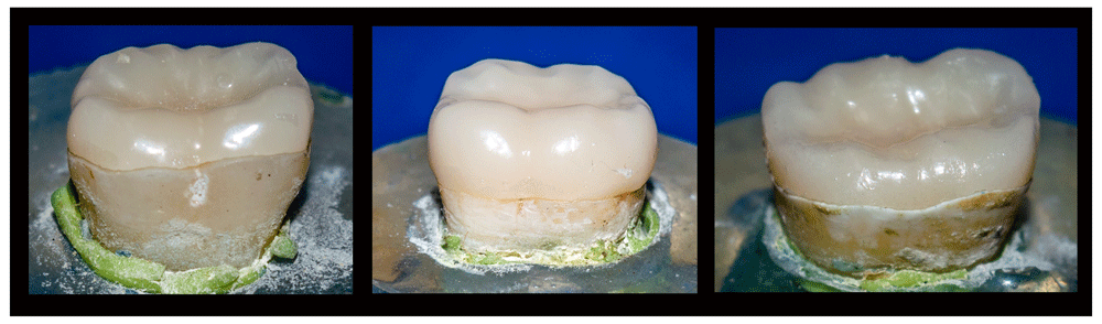
Left, Group I; middle, Group II; right, Group III.
Fatigue resistance test
Step-stress accelerated life testing (SSALT)
Cyclic failure loads were established in an electric machine (Instron, Model 3345, Instron, USA) using a compressive mode of load applied occlusally, dynamically loaded under water in a chamber at 35-37°C to simulate the intraoral conditions at a frequency of 2 Hz, where the sample is subjected to a prescribed number of cycles at each of a sequence of increasing stress levels, until failure of the sample. Starting with a stress level below the anticipated material’s fatigue limit (30–60% of single load to failure)13 is chosen.
The sample is then tested at the mild profile stress level of (698 N) (30% of single load to failure mean value recorded for zirconia-reinforced lithium silicate)14 until either failure occurs or the run-out at a previously set number of cycles (5000 cycles) is achieved. If failure occurs, the stress level and the number of cycles is recorded in Newtons. The failure was manifested by an audible crack and confirmed by a sharp drop in the load-deflection curve. If run-out occurs, the stress level is increased by a preselected stress increment and the same sample is run again at the new stress level15. This procedure is continued until the specimen does fail. Where load is stepped at (100 N per 5000 cycles) run as 700, 800, 900, 1000, 1100, 1200, 1300, 1400, 1500, 1600 and 1700 N at a maximum of 5000 cycles each.
The maximum fatigue strength supported by each specimen was calculated according to the equation16
Where 𝜎e is the maximum fatigue strength corresponding to NLife cycles, 𝜎0 is the previous maximum fatigue stress that did not result in failure, Δ𝜎 is the step increase in maximum fatigue stress, NFail the cycles to failure at the fatigue stress and the NLife defined cyclic fatigue life.
Examination of fractured samples. Fractographic analysis was performed to detect the modes of failure of the samples and the retrievability of remaining tooth structure. This analysis was done for all samples using high-performance Leica MZ6 Stereomicroscope (Meyer instruments. USA) with 8:1 zoom indicating areas of interest for further examination under a scanning electron microscope (SEM; Model Quanta 250 Field Emission Gun, FEI company., the Netherlands) attached with EDX Unit (Energy Dispersive X-ray Analyses), with accelerating voltage 30 K.V., 50X and 200X magnification and resolution of 1 nm. The samples were clean and conductive, and no coating was required since the LowVac mode was used. The specimens were attached to the specimen holder using any suitable SEM vacuum-quality adhesive.
3D scanning. Three samples of the fifty-four samples that were constructed for the fatigue resistance test (one for each group) and their corresponding Occlusal veneers were scanned before cementation to produce models with real geometrical measures. 3D reconstruction from Cone beam computed tomography (CBCT) data was found to be accomplished with a high linear, volumetric, and geometric accuracy17. A CBCT scanner (Next Generation iCAT scanner; Kaltenbach & Vaigt Gmb, Germany) was used to obtain CBCT images in this research.
After acquisition, data were exported and transferred in DICOM format. Mimics software (version 17; materialize, Belgium) was used for segmenting the scanned objects into separate elements. Definitive threshold was assigned to select certain tissue and its volumetric model was automatically calculated. The produced STL files were viewed and assembled on Meshmixer software version 3.3.15 (Autodesk, Inc, USA) where mesh improvement and refinement was carried out.
Reverse engineering and assembly. Reverse-engineering was performed using NX software v10 (Siemens) (FreeCAD is an open access alternative). The generated 3D CAD geometry of each element was then assembled using Solidworks software 2017 (Dassault Systems SolidWorks Corporation) (FreeCAD is an open access alternative) to produce three models one for each group (Figure 4).
The process of finite element analysis was carried out by ANSYS R16.2 software (ANSYS, Canonsburg, PA, USA) (OpenFOAM is an open access alternative). It included three phases: the pre-processing, processing and post-processing phases.
In the pre-processing phase, the type of element was defined as Solid 10 node 187. It was presumed that the characteristics of all materials were isotropic, homogenous, and linear elastic. Each material's characteristics (elasticity module and Poisson ratio) were gathered and uploaded to ANSYS R16.2 software (Table 2). A perfect bonding (rigid) condition with no slip interface conditions between the components. During this process each model was divided into small parts called elements, connected at points called nodes to form a mesh structure. Parabolic tetrahedral solid elements were used to form a fine solid mesh. The overall number of elements and nodes was recorded (Table 3).
| Material | Modulus of elasticity (GPa) | Possion’s ratio |
|---|---|---|
| Enamel | 84.119 | 0.3019 |
| Dentin | 18.619 | 0.3119 |
| Periodontal ligaments | 0.000068920 | 0.4520 |
| Cancellous bone | 1.3720 | 0.3020 |
| Cortical bone | 13.720 | 0.3020 |
| Pulp | 0.00221 | 0.4521 |
| Zirconia-reinforced lithium silicate Vita Suprinity | 7022 | 0.2123 |
| Rely X Ultimate Resin Cement | 7.712 | 0.3324 |
| Model | Element | Node |
|---|---|---|
| Group I | 493980 | 706990 |
| Group II | 795240 | 1147980 |
| Group III | 533700 | 935780 |
In the processing phase, following the creation of the 3D meshes, a zero-displacement boundary condition was set at all nodes of the cortical bone that were confined in X, Y, and Z directions. The occlusal surface of the occlusal veneer was loaded by a 500 N 3D ball model at the inner inclines of the mesiobuccal, distobuccal and mesiolingual cusps18.
In the post-processing phase, the output of the processing phase was displayed as graphical and numeric outputs in the form of von Mises Stress25,26 The von Mises criterion is a formula for calculating whether the stress combination at a given point will cause failure. If the von Mises Stress exceeds the yield stress, then the material is considered to be at the failure condition. Outputs were also displayed as total deformation25, physical deformations that can be calculated on and inside a part or an assembly, and Maximum Equivalent Stress Safety Tool25, a particular combination of principal stresses cause failure if the maximum equivalent stress in a structure equals or exceeds a specific stress limit.
Data from the three groups were gathered, arranged and analysed using IBM SPSS, version 21 (SPSS, Chicago, IL, USA). One-way ANOVA was used to determine whether there was statistical difference between the three groups. Survival rates (%) were compared by chi square test. Statistical analysis was performed using Graph-Pad Instat statistics software (version 3.06) for Windows. The data obtained using the accelerated fatigue test were visualised using a Kaplan Meier survival curve to cycles and life table for load to compare fatigue resistance of the groups. P values ≤0.05 were considered to be statistically significant in all tests.
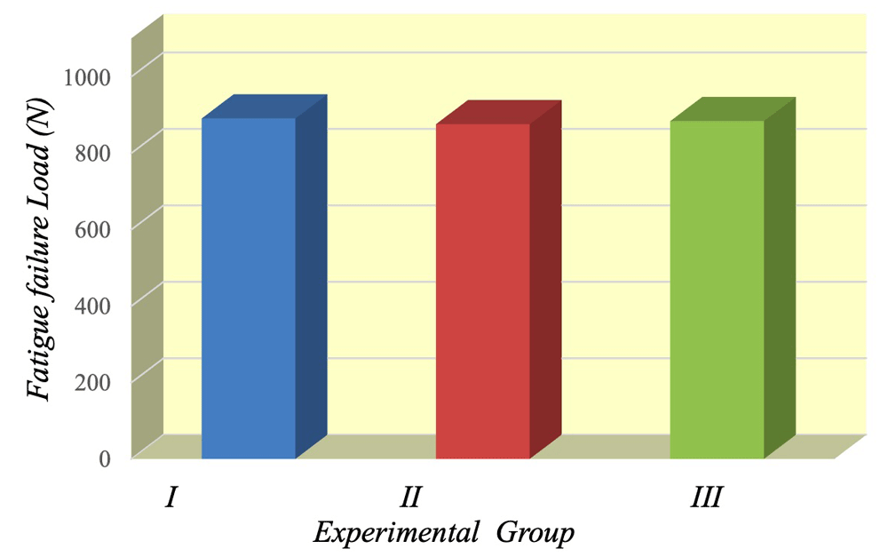
It was discovered that the largest mean±SD values were registered for Group II (circumferential preparation); (890.57±211.53 N) followed by Group I (conventional preparation) mean±SD values; (883.54±135.91 N) whereas, the smallest mean±SD values were recorded for Group III (intracoronal extension) (875.57±143.52 N). As indicated by ANOVA test the difference among groups was statistically non-significant (p=0.9814). Raw fatigue resistance data, alongside all other raw results, are available as Underlying data27.
Descriptive statistics of fatigue failure load test results; mean values, standard deviation (SD) and confidence intervals (low and high) for all groups are summarized in Table 4 and depicted graphically in Figure 5.
| Variables | Mean | Std. Deviation | 95% Confidence Interval | ANOVA | |||
|---|---|---|---|---|---|---|---|
| Low | High | F | P value | ||||
| Experimental group | Group I | 883.54 | 135.91 | 780.76 | 986.32 | 0.02 | 0.9814 |
| Group II | 890.57 | 211.53 | 746.51 | 1034.6 | |||
| Group III | 875.57 | 143.52 | 780.15 | 970.99 | |||
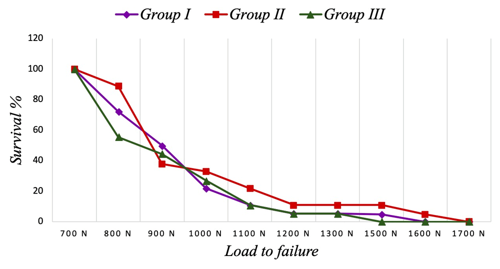
Survival rate. Survival rate of samples recorded for all experimental groups as function of step load was tabulated and graphically drawn (Table 5, Figure 6).
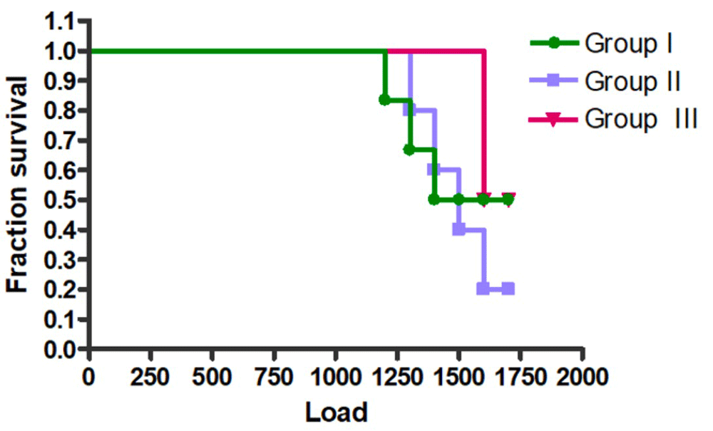
Kaplan-Meier survival curve. Analysis using Kaplan-Meier survival curves, through the log-rank test, showed non-significant differences between groups (Figure 7). The survival performance was best for Group II and worst for Group III.
Fractographic analysis of fractured samples
Mode of failure
Fracture behaviour of the samples was classified in to three modes and described in Table 6 as observed by stereomicroscope (Figure 8) and SEM (Figure 9), where retrievable survival of the remaining tooth structure is classified as modes I and II and irretrievable survival of remaining tooth structure: mode III. Results of each group are tabulated (Table 7) and classified as retrievable and irretrievable (Table 8).
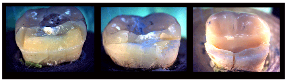
Left, Mode I; middle, Mode II; right, Mode III.
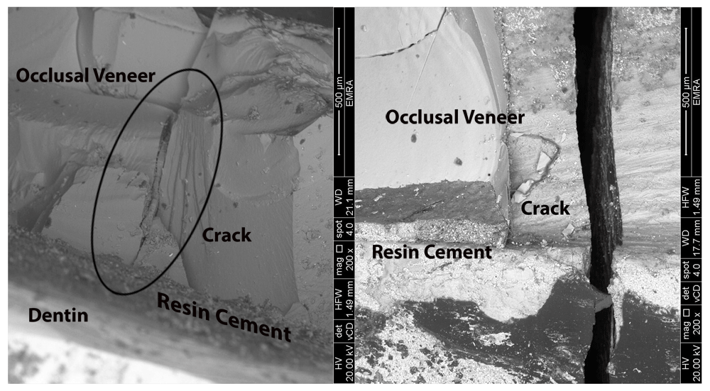
Left, retrievable survival (crack limited to veneer); right, irretrievable survival (crack beyond the veneer).
| Mode of failure | Number of samples of Group I | Number of samples of Group II | Number of samples of Group III |
|---|---|---|---|
| Mode I | 8 | 14 | 4 |
| Mode II | 5 | 2 | 2 |
| Mode III | 5 | 2 | 12 |
The FEA revealed stresses at every node in each model. These results were displayed as stress contours overlaid on the original model. The calculated numeric data of stress, deformation and safety factor in the models were transformed into color graphics.
Equivalent (von Mises) stress. For each group, the von- Mises stress values of different areas were calculated and compared (Table 9). The stress distribution values were generally found to be low and within the safe range except at certain areas.
Asterisk marks the highest value in each group.
Total deformation. The maximum value of total deformation in Group I was 0.035 mm, located on the lingual half of the occlusal veneer tooth complex. The maximum value in Group II was 0.03785 mm, equally distributed over the occlusal veneer tooth complex. In Group III, the maximum value was 0.04 mm located on the distal half of the occlusal veneer tooth complex as well as distal half of root (Figure 10).
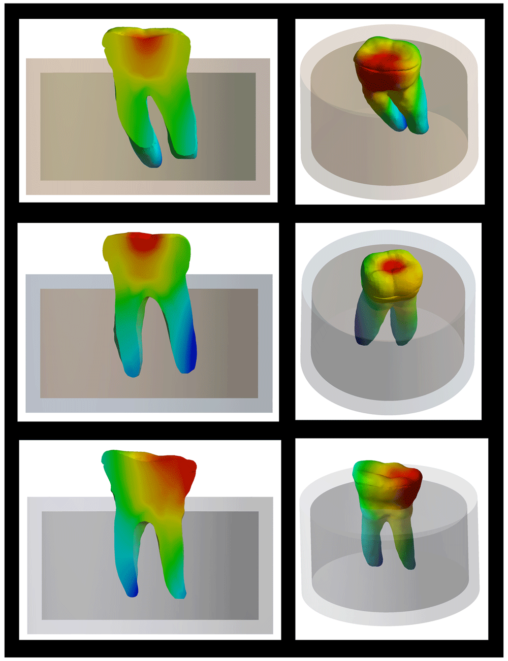
Top, Group I; Middle, Group II; Bottom, Group III.
Safety factor. In the three groups there was high safety factor where the maximum equivalent stress was less than the stress limit. Where the lowest safety factor recorded (Figure 11, denoted by the red color) for Group I (0.2135) was at the buccal neck of the tooth, for Group II (0.25126) it was at the neck of the tooth and for Group III (0.19275) it was at the lingual neck of the tooth.
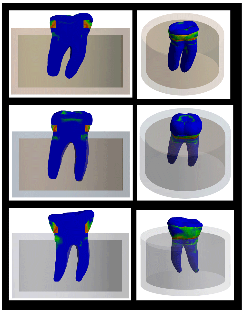
Top, Group I; Middle, Group II; Bottom, Group III.
The stress distribution among different layers of the model differed as well as areas of total deformation. This could be correlated with the fracture behavior of samples of the three groups as follows:
Group I (planar occlusal veneer preparation design)
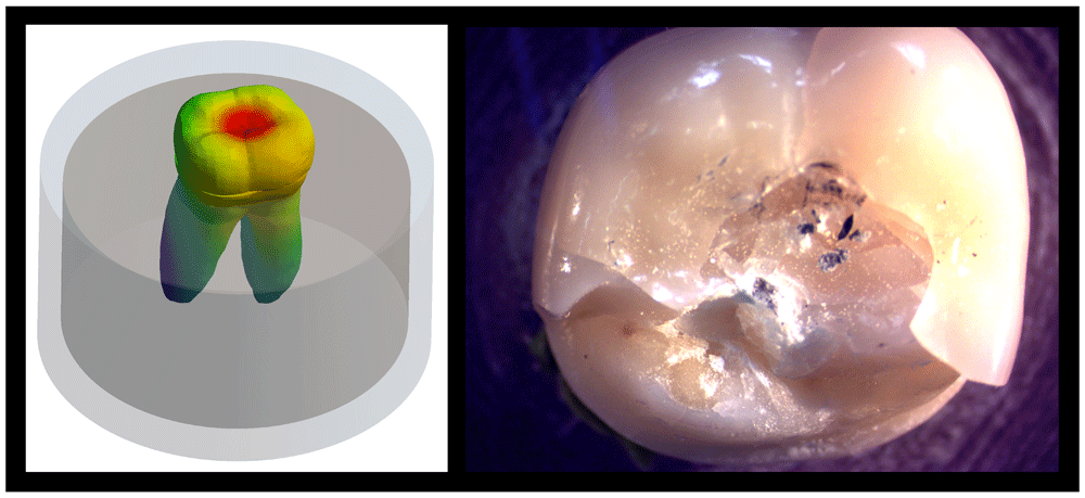
This preparation design delivers almost equivalent amounts of stresses to the occlusal veneer and to axial walls of the tooth. Where stresses are concentrated towards lingual, mesial and distal areas of occlusal veneer tooth complex, with maximum stresses concentrated at lingual half of occlusal veneer and tooth. The maximum total deformation was observed at lingual and mesial areas of occlusal veneer- tooth complex.
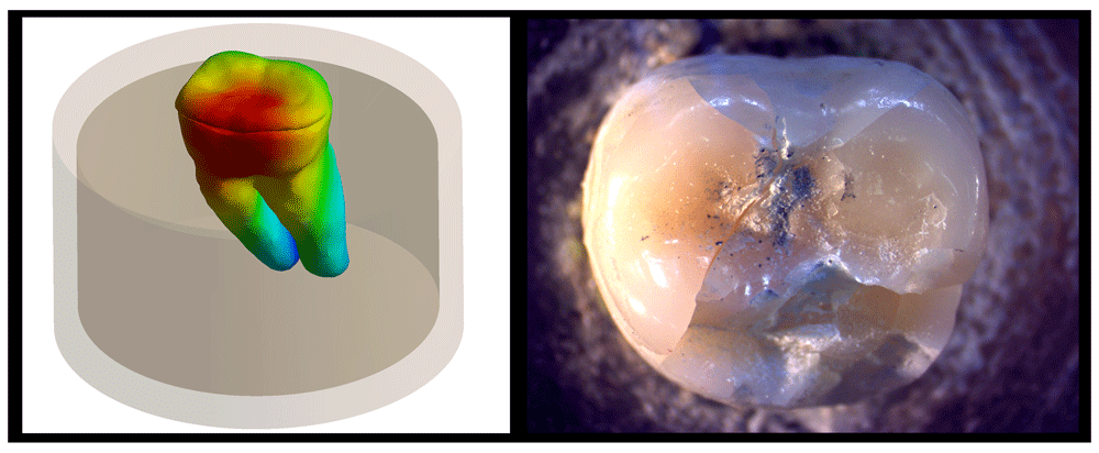
Upon observation of fracture behavior of Group I samples of fatigue resistance, the failure in most of the samples occurred at lingual and mesial regions of restoration which fails early upon dynamic loading causing enamel fracture at underlying axial wall (Figure 12).
Group II (circumferential occlusal veneer preparation design)
This preparation design equally distributed stresses over the tooth where maximum stresses are observed at the occlusal veneer center and the lingual half of the occlusal veneer and tooth structure. The maximum total deformation was equally absorbed by the entire occlusal veneer shielding the underlying tooth structure.
Upon observation of fracture behavior of most of group II samples of fatigue resistance, the failure in most of the samples occurred at the lingual half of the occlusal veneer not including the tooth in any form of fracture or cracking (Figure 13).
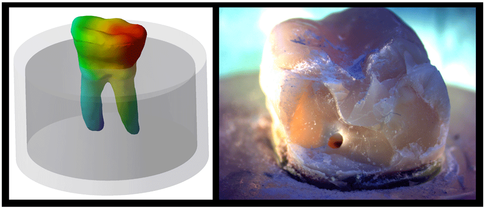
Group III (intracoronal cavity occlusal veneer preparation design)
This preparation design delivers less stress to the occlusal veneer and transfers larger stresses to the center of the tooth and cervical area, concentrated towards the lingual and distal areas, with maximum stresses at the center of occlusal veneer. The maximum total deformation was observed at the distal and lingual portions of the occlusal veneer-tooth complex reaching as far as cervical area and the root.
Upon observation of fracture behavior of group III samples of fatigue resistance, the failure of the majority of the samples occurred along the lingual and distal portions of the occlusal veneer- tooth complex extending to the tooth cervical area (Figure 14).
Treatment of patients with advanced wear is complicated. The huge complications include: possible wear compensation by tooth eruption (preserving vertical dimension) and further reduction of sound tooth structure to restore worn teeth, where there’s a broad variation in the needed amount of reduction for different restorations4.
In cases of worn dentition, conventional full crowns can be manufactured. However, in order to obtain a feasible and esthetic restoration, application of such full-coverage crowns usually involve the removal of additional tooth structure. In patients already experiencing significant loss of tooth tissue, further destruction may compromise the long-term viability of the teeth28.
More conservative approaches include the use of occlusal veneers, which are extracoronal restorations that require a minimalistic preparation driven by interocclusal clearance and conservative considerations2.
The root submergence material should replicate the ability of bone to absorb masticatory force and in return sustain compressive and tangential force in a fracture resistance test9. Gonzalez et al.29 confirmed the significance of inclusion of artificial periodontal ligament in fatigue resistance research.
Planar occlusal veneer preparation design have gained a huge reputation over the years as a conservative minimally invasive treatment in severely worn dentition cases, as affirmed in studies by Magne et al.30, Clausen et al.31, Sasse et al.32, Al Akhali et al.33 and Yazigi et al.11. Modifications to this design seek to improve bonding strength to different substrates, to make use of an intracoronal cavity or a previously restored tooth31,32 and to improve fracture resistance through cuspal coverage33. Maximum chewing forces in the posterior molar region range between 200 to 540 N. In bruxers, these values dramatically increase to the range of 800 N34.
According to Preis et al.6 ZLS crowns showed good long-term resistance to fatigue during ageing simulation. The high fracture stability of the ZLS crowns after thermal cyclic mechanical loading was comparable to lithium disilicate.
The evaluation technique of fatigue resistance used in this research (accelerated fatigue over one day for each specimen), initially implemented by Fennis et al.35 and used by Magne and Knezevic36 in several experiments, provides a fair equilibrium between single load-to-failure testing and more advanced fatigue testing (minimum 1,000,000 cycles). The fatigue testing was carried out in water at a frequency of 2 Hz among the bounds of chewing frequency range (0.6-2.3 Hz)34.
Following the mild step-stress profiles that the universal testing machine could deliver (a maximum fatigue load of 1700 N)37. In this research, a stepped load protocol with 5,000 cycles in each step and a load increment of 100 N was followed to assess the fatigue resistance of ceramic occlusal veneers.
3D FEA was conducted in the current study to assess the distribution of stresses among ceramic occlusal veneers with different preparation configurations on molar teeth. In the posterior region, fixed restorations should withstand an approximately 500 N occlusal load7,38. Therefore, 500 N was the load of choice in our research.
In the current study the highest mean±SD values were recorded for Group II (circumferential finish line); (890.57±211.53 N) followed by Group I (conventional) (Planar preparation) mean±SD values; (883.54±135.91 N) while the lowest mean±SD values were recorded for Group III (intracoronal cavity extension) (875.57±143.52 N). Statistically, the distinction between groups was not significant.
Since that according to Varga et al.39 the mean maximum voluntary molar biting force was found to be 777.7 ± 78.7 N in males and 481.6 ±190.42 N in females. The present research shows that the average fatigue limit of 890 N for ZLS is powerful enough to resist repeated occlusal forces in molar restorations.
Despite the fact that the occlusal veneers in all three groups in this study had comparable and clinically acceptable fatigue resistance, their modes of failure were different. The modes of failure were classified as resulting in either retrievable or irretrievable survival of remaining tooth structure. A retrievable fracture is above the CEJ, meaning that in case of fracture, the tooth can be restored with some modifications in the preparation design, while an irretrievable fracture extends below the CEJ and the tooth is likely to be extracted40.
A thorough fractographic assessment demonstrated the similarity and uniqueness of the fracture pattern among the tested specimens in each group, where Group I showed a majority of teeth undergoing occlusal veneer fracture with intact tooth structure or retrievable survival of remaining tooth structure, Group II showing a majority of teeth undergoing fractured occlusal veneer with intact tooth structure and Group III showing majority of teeth undergoing catastrophic fracture of both occlusal veneer and tooth structure, which signifies the validity and reproducibility of the current testing method as confirmed by Borba et al.41.
Among the three tested occlusal veneer preparation designs, the modified intracoronal extension design resulted in a majority of teeth undergoing catastrophic fracture of occlusal veneer and tooth structure (irretrievable survival), while for planar and circumferential approaches, the majority of failures observed were confined to occlusal veneers, sparing underlying tooth, and few showed retrievable survival of remaining tooth structure. This denotes that the planar and circumferential finish line occlusal veneer preparation designs resulted in better distribution of stresses on the restoration-tooth complex, and thus more favourable fracture behavior.
In this study, the Kaplan-Meier analysis showed that planar occlusal veneers exhibited failures at fatigue loads that were not significantly different from those exhibited by circumferential preparation or intracoronal extension modified occlusal veneer designs. Previously published results by Bitter et al.42 showed higher fracture resistance of MOD-inlays compared to partial or total onlays using a central load application. The 3D finite element analysis data recorded showed that all three occlusal veneer preparation designs yielded low von-Mises stresses within the factor of safety of the model, with lesser safe values at the cervical enamel region. Among the three tested occlusal veneer preparation designs the modified intracoronal extension occlusal veneer preparation design showed the highest equivalent von Misses stress values in dentin, enamel and Occlusal veneer–tooth complex. This was in agreement with Dejak et al.20 who proved that bonded inlays strengthen the prepared teeth but fail to restore their inherent resistance to failure. It was observed that the occlusal veneers of circumferential finish line preparation design sustained the maximum stress values when compared to occlusal veneers of planar and intracoronal extension designs, shielding the underlying tooth structure from fracture.
The current findings are in agreement with research by Hamdy et al.43, in which bonded overlays presented a benign distribution of functional occlusal forces, demonstrating some protective impact of these restorations against irretrievable fractures. Yamanel et al.44 confirmed that the onlay (cuspal coverage) design protected the tooth structures more efficaciously than the inlay (intracoronal extension) design.
From the researcher’s point of view, the extension of the planar preparation design on to axial surfaces to include cuspal coverage by circumferential finish line preparation in Group II proved beneficial in stress distribution uniformity among occlusal veneer and tooth, sheltering the tooth structure from deleterious effect of excessive occlusal loads as confirmed in the FEA simulation, resulting in failure of occlusal veneer always sparing the underlying tooth structure intact as also concluded in a study by Yamanel et al.44 The intracoronal extension of the occlusal veneer preparation in Group III resulted in a wedging action transferring the great majority of occlusal stresses to cervical area of tooth as confirmed by FEA simulation, resulting in the most catastrophic failures (irretrievable survival) sustained by any of the three designs, and as confirmed in a study by Yamanel et al.44.
This study has some limitations, it assumed that all the materials were linearly elastic, homogeneous, and isotropic, and that the bonds between the ceramic occlusal veneers and tooth structure were ideal and that teeth are only subjected to static load in Finite Element Analysis simulation. The outcomes of the present study can only predict the clinical behaviour, and additional clinical testing is advised.
Choosing the occlusal veneer preparation design to restore worn molars may influence the stresses within the crown-tooth system. It is essential to guarantee that the occlusal veneer preparation design offers the already worn tooth sufficiently conservative and non-invasive preparation to be able to withstand occlusal forces and, in case of fracture, the remaining tooth structure is not placed at risk and is considered restorable.
This research disclosed that planar and circumferential finish line occlusal veneer preparation designs had elevated incidences of restorable fractures relative to intracoronal extension occlusal veneer preparation design. Therefore, clinicians may choose the planar and the circumferential designs to improve the clinical performance and longevity of the restored worn molars.
1. In vivo studies should be conducted to compare the behavior of the three tested designs.
2. A research project to measure the difference in retention between the planar, circumferential finish line and intracoronal extension occlusal veneer preparation designs should be conducted.
3. Other preparation designs that include a missing cusp with differently leveled finish line should be designed.
1. All the occlusal veneers survived above the normal biological range of masticatory forces, and most failures in tooth structure during fatigue may have had the potential to be restored (except for the occlusal veneer preparation design with intracoronal extension).
2. All three occlusal veneer preparation designs showed stress values within the safety factor when subjecting the models to the average biting force, with more stresses being concentrated at the cervical enamel.
3. Planar and circumferential preparation occlusal veneer preparation designs showed more favorable failure behavior as compared to intracoronal extension occlusal veneer preparation design based on the fractographic and 3D finite element analyses.
Open Science Framework: Fatigue Resistance and 3D Finite Element Analysis of Machine Milled Ceramic Occlusal Veneers with New Preparation Designs Versus Conventional Design. “In-Vitro Study”. https://doi.org/10.17605/OSF.IO/57RQY27.
This project contains the following underlying data:
Axial Walls FEA.xlsx
Cervical Area FEA.xlsx
Fatigue Resistance Test.xlsx
Group II FEA.xlsx
Group I FEA.xlsx. Group III FEA.xlsx is also available
Occlusal Surface FEA.xlsx
Occlusal veneer and occlusal dentin Centers.xlsx
SEM raw.doc
Data are available under the terms of the Creative Commons Zero "No rights reserved" data waiver (CC0 1.0 Public domain dedication).
| Views | Downloads | |
|---|---|---|
| F1000Research | - | - |
|
PubMed Central
Data from PMC are received and updated monthly.
|
- | - |
Provide sufficient details of any financial or non-financial competing interests to enable users to assess whether your comments might lead a reasonable person to question your impartiality. Consider the following examples, but note that this is not an exhaustive list:
Sign up for content alerts and receive a weekly or monthly email with all newly published articles
Already registered? Sign in
The email address should be the one you originally registered with F1000.
You registered with F1000 via Google, so we cannot reset your password.
To sign in, please click here.
If you still need help with your Google account password, please click here.
You registered with F1000 via Facebook, so we cannot reset your password.
To sign in, please click here.
If you still need help with your Facebook account password, please click here.
If your email address is registered with us, we will email you instructions to reset your password.
If you think you should have received this email but it has not arrived, please check your spam filters and/or contact for further assistance.

Comments on this article Comments (0)