Keywords
stroke, IL-1Ra, immunoglobulins, complement, infection
stroke, IL-1Ra, immunoglobulins, complement, infection
Blocking the actions of the inflammatory cytokine interleukin-1 (IL-1) using a highly selective IL-1 receptor antagonist (IL-1Ra) reduced injury and improved outcome in multiple experimental animal models of cerebral ischemia and is in ongoing clinical stroke trials1–4. The inflammatory-modifying properties of IL-1Ra may confer protective effects to the brain after stroke; however, due to its potential for immunosuppression, it may also compromise systemic immune responses important for defence against infection. Systemic immune dysregulation is particularly important to consider in the context of stroke as patients are highly susceptible to infection, which likely involves roles for stroke-induced impairments in some immune functions5.
We have previously shown deficits in early antibody responses, particularly IgM, associated with innate-like B cells in both experimental animals and stroke patients, which may contribute to post-stroke infection susceptibility6. IL-1β is reported to induce IgM production in innate-like B cells7; therefore, treatment with IL-1Ra may inhibit these important anti-microbial effects. We assessed whether markers associated with antibody-mediated antibacterial defences were compromised in patients treated with IL-1Ra after stroke. In summary, our data suggests treatment with IL-1Ra is unlikely to aggravate antibody-associated immune function deficits induced by stroke.
This study involved tertiary analysis of plasma samples taken from a randomised, placebo-controlled phase II trial originally designed to determine the safety and biological activity of intravenous (IV) IL-1Ra4. The online clinical trials registries ClinicalTrials.gov and ISRCTN went live online during the year 2000, at which time online trial registration was a relatively new recommendation. The original IV IL-1Ra trial was set-up in 2000 and commenced February 2001 and therefore this trial was not officially registered. Ethical approval for reanalysis of the samples was obtained through the Health Research Authority National Research and Ethics Service Committee (16/NW/0853).
Participants and study procedures. In brief, patients ≥ 18 years of age with a clinical diagnosis of stroke within six hours of stroke onset were eligible. Exclusion criteria included National Institutes of Health Stroke Scale (NIHSS) score of ≤ 4, pre-stroke modified Rankin Scale (mRS) score of ≥ 4 or rapidly improving neurological deficit. Patients were randomly assigned to treatment with recombinant methionylated human IL-1Ra (n=17) or placebo (n=17) stratified by age (< 70 and ≥ 70 years), baseline stroke severity (NIHSS score 4–9, 10–20, ≥ 21) and time since stroke onset (< 4 or ≥ 4 h), but not by sex. IL-1Ra was initially administered as an IV loading dose of 100 mg over 60 seconds, followed by 72 hours of consecutive infusions at 2 mg/kg/h. Full patient baseline characteristics and stratification of groups are provided as Extended data8.
Non-stroke control patients (n=13) of a similar age range with no previous history of stroke or transient ischemic attack were also recruited. Control patients were living independently at home, free of infection and able to provide written, informed consent. Controls were matched to stroke patients (six to patients receiving IL-1Ra and seven to patients receiving placebo) on a basis of age (±5 years), sex and degree of atherosclerosis, which was determined using a non-invasive assessment of either ankle-brachial pressure or carotid atherosclerosis using Doppler, as previously described9.
Blood sampling. Venous blood samples were collected prior to the initiation of treatment (admission), at the next 9 am time point (if admission was before 7 am or after 11 am), and then at 9 am at 24 hours, 2 days, 3 days, 4 days and at 5–7 days after stroke, into tubes containing a final concentration of 10 μg/ml pyrogen-free heparin and wrapped in cool packs. Control patients were sampled at 9 am and also at matched patient admission time (two hours) if this was not between 7 am and 11 am. Samples were centrifuged one hour after collection at 2000 xg for 30 min at 4°C. Plasma was separated and frozen in aliquots at -70°C until further analysis.
Immunoglobulins and complement components were measured in plasma samples using MILLIPLEX® multiplex assays. Patient details were blinded from samples and coded samples were randomised across plates for analysis. The MILLIPLEX®MAP Human Isotyping Magnetic Bead Panel- Isotyping Multiplex Assay (HGAMMAG-301K-06, Merck Millipore Corporation, Billerica, MA, USA) was used to measure IgG1, IgG2, IgG3, IgG4, IgA and IgM. MILLIPLEX®MAP Human Complement Panel 1 (HCMP1MAG-19K, Merck Millipore Corporation) was used to measure C2, C4b, C5, C9, Mannose-binding lectin (MBL), Factor D (Adipsin) and Factor I. Many samples had concentrations of Factor D and Factor I below the detection range of the standard curve and so results for these analytes are not reported. MILLIPLEX®MAP Human Complement Panel 2 (HCMP2MAG-19K, Merck Millipore Corporation) was used to measure C1q, C3, C3b/ iC3b, C4, Factor B, Properdin and Factor H. Samples were assayed as singlets and all samples, standards and quality controls were prepared in accordance with the manufacturer’s instructions. Samples were incubated with beads on a plate for one hour (isotyping assay) or overnight (complement assays) at 4°C and washes carried out using a magnetic plate washer. Plates were analysed using a Magpix™ Luminex® machine and Luminex xPonent® software version 4.2, with a sample volume of 50 ml per well and a minimum of 50 events counted per sample.
Noradrenaline was measured in plasma samples using a Noradrenaline ELISA kit (BA E-5200; LDN®, Nordhorn, Germany). Patient details were blinded from samples and coded samples were randomised across plates for analysis. Samples were assayed as singlets and all samples, standards and quality controls were prepared in accordance with the manufacturer’s instructions, where noradrenaline is extracted from plasma using a cis-diol-specific affinity gel, acylated, enzymatically converted and then measured by ELISA. Optical density at 450 nm was measured using an MRX microplate Reader (Dynatech Labs, Chantilly, VA).
All immunoglobulin and complement components were measured in μg/ml and the D’Agostino and Pearson omnibus test was used to determine Gaussian distribution of sample data. As data were non-normally distributed, sample values were log10-transformed. As the precise kinetics of individual patient responses may vary, the maximal and minimal concentrations of each mediator in the first seven days after stroke were compared to non-stroke controls. Maximal and minimal concentrations from IL-1Ra-treated and placebo-treated stroke patients and non-stroke controls were compared by one-way ANOVA with Bonferonni correction. Noradrenaline concentrations were measured in ng/ml and the D’Agostino and Pearson omnibus test was used to confirm Gaussian distribution of sample data. Maximal and minimal noradrenaline concentration from IL-1Ra-treated and placebo-treated stroke patients and non-stroke controls were compared by one-way ANOVA with Bonferonni correction. Data analysis was performed using GraphPad Prism 6.0 statistical analysis software and for all experiments, values of P ≤ 0.05 were accepted as statistically significant.
An earlier version of this article can be found on bioRxiv (https://doi.org/10.1101/587881).
Immunoglobulin M (IgM) is the predominant immunoglobulin isotype associated with early B cell antibody responses to infection by innate-like B cells, which we have previously shown to be depleted after experimental stroke in mice6,10,11. Lower minimum concentrations of IgM were measured after stroke in comparison to non-stroke controls, and no difference was found between placebo and IL-1Ra treated patients. (Figure 1A)12. Maximum IgM concentrations in the first seven days after stroke were also assessed and did not significantly differ in IL-1Ra or placebo treated patients in comparison to non-stroke controls (Supplementary Figure 1A, Extended data)8. This indicates that the reduced minimum IgM concentration measured over the first seven days reflects an actual reduction in circulating IgM in stroke patients and is not an artefact of increased variance in IgM concentration after stroke.
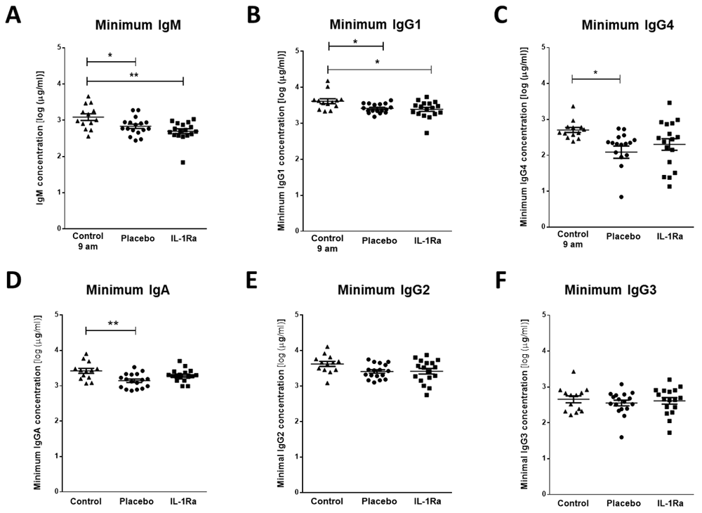
(A) Minimum IgM concentration measured in the first seven days after stroke was lower in both placebo and IL-1Ra treated patients in comparison to healthy controls. Data show mean ±SD, * P<0.05, ** P<0.01, one-way ANOVA with Bonferonni correction. (B) Minimum concentration of IgG1 and measured in the first seven days after stroke was reduced in both placebo and IL-1Ra treated patients in comparison to healthy controls. Minimum IgG4 (C) and IgA (D) concentrations were reduced in placebo-treated stroke patients in comparison to healthy controls. There was no significant difference between placebo-treated and IL-1Ra-treated stroke patients. No significant difference in IgG2 (E) and IgG3 (F) concentration was detected between placebo-treated and IL-1Ra-treated stroke patients in comparison to healthy controls. Data show mean ±SD, * P<0.05; ** P<0.01; one-way ANOVA with Bonferonni correction.
Minimum IgG1 concentration was significantly reduced in both placebo-treated and IL-1Ra-treated stroke patients in comparison to non-stroke controls (Figure 1B)12. Minimum IgG4 (Figure 1C) and IgA (Figure 1D) concentrations were significantly reduced in placebo-treated stroke patients only. However, there was no significant difference in these immunoglobulins between placebo-treated and IL-1Ra-treated patients. Minimum concentrations of IgG2 (Figure 1E) and IgG3 (Figure 1F) were not significantly altered in IL-1Ra or placebo treated patients in comparison to non-stroke controls. Maximal circulating concentrations of all immunoglobulin isotypes measured in the first seven days after stroke were also compared to non-stroke controls and no significant differences were measured in any immunoglobulin isotypes (Supplementary Figure 1B-F, Extended data)8.
As complement components are directly associated with the antibacterial functions of immunoglobulins, we investigated stroke-induced changes in circulating complement components and if any changes observed were further influenced by treatment with IL-1Ra. Stroke induced a significant reduction in the minimum concentrations of C3b/ iC3b (Figure 2A), C3 (Figure 2B), C4 (Figure 2C), Factor H (Figure 2D) and Properdin (Figure 2E) measured in the first seven days after stroke in both placebo and IL-1Ra treated patients in comparison to non-stroke controls12. Maximum circulating concentrations of these complement components measured in the first seven days after stroke were also compared to non-stroke controls and no significant differences were seen (Supplementary Figure 2A–E, Extended data)8.
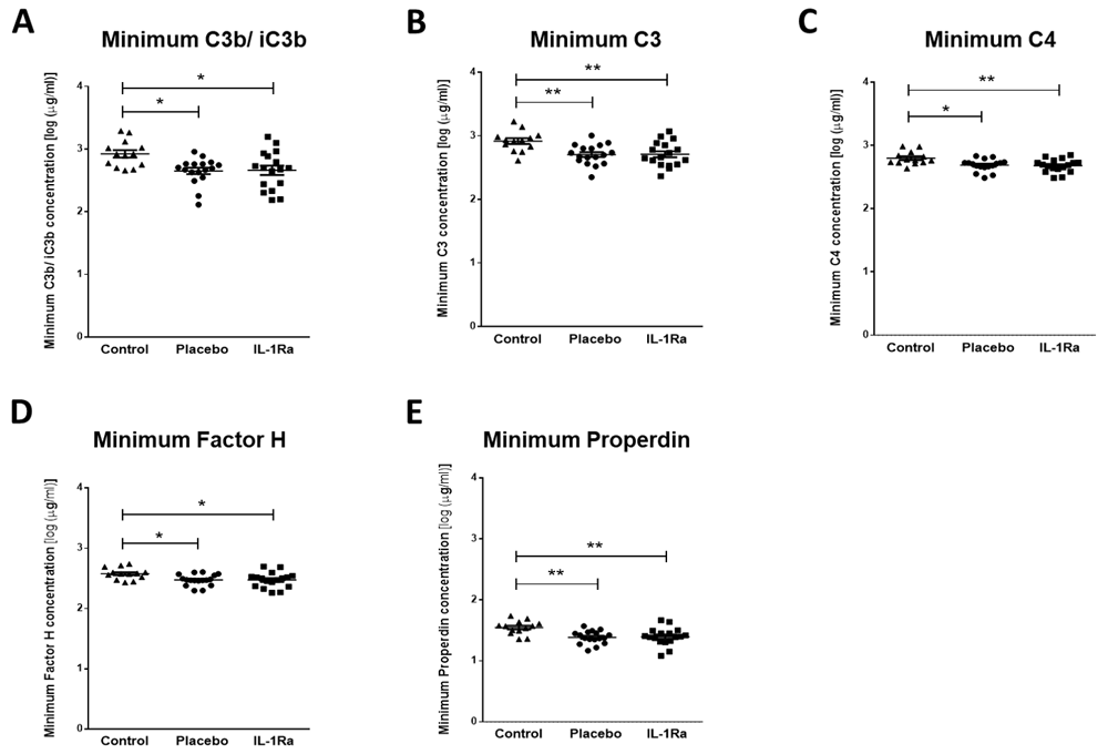
Minimum concentrations of (A) C3b/ iC3b, (B) C3, (C) C4, (D) Factor H and (E) Properdin were measured in the first seven days after stroke were reduced in both placebo and IL-1Ra treated patients in comparison to healthy controls. Data show mean ±SD, * P<0.05; ** P<0.01; one-way ANOVA with Bonferonni correction.
In contrast, stroke induced a significant increase in maximal circulating concentrations of C1q (Figure 3A), C5 (Figure 3D) and C9 (Figure 3E) in both IL-1Ra and placebo treated patients measured in the first seven days after stroke in comparison to non-stroke controls. Maximum concentrations of C2 (Figure 3B) and C4b (Figure 3C) were increased in placebo-treated patients only12. However, no significant difference was apparent between placebo treated and IL-1Ra treated patients for these factors, suggesting IL-1Ra treatment exerts no effects additional to stroke. Minimum concentrations of these complement components measured in the first week after stroke were also compared to non-stroke controls and no significant differences were seen (Supplementary Figure 3A–E, Extended data)8.
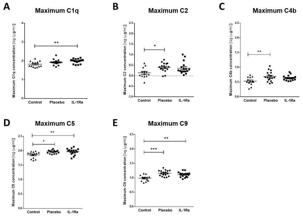
Maximum concentrations of (A) C1q, (B) C2, (C) C4b, (D) C5 and (E) C9 were measured in the first seven days after stroke were increased in both placebo and IL-1Ra treated patients in comparison to healthy controls. Data show mean ±SD, * P<0.05; ** P<0.01; one-way ANOVA with Bonferonni correction.
Minimal and maximal levels of Factor B, MBL and C5a measured in the first week after stroke were also compared to non-stroke controls. Concentrations of Factor B (Figure 4A, B) and MBL (Figure 4C, D) were not significantly altered by stroke or by treatment with IL-1-Ra12.
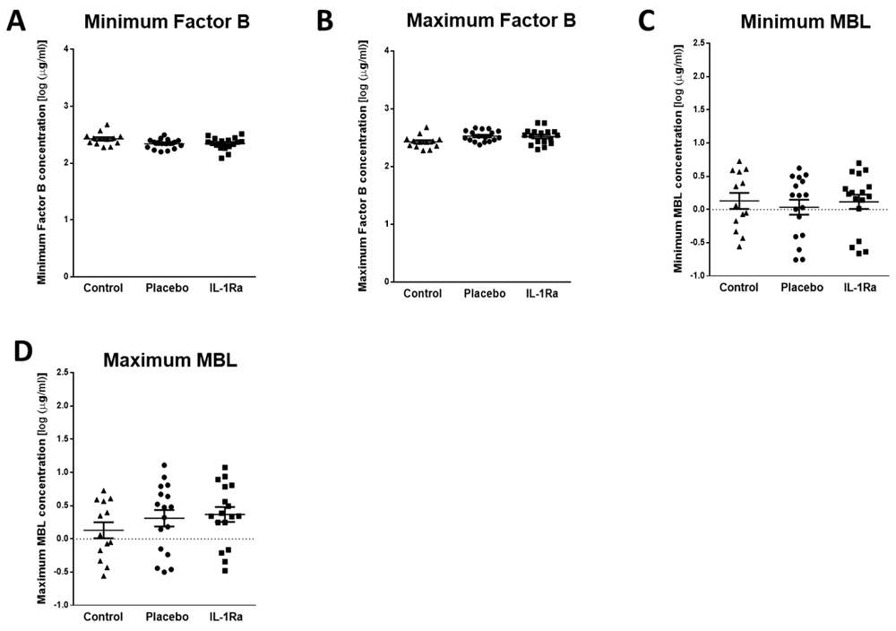
Minimal and maximum concentrations of (A, B) Factor B and (C, D) mannose-binding lectin (MBL) were measured in the first seven days after stroke were unchanged in both placebo and IL-1Ra treated patients in comparison to healthy controls. Data show mean ±SD, one-way ANOVA with Bonferonni correction.
Splenic noradrenaline levels are increased after experimental stroke and may be toxic to IgM producing B cells6. Maximum noradrenaline concentration measured in the first seven days after stroke was increased in both placebo and IL-1Ra treated patients in comparison to non-stroke controls (Figure 5A)12. Treatment with IL-1Ra had no additional effect on noradrenaline concentration when compared to placebo. Minimum noradrenaline concentration in the first seven days after stroke was also measured and was not significantly different to non-stroke controls (Supplementary Figure 4, Extended data) or affected by IL-1Ra treatment8.
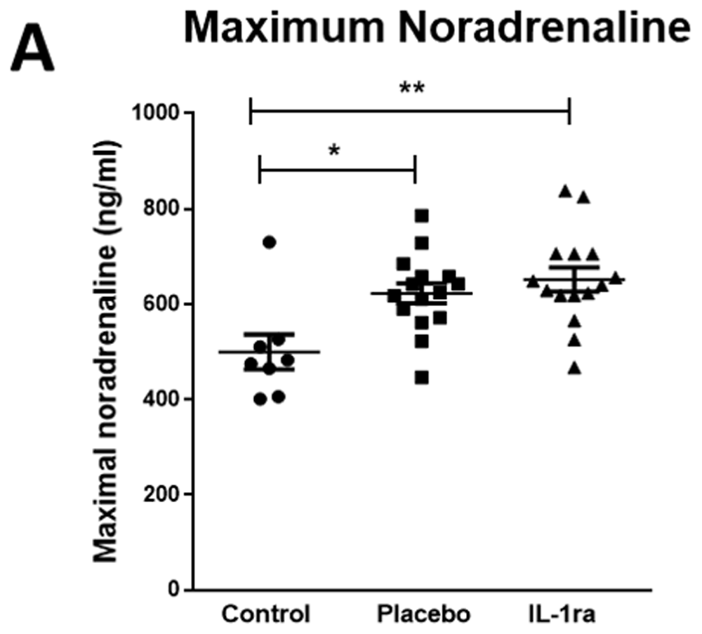
Maximal noradrenaline concentration measured in the first seven days after stroke was significantly higher in both placebo and IL-1Ra treated patients in comparison to controls. Data show mean ± SD *P<0.05; **P<0.01; one-way ANOVA.
In this study, we assessed if markers associated with antibody-mediated antibacterial defences were compromised in patients treated with IL-1Ra after stroke. Plasma IgM, IgG1, IgG4 and IgA immunoglobulin concentrations were reduced after stroke and this was not further altered by treatment with IL-1Ra. Assessment of complement components indicated induction of the classical pathway of complement activation after stroke but inhibition of the alternative pathway without modulation by IL-1Ra. Plasma noradrenaline was increased after stroke and also not influenced by treatment with IL-1Ra. These data suggest that treatment with IL-1Ra is unlikely to aggravate antibody-associated immune function deficits induced by stroke.
The IL-1 family of cytokines play a critical role in host defence to pathogens by signalling to a variety of host cells to induce downstream effects including, but not limited to, pro-inflammatory cytokine and chemokine production, immune cell recruitment and upregulation of vascular adhesion molecules13,14. However, in conditions of sterile inflammation and tissue injury, such as stroke, these effects can aggravate primary tissue damage and impair injury repair mechanisms. Blocking IL-1 signalling has shown improved outcome in both experimental animal and patient stroke studies1,4,15. However, the immunosuppressive effects of blocking IL-1 signalling after stroke may additionally inhibit systemic responses to infection, further increasing the risk of infection in patients who are already immune compromised16,17. Indeed, meta-analysis studies have shown an increased risk of serious infection in rheumatoid arthritis patients treated for prolonged periods with the IL-1 blocking drug anakinra16. However, as of yet this has not been observed in stroke patients, potentially reflecting differences in the duration of treatment. No statistically significant differences in infection incidence were seen between IL-1Ra and placebo treated patients in this study, with 5/17 IL-1Ra treated patients experiencing infection between admission and day seven and 4/17 infections in placebo treated patients. Consistent with this pattern, we have shown here that relatively short duration of treatment with IL-1Ra after acute stroke did not further affect stroke-induced changes to circulating immunoglobulin, complement or noradrenaline concentrations and is therefore unlikely to further compromise immune defence against infection through reducing the availability of these antibacterial mediators.
IL-1 cytokine family members are reported to have variable effects on B cell antibody production. IL-1β was reported to be important for the rapid production of anti-bacterial IgM by innate-like B cells important for early containment of infection prior to the induction of adaptive immune responses7,18. This would suggest treatment with IL-1Ra after stroke could further compromise the early production of IgM in innate-like B cells which are already known to be reduced in number after stroke6. However, this effect of IL-1Ra on IgM concentrations was not seen. We know that experimental stroke results in a significant loss of many populations of B cells and associated IgM6; therefore, it is possible that the effects of the stroke itself on B cells overwhelm any additional effects of cytokines that could moderately enhance or inhibit immunoglobulin production. Furthermore, we do not know if remaining B cells are functionally impaired and therefore able to respond to IL-1β signalling as they would under normal homeostatic conditions. We have previously reported that stroke is associated with reduced circulating IgM concentrations in comparison to non-stroke controls6, an effect reproduced here. Further studies will be required to determine if IgM, or any of the mediators assessed in this study, would be useful as biomarkers to determine which patients are likely to develop infection after stroke.
We have shown for the first time that circulating IgG1, IgG4 and IgA concentrations were reduced in the first seven days after stroke in comparison to non-stroke controls. This is in agreement with previous data showing that pan-IgG concentrations were reduced in patients after stroke, although subclasses of IgG were not assessed in that study and no reduction in IgA was found at the seven day time point assessed19. IgA is the most predominant immunoglobulin isotype at mucosal surfaces including the respiratory tract and is crucial for antibacterial protection at these sites20. Given the early reduction of IgA in placebo-treated stroke patients, determining the effect of stroke on IgA-producing B cells at infection susceptible sites, such as the lung mucosa, could further elucidate if this has an important role in post-stroke infection susceptibility.
In contrast to the short half-life of IgA and IgM20–22, the half-lives of IgG1 and IgG4 are reported to be 21 days and therefore, an early reduction in IgG concentration is not compatible with a lack of de novo production after stroke due to loss of B cells23. Previous studies have suggested that reduced total-IgG after stroke may be associated with increased loss or catabolism of IgG, which could account for reductions in concentration occurring more rapidly than its natural half-life24. An alternative explanation could be that reduced IgG concentration is indicative of vascular risk factors and inflammatory changes preceding stroke that are associated with stroke risk. However, control patients in this study were matched for risk factors including their degree of atherosclerosis and would be expected to show similar changes to stroke patients if these were associated with risk factors. Understanding that the kinetics of an individual immunoglobulin subset changes both preceding and as a result of stroke, and their associations with post-stroke infections, could be invaluable in providing new therapeutic targets to reduce incidence of infection and improve outcome in patients.
The complement system has a crucial role in enhancing humoral immune defence and protecting from bacterial infection via interactions with both the innate and adaptive immune systems25. As activation of complement is closely associated with efficient immunoglobulin-mediated clearance of pathogens, we determined whether these pathways were compromised by stroke. We have assessed, for the first time, individual concentrations of multiple complement components covering all pathways of complement activation after stroke. These exploratory data suggest there are no overall deficits in complement activation after stroke. Complement activation pathways converge at multiple points; however, their initial activation mechanisms are distinct. The classical complement pathway is activated when IgM or IgG immune complexes bind to C1 (composed of C1q, C1r and C1s)25,26. Maximum circulating concentration of complement components associated with the classical and lectin pathways of activation, C1q, C2 and C4b, and end stage mediators common to all pathways, C5 and C9, were increased in the first seven days after stroke in comparison to non-stroke controls. As concentrations of MBL itself was not significantly altered by stroke, this suggests the classical complement pathway is specifically activated after stroke.
In contrast, the alternative pathway of complement activation is initiated by microbial cell surfaces and polysachharide antigen and results in a cascade that generates C325,26. Complement components that were significantly downregulated after stroke, C3b/ iC3b, C3, Factor H (fH) and Properdin, are more associated with the alternative pathway of complement activation, suggesting that the alternative pathway is suppressed. These data are in agreement with previous studies investigating systemic C-reactive protein, C3c and C4 complement concentrations in the serum of patients 24 h after ischemic stroke, which concluded that the classical pathway of complement activation was activated in the first 24 h after ischemic stroke, whereas C3c, associated with the alternative pathway, was reduced27,28. The roles of individual pathways of complement activation in infection susceptibility after stroke remains to be determined, but these data suggest overall deficits in complement concentration are unlikely to contribute to reduced antibody-mediated clearance of pathogens that may occur after stroke, further supporting reduced circulating immunoglobulins as an important influence on infection susceptibility.
In this study, circulating noradrenaline concentrations measured in the first week after stroke were increased in comparison to non-stroke controls but were not influenced by treatment with IL-1Ra. This is in agreement with previous studies showing activation of the sympathetic nervous system in both stroke and subarachnoid haemorrhage patients that resulted in increased plasma noradrenaline concentrations that persisted up to 10 days29–31. Our previous studies have shown that after experimental stroke, activation of the sympathetic nervous system and release of noradrenaline within the spleen is toxic to resident B cells and preventing noradrenaline signalling using the β-blocker propranolol prevented B cell and IgM loss and resulted in reduced infectious burden6. The cytokine IL-1β is also increased in the spleen after stroke and is reported to activate peripheral nerves, including the splenic nerve, and increase production of splenic noradrenaline32,33. However, blockade of IL-1β signalling did not alter circulating concentrations of noradrenaline after stroke.
In summary, we have shown that treatment with IL-1Ra after stroke does not affect circulating concentrations of immunoglobulins, complement components or noradrenaline and is therefore unlikely to further increase patient susceptibility to infection via pathways in which these mediators are key participants. This is in agreement with data from IL-1Ra Phase II trials, in which treatment of stroke patients with IL-1Ra did not aggravate incidence of infection4,12. These data suggest that blocking IL-1 in a stroke context may not be concerning from the perspective of increasing infection risk in patients. Additionally, the reductions in circulating immunoglobulin concentrations detected after stroke in this study further support that antibody mediated immune defence may be an important therapeutic target to reduce the burden of infection after stroke.
Edinburgh Data Share: Interleukin-1 receptor antagonist treatment in acute ischaemic stroke does not alter systemic markers of anti-microbial defence. https://doi.org/10.7488/ds/257012
This project contains the following underlying data:
- Full noradrenaline data by patient and timepoint.xlsx (noradrenaline concentrations in ng/ml)
- Full complement data by patient and timepoint.xlsx (complement concentrations in μg/ml)
- Full immunoglobulin data by patient and timepoint.xlsx (immunoglobulin concentrations in μg/ml)
- Figure 1A dataset.pzf – Figure 5A dataset.pzf (raw data underlying Figure 1–Figure 5 in GraphPad Prism format)
- Figure 1 full data excel format.xlsx (minimum concentrations of immunoglobulins in μg/ml)
- Figure 2 full data excel format.xlsx (minimum concentrations of complement components C3b/iC3b, C3, C4, Factor H and Properdin in μg/ml)
- Figure 3 full data excel format.xlsx (maximum concentrations of complement components C1q, C2, C4b, C5 and C9 in μg/ml)
- Figure 4 full data excel format.xlsx (concentrations of complement components Factor B and MBL in μg/ml)
- Figure 5 full data excel format.xlsx (maximum concentrations of noradrenaline in ng/ml)
Edinburgh Data Share: Supplementary Figures: Interleukin-1 receptor antagonist treatment in acute ischaemic stroke does not alter systemic markers of anti-microbial defence. https://doi.org/10.7488/ds/25458
This project contains the following extended data:
- Supplementary Figure 1.tif (figure showing maximum concentrations of immunoglobulin)
- Supplementary Figure 2.tif (figure showing maximum concentrations of complement components C3b/iC3b, C3, C4, Factor H and Properdin)
- Supplementary Figure 3.tif (figure showing minimum concentrations of complement components C1q, C5, C9, C2 and C4b)
- Supplementary Figure 4.tif (figure showing minimum concentrations of noradrenaline)
- Supplementary Table 1.tif (table of baseline characteristics of stroke patients and stratification of groups)
- Data Supplementary Fig 1.xlsx - Data Supplementary Fig 4.xlsx (data underlying Supplementary Figures 1-4)
- Data Supplementary Fig 5.xlsx (Supplementary Table 1 in spreadsheet format)
Data are available under the terms of the Creative Commons Attribution 4.0 International license (CC-BY 4.0).
| Views | Downloads | |
|---|---|---|
| F1000Research | - | - |
|
PubMed Central
Data from PMC are received and updated monthly.
|
- | - |
Provide sufficient details of any financial or non-financial competing interests to enable users to assess whether your comments might lead a reasonable person to question your impartiality. Consider the following examples, but note that this is not an exhaustive list:
Sign up for content alerts and receive a weekly or monthly email with all newly published articles
Already registered? Sign in
The email address should be the one you originally registered with F1000.
You registered with F1000 via Google, so we cannot reset your password.
To sign in, please click here.
If you still need help with your Google account password, please click here.
You registered with F1000 via Facebook, so we cannot reset your password.
To sign in, please click here.
If you still need help with your Facebook account password, please click here.
If your email address is registered with us, we will email you instructions to reset your password.
If you think you should have received this email but it has not arrived, please check your spam filters and/or contact for further assistance.
Comments on this article Comments (0)