Keywords
Left ventricular hypertrophy, geometric patterns of left ventricular remodeling, concentric remodeling, concentric hypertrophy, eccentric hypertrophy
Left ventricular hypertrophy, geometric patterns of left ventricular remodeling, concentric remodeling, concentric hypertrophy, eccentric hypertrophy
The left ventricle (LV) remodels as a response to cardiovascular disease and myocardial injury. Characterized by an increase in LV myocardial mass, left ventricular hypertrophy (LVH) is an established predictor of poorer cardiovascular outcomes1.
Four classical geometric patterns of LV remodeling have been defined based on LV mass and relative wall thickness: normal, concentric remodeling, concentric and eccentric hypertrophy2. This convenient approach of characterizing LV remodeling has been studied across various patient populations, including patients with coronary artery disease, aortic stenosis, hypertensive heart disease and community-based general populations3–6. Whilst some studies demonstrated prognostic associations with these patterns of LV remodeling, others have not. Knowledge of remodeling patterns (concentric and eccentric hypertrophy) provided particularly limited incremental prognostic information beyond LVH1,7–10.
In this study, we aim to conduct a comprehensive systematic review and network meta-analysis to examine the characteristics and prognosis associated with the four conventional geometric patterns of LV remodeling.
A comprehensive literature search was performed on MEDLINE/PubMed (1946 onwards), Embase (1974 onwards) and the Cochrane Library (1996 onwards) until January 2019. Full text publications evaluating the four conventional LV geometry patterns (normal geometry, concentric remodeling, concentric and eccentric hypertrophy) and prognosis were included. The basic search protocol and specific terms used in the search strategy are available as Extended data11. We conducted the literature search using Medical Subject Headings or Emtree, and free text terms. There were no restrictions on language.
Two investigators (Q.Z. and G.L.) independently searched for eligible studies based on the pre-defined eligibility criteria. Full-text studies that compared the prognosis of the four conventional LV geometry patterns (i.e. normal geometry, concentric remodeling, concentric and eccentric hypertrophy) were included. We excluded publications in non-adult populations, case reports, commentaries, abstracts, letters-to-editors and review articles. The bibliography in the identified publications and review articles were also reviewed.
The following data were extracted in duplicates by the two investigators (Q.Z. and G.L.) from the included studies: (1) study characteristics (publication year and patient population); (2) baseline characteristics (mean age, sex distribution, and proportion of patients with hypertension, coronary artery disease, diabetes and other significant risk factors); (3) the four LV remodeling patterns; and (4) adverse prognosis defined as all-cause mortality. Eligible studies that did not report all-cause mortality as an end-point were still included to examine clinical characteristics associated with geometric patterns of LV remodeling. Any disagreements were resolved by discussion with a third investigator (C.W.L.C.). In publications with survival curves, the cumulative survival rates were estimated by digitizing the plots (WebPlotDigitizer version 3.9, Austin, Texas, USA).
Two investigators (C.W.L.C. and Z.Q.) independently appraised the quality of each study using the Quality In Prognosis Studies tool12. Six domains (study participation, study attrition, prognostic factor measurement, outcome measurement, study confounding; and statistical analysis and reporting) were evaluated to assess the risk of bias in the prognostic studies. In each of the six domains, the risk of bias was classified as “low”, “moderate” or “high”.
A network geometry of the four LV remodeling patterns was constructed. Each node represented a remodeling pattern and its size was weighted by the number of individuals in that group. The connecting line between two nodes denoted direct comparison and its thickness reflected the number of studies included.
The random-effects meta-regression models were used to measure the impact of baseline characteristics on the effect size of the outcome. The risk ratio (RR) of each LV remodeling group was estimated and reported in the study. To rank the prognosis of all the geometric patterns, we used surface under the cumulative ranking (SUCRA) values13. Rank probabilities of all the groups were first estimated, then followed by a step function to summarize the cumulative ranking for estimating the SUCRA values of each group, ranging from 0 to 100%. Larger SUCRA values indicated better prognosis.
Both node-splitting and inconsistency modeling were used to test the consistency assumption. The former method involved fitting a series of node-splitting models, one model for each group pair in which there was direct and indirect comparisons. In the latter method, an inconsistency model was fitted and the global Wald test would determine if significant inconsistency was present14. Statistical analyses were performed using Stata/MP Version 13 (StataCorp., College Station, Texas, USA), with the network and network graphs package.
From an initial 257 publications, 22 echocardiographic studies of diverse cardiovascular diseases satisfied inclusion/exclusion criteria and were included in this study (Figure 1)6–10,15–31. The thresholds used to define LVH and increased concentricity were heterogeneous across the studies (Table 1).
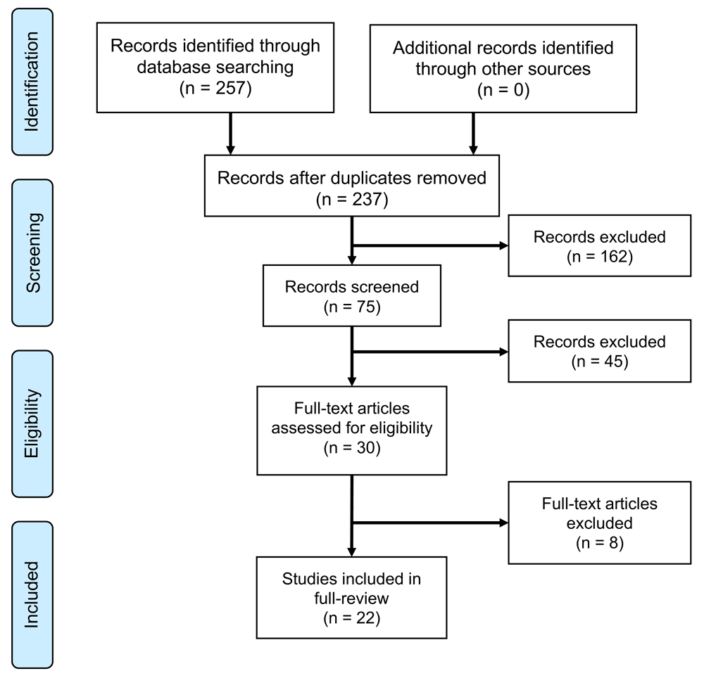
The systematic review and network meta-analysis was conducted according to the guidelines recommended by PRISMA.
| Reference | Population | Patients (n) | Females (n) | Age (years) | Definition of left ventricular hypertrophy | Definition of increased concentricity | Follow-up duration (years) | |
|---|---|---|---|---|---|---|---|---|
| Males (g/m2) | Females (g/m2) | |||||||
| Beger 2011 | Coronary artery disease | 973 | 251 | 66.8 | 102 | 88 | Posterior Wall thickness; 11mm | 4.9 |
| Verma 2008 | Coronary artery disease | 603 | 192 | 65.6 | 115 | 95 | RWT; 0.42 | 2.1 |
| Ghali 1998* | Coronary artery disease | 446 | 201 | 56.9 | 131 | 100 | RWT; 0.45 | 9.0 |
| Shigematsu 1998 | Hypertension | 77 | 25 | 57.0 | 125 | 125 | RWT; 0.44 | Not stated |
| Gerdts 2008 | Hypertension | 937 | 388 | 65.5 | 116 | 104 | RWT; 0.43 | 4.8 |
| Fabiani 2017 | Hypertension | 749 | 325 | 62.0 | 115 | 95 | RWT; 0.42 | 3.7 |
| Verdecchia 1996 | Hypertension | 274 | 37 | 53.0 | 125 | 125 | RWT; 0.45 | 3.2 |
| Kohara 1999 | Hypertension | 150 | 78 | 58.3 | 118 | 108 | RWT; 0.41 | Not stated |
| Krumholz 1995 | General Population | 3209 | 1813 | 57.0 | 143 | 102 | RWT; 0.45 | 7.7 |
| Lieb 2014 | General Population | 4492 | 216 | 53.3 | 207g | 170g | RWT; Males: 0.419; Females: 0.435 | 4.0 |
| Gardin 2001 | General Population (>65 years old) | 2506 | 1622 | - | >95th percentile | >95th percentile | RWT; 0.48 | 6.0 |
| Milani 2006 | Patients EF>50% | 35, 602 | 18, 869 | 60.0 | 116 | 104 | RWT; 0.43 | 3.2 |
| Lavie 2006 | Patients EF>50% (>70 years old) | 9771 | 5569 | 77.5 | 116 | 104 | RWT; 0.43 | 3.1 |
| Lavie 2009 | Patients EF>50% (>70 years old) | 8088 | 4564 | 77.0 | 116 | 104 | RWT; 0.43 | 3.1 |
| Ghali 1998* | Patients EF>45% | 542 | 347 | 54.0 | 131 | 100 | RWT; 0.45 | 9.0 |
| Katz 2013 | HFpEF | 402 | 251 | 62.8 | 48g/m2.7 | 44g/m2.7 | RWT; 0.42 | 1.0 |
| Apostolaskis 2014 | Atrial fibrillation | 2433 | 1058 | 69.0 | 115 | 95 | RWT; 0.42 | 3.5 |
| Shah 2014 | Atrial Fibrillation | 1088 | 496 | 69.1 | 115 | 95 | RWT; 0.42 | 6 |
| Debry 2017 | Aortic stenosis | 331 | 150 | 73.0 | 115 | 95 | RWT; 0.42 | 3.1 |
| Capoulade 2017 | Aortic stenosis | 747 | 426 | 69.0 | 49g/m2.7 | 47g/m2.7 | RWT; 0.42 | 6.4 |
| Rymuza 2017 | Aortic stenosis (TAVI) | 208 | 107 | 79.4 | 115 | 95 | RWT; 0.42 | 1.5 |
| Paoletti 2016 | Chronic kidney disease | 445 | 222 | 64.0 | 131 | 100 | RWT; 0.45 | 5.9 |
| Park 2018 | Ischemic strokes | 2069 | 787 | 65.5 | 115 | 95 | RWT; 0.42 | 3.1 |
Of the 76,142 individuals pooled from the 22 studies (50.1% males; 64.4±7.9 years), 49.7% had normal geometry; and 31.1%, 10.5% and 8.7% had concentric remodeling, concentric and eccentric hypertrophy, respectively. The proportion of females with concentric and eccentric hypertrophy was high (40–45%). Compared to the other geometric patterns, concentric hypertrophy was associated with the highest prevalence of cardio-metabolic risk factors and cardiovascular diseases. Eccentric hypertrophy was associated with a high prevalence of atrial fibrillation and low LV ejection fraction (Table 2).
Most of the studies demonstrated low risk of bias in the six domains examined (Table 3). The network geometry of LV remodeling patterns was constructed in Figure 2. Concentric remodeling was associated with higher all-cause mortality compared to normal geometry (RR 1.56 [95% confidence interval (CI): 1.31 to 1.85]), and a lower mortality risk compared to concentric hypertrophy (RR 0.79 [95% CI 0.67 to 0.93]). The mortality risk of concentric remodeling was similar compared to eccentric hypertrophy (RR 0.91 [95% CI 0.76 to 1.09]) (Table 4).
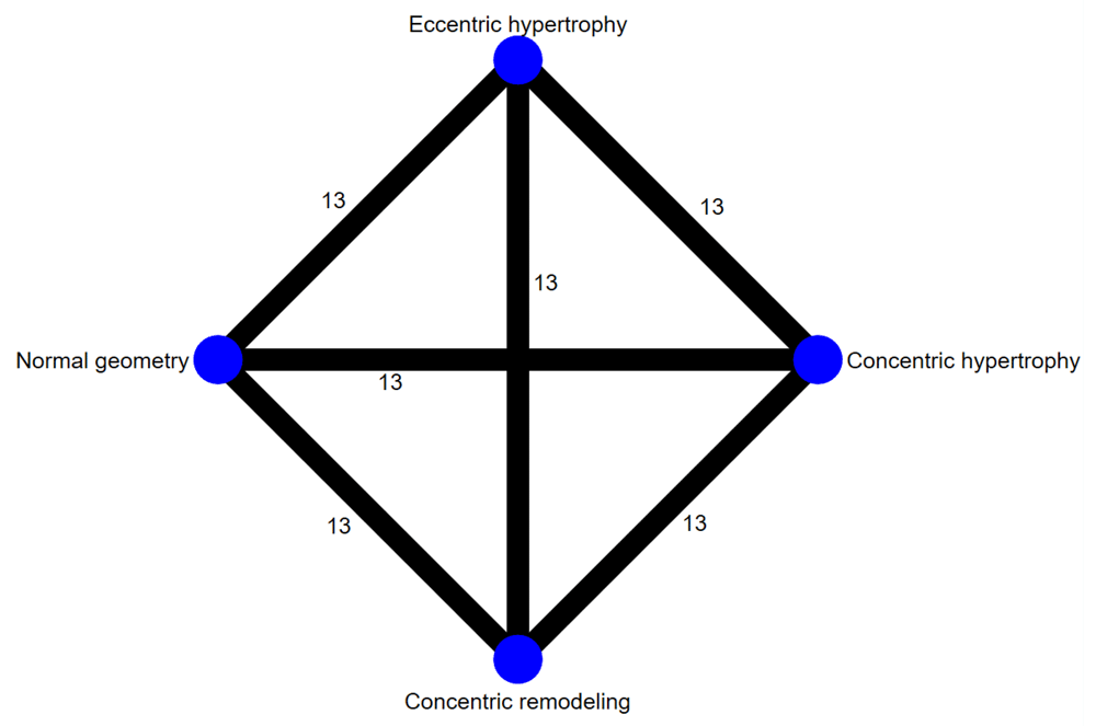
The numbers on the connecting lines denote the studies included for direct comparison.
Results presented in risk ratio and corresponding 95% confidence interval.
| Reference group | ||||
|---|---|---|---|---|
| Normal Geometry | Concentric Remodeling | Concentric Hypertrophy | Eccentric hypertrophy | |
| Normal Geometry | 1.00 | 0.64 [0.54, 0.76]* | 0.51 [0.43, 0.60]* | 0.59 [0.49, 0.70]* |
| Concentric Remodeling | 1.56 [1.31, 1.85]* | 1.00 | 0.79 [0.67, 0.93]* | 0.91 [0.76, 1.09] |
| Concentric Hypertrophy | 1.97 [1.63, 2.39]* | 1.27 [1.08, 1.49]* | 1.00 | 1.15 [0.97, 1.36] |
| Eccentric Hypertrophy | 1.71 [1.43, 2.04]* | 1.10 [0.92, 1.31] | 0.87 [0.73, 1.03] | 1.00 |
Compared to normal geometry, concentric hypertrophy was associated with highest risk of all-cause mortality (RR 1.97 [95% CI 1.63 to 2.39]; Table 4). The confidence limits overlapped with eccentric hypertrophy (RR 1.71 [95% CI 1.43 to 2.04]). Moreover, the mortality risk of concentric hypertrophy was not significantly increased compared to eccentric LVH (RR 1.15 [95% CI 0.97 to 1.36]). Based on the SUCRA values, the geometric patterns ranked from best to worst prognosis were: normal geometry, concentric remodeling, eccentric hypertrophy and concentric hypertrophy (Figure 3).
A, normal geometry; B, concentric remodeling; C, concentric hypertrophy; D, eccentric hypertrophy.
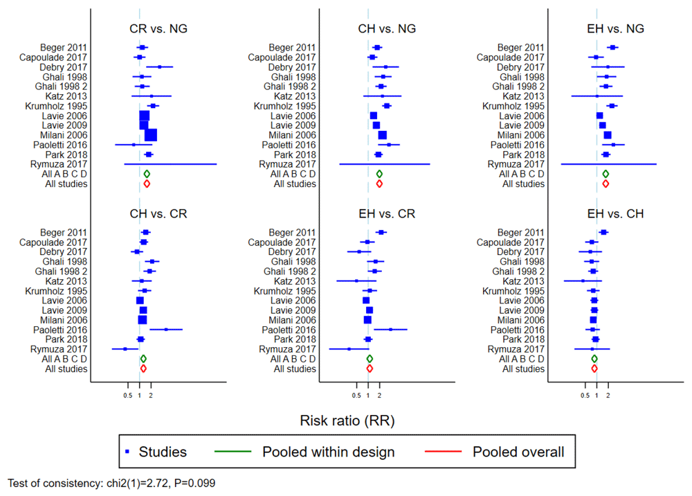
Results from both node-splitting method and inconsistency model showed no evidence on the violation of consistency assumption between direct and indirect comparisons. Specifically, the pooled estimates between models of consistency (red diamonds) and inconsistency (green diamonds) were identical because all the studies included the four remodeling patterns (Figure 4).
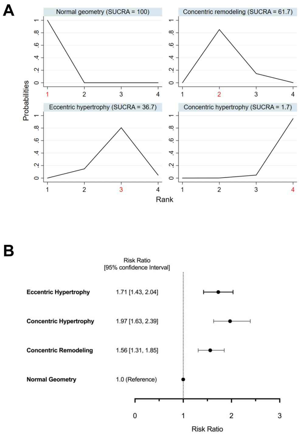
(a) Rank probabilities of effectiveness and SUCRA scores; and (b) prognosis of the four geometric patterns of left ventricular hypertrophy.
In this systematic review and network meta-analysis of 22 echocardiographic publications (n=76,133 individuals), we report the characteristics and prognosis associated with the different patterns of LV remodeling. The study populations were heterogeneous and, importantly, the definitions used to classify the geometric patterns were not uniform. Concentric hypertrophy is associated with the highest prevalence of cardiometabolic risk factors and diseases. Eccentric hypertrophy is associated with a high prevalence of atrial fibrillation and the lowest LV ejection fraction. Although concentric hypertrophy is associated with the highest risk of all-cause mortality, the risks overlapped with eccentric hypertrophy. Eccentric hypertrophy has a similar mortality risk compared to concentric remodeling.
The pathophysiology of LVH has been well described and studied for the past 50 years32,33. Cardiac hypertrophy is initially an adaptive response to the wall stress according to the Law of LaPlace. Ultimately, cardiac decompensation occurs as a consequence of myocyte death and myocardial fibrosis34,35. Whilst geometric patterns of LV remodeling are clinically meaningful to describe the hypertrophic response due to mechanical stress from either pressure (concentric hypertrophy) or volume overload (eccentric hypertrophy), it may not adequately identify the transition point where adaptive hypertrophy decompensates (Figure 5). This transition point before cardiac decompensation occurs is an important potential risk marker to target more intensive management and closer surveillance. In this study, we have demonstrated that both concentric and eccentric hypertrophy were associated with similar risks of increased all-cause mortality. These observations may suggest that the risk of adverse prognosis is increased once LVH develops, regardless of geometric patterns. It may also suggest that some patients with concentric or eccentric LVH may be in the compensated phase and begets the question of whether there are other strategies to identify high-risk LVH phenotypes.
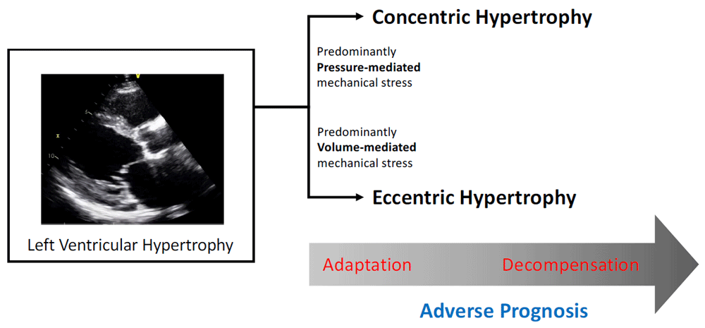
Geometric patterns of left ventricular remodeling are useful to identify the mechanisms of mechanical stress; but may not adequately identify the transition between myocardial adaptation and decompensation.
To address the complex interaction between LV dilatation and myocardial thickening in the pathophysiology of LVH, several studies have recently examined an expanded four-group LVH classification: dilated/non-dilated concentric hypertrophy and dilated/non-dilated eccentric hypertrophy36–40. In this proposed four-group LVH classification, dilated concentric hypertrophy was associated with the worst prognosis and non-dilated eccentric hypertrophy had the most favourable profile36–39. However, more guidance is needed before this complex classification can be integrated into routine clinical practice. Recently, we have developed the remodeling index (RI), based on a biophysical model of Laplace’s Law. The RI integrates LV volume and myocardial thickening into a single measurement41. We further demonstrated that hypertensive LVH patients with abnormally low RI (suggestive of excessive myocardial thickening relative to LV dilatation) had increased myocardial fibrosis, elevated circulating markers of myocardial injury and wall stress; and in a small number of patients with dilated cardiomyopathy, an abnormally high RI (suggestive of excessive LV dilatation relative to myocardial thickening) was associated with adverse cardiovascular events41. The prognostic value and clinical utility of this index are currently being examined in a large cohort of hypertensive patients (ClinicalTrials.gov identifier: NCT02670031).
These emerging data support the notion that cardiac remodeling in hypertrophy is heterogeneous and complex; and the conventional geometric patterns of LV remodeling is not adequate to risk-stratify patients with LVH.
The study populations included in the meta-analysis were heterogeneous. It is possible that the conventional remodeling patterns has incremental prognostic value in certain cardiac conditions. Unfortunately, the limited number of studies precluded stratified analyses to examine the prognostic value of LV geometric patterns in the different cardiovascular conditions. The definitions used for classifying geometric patterns were not consistent across the different studies. This is concerning and reinforces the necessity to apply consensus definitions in future studies2.
Concentric and eccentric hypertrophy are associated with increased and similar all-cause mortality. Possible explanations for these observations include the heterogeneous populations, inconsistent definitions used in the classification and the inherent limitations of the conventional patterns of LV geometry to adequately risk stratify LVH. Well-validated novel approaches to improve risk stratification of LVH should be explored in future research.
All data underlying the results are available as part of the article and no additional source data are required.
Open Science Framework: Prognosis associated with geometric patterns of left ventricular remodeling: systematic review and network meta-analysis. https://doi.org/10.17605/OSF.IO/3CJMW11.
This project contains the following extended data:
Open Science Framework: PRISMA checklist for “Prognosis associated with geometric patterns of left ventricular remodeling: systematic review and network meta-analysis”. https://doi.org/10.17605/OSF.IO/3CJMW11.
Data are available under the terms of the Creative Commons Zero "No rights reserved" data waiver (CC0 1.0 Public domain dedication).
| Views | Downloads | |
|---|---|---|
| F1000Research | - | - |
|
PubMed Central
Data from PMC are received and updated monthly.
|
- | - |
Provide sufficient details of any financial or non-financial competing interests to enable users to assess whether your comments might lead a reasonable person to question your impartiality. Consider the following examples, but note that this is not an exhaustive list:
Sign up for content alerts and receive a weekly or monthly email with all newly published articles
Already registered? Sign in
The email address should be the one you originally registered with F1000.
You registered with F1000 via Google, so we cannot reset your password.
To sign in, please click here.
If you still need help with your Google account password, please click here.
You registered with F1000 via Facebook, so we cannot reset your password.
To sign in, please click here.
If you still need help with your Facebook account password, please click here.
If your email address is registered with us, we will email you instructions to reset your password.
If you think you should have received this email but it has not arrived, please check your spam filters and/or contact for further assistance.
Comments on this article Comments (0)