Keywords
epistaxis, juvenile angiofibroma, haemophilia a, male adolescents, nasal endoscopy, nasal surgical procedures, computed tomography angiography
epistaxis, juvenile angiofibroma, haemophilia a, male adolescents, nasal endoscopy, nasal surgical procedures, computed tomography angiography
Juvenile angiofibroma (JA) is a benign vascular tumour accounting for 0.5% of all head and neck neoplasms1. It occurs almost exclusively in adolescent males nine to 19-years-old, with a mean age at diagnosis of 15 years2. The clinical presentation involves unilateral epistaxis, nasal obstruction, and an intranasal mass. Epistaxis may be profuse and require nasal packing, vasopressors, antifibrinolytics and transfusions, even in individuals with normal bleeding parameters. However, with haemophilia, such epistaxis is more difficult to control and can be life-threatening. To our knowledge, only one case of JA in a haemophiliac has been reported in the English literature3. We report another case here.
An 18-year-old male Filipino adolescent was referred to the Department of Otorhinolaryngology of the Philippine General Hospital for recurrent epistaxis. Previously diagnosed with severe haemophilia A at age 16, he initially presented with recurrent right nasal congestion and an episode of predominantly right-sided epistaxis described as sudden and profuse, amounting to 1500 ml. At that time, he was admitted to a provincial hospital and received blood and cryoprecipitate transfusions. Following discharge, epistaxis of 100 ml recurred almost daily, requiring nasal packs, repeated hospitalizations of one to two weeks in duration, and transfusions. Cryoprecipitate was often used to control the bleeding since plasma-derived Factor VIII (pFVIII) was seldom available due to shortage of supply and cost. His past history also included hemarthroses and gum bleeding since early childhood, but his symptoms were initially ignored and later only attributed to haemophilia although nasal congestion gradually progressed to obstruction.
After two years of such management, nasal endoscopy performed for the first time by a visiting otorhinolaryngologist revealed a right intranasal mass. He was referred to our institution and admitted with an impression of JA (Radkowski IA) and severe haemophilia A. Following admission, he suffered from hypovolemic shock several times due to difficulty in acquiring blood, cryoprecipitate and Factor VIII. With previous Factor VIII Assay levels less than 1%, 1900 units of Factor VIII concentrate were empirically administered (calculated by weight) to raise levels to normal. His condition was compounded by development of Factor VIII antibodies because of previous, repeated cryoprecipitate transfusions in a suboptimal health-care setting. Although his baseline inhibitor titre had been negative, the preoperative inhibitor titre following multiple transfusions with various blood products was positive 3.5 Bethesda units (BU), necessitating pre-, intra- and post-operative transfusion with recombinant Factor VII (rFVIIa) in addition to higher doses of Factor VIII. Unfortunately, rFVIIa only became available post-operatively.
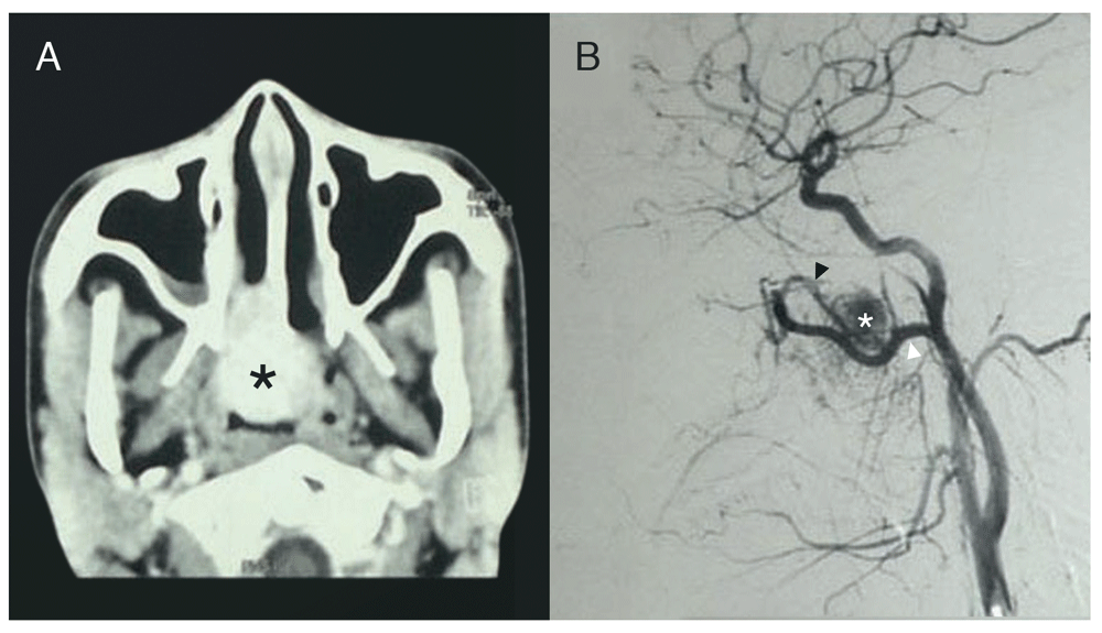
Contrast-enhanced computed tomography (CT) scans showed a hyperdense right intranasal mass corroborated by preoperative embolization angiography as an intranasal vascular tumour supplied by the right sphenopalatine artery and internal maxillary artery (IMA) (Figure 1A and 1B). The vast majority (90%) of the blood supply arose from distal sphenopalatine branches of the right IMA, while the remaining 10% came from both ascending pharyngeal arteries (Figure 1B).

A. CECT Scan showing enhancing nasopharyngeal mass (asterisk) and B. Angiography showing vascular tumour (asterisk) supplied by sphenopalatine (black arrowhead) and internal maxillary (white arrowhead) arteries. Adobe Photoshop CC 20.01 release was used to erase identifying patient details, remove pixelated areas from black background, and enhance contrast to sharpen image (applied to entire image).
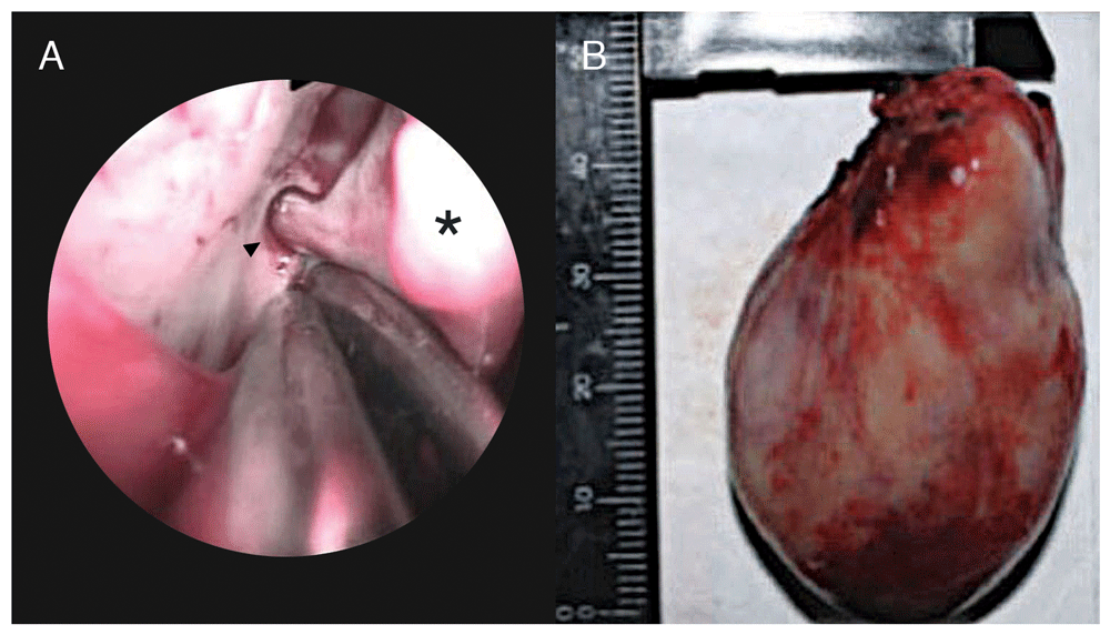
Within 24 hours post-embolization, the patient underwent endoscopic surgery under general endotracheal anaesthesia with Sevoflurane. Factor VIII was given before, during, and after surgery, with recombinant Factor VII added post-operatively. Intraoperatively, a fleshy, vascular 4.7 × 3.2 × 2.7 cm mass was seen arising from the right sphenopalatine foramen. The sphenopalatine artery was cauterized and ligated, and the mass was delivered trans-orally (Figure 2A and 2B). Intraoperative blood loss was 300cc and post-operative bleeding was negligible. In total, the patient received 39,500 units of commercially available pFVIII, 24 mg of rFVIIa, 22 units of packed red blood cells (PRBC), 301 units of cryoprecipitate, 1 unit of whole blood and 3 units of fresh frozen plasma (FFP). Final haematoxylin-eosin stained histopathology findings showed endothelium-lined capillaries with absent smooth muscle cells in a fibrous stroma, consistent with JA. The patient was discharged after two months in hospital and has followed up regularly, with no evidence of tumour on nasal endoscopy and no recurrence of nasal obstruction or epistaxis reported by the patient for seven years. He has completed a vocational course at college and is well. Figure 3 summarizes the timeline.

A. Intraoperative endoscopic view of the sphenopalatine artery (black arrowhead) supplying the mass (black asterisk) and B. Gross specimen measuring 4.7 × 3.2 × 2.7 cm. Adobe Photoshop CC 20.01 release was used to erase identifying patient details and sharpen the image (applied to entire image 2B).
To our knowledge, there is only one previous case of JA and concomitant haemophilia in the English literature, twice reported by Ozturk et al. in 19993 and by Celiker et al. in 20044. In their case, the preliminary diagnosis of JA was confirmed by biopsy at a different medical centre, where massive haemorrhage jeopardized the patient’s life. On referral to their institution, preoperative embolization, surgical excision, and adequate Factor VIII replacement saved the patient4.
Similarly, significant risk to our patient’s life was posed by delayed diagnosis from hasty attribution of epistaxis to haemophilia alone, and not the possibility of a vascular tumour such as JA. Per haemophilia management guidelines, the long history of “spontaneous bleeding into joints or muscles” in our patient corresponded to the baseline Factor VIII assay clotting factor level of “<1 IU/dL or <1% of normal” seen in severe haemophilia5. While recent-onset of bleeding from “mucous membranes in the mouth, gums, nose, and genitourinary tract” was serious, massive bleeding with “neck/throat” involvement was “life-threatening.” This degree of epistaxis should not have been expected in patients with haemophilia A alone, where major bleeding from these areas only occurs 5–10% of the time5. Moreover, the symptom of nasal obstruction was long-overlooked. Unfortunately, two full years passed before the underlying tumour was discovered.
Current guidelines5 advise otolaryngologist referral only for “persistent or recurrent” epistaxis, but the emphasis in this recommendation is for control of bleeding only and not to investigate a different underlying cause such as JA. Our experience demonstrates that vascular lesions causing epistaxis may remain undetected when presumptively attributed to pre-existing bleeding disorders and are likely to remain undetected unless sought.
In conclusion, although guidelines do not mention vascular lesions such as JA, a high index of suspicion should be maintained in adolescent males with epistaxis and nasal obstruction. Clinicians should carefully assess the cause of epistaxis in any patient with a bleeding disorder, and direct visualization of the source should be attempted (and verified by ancillary diagnostic techniques such as imaging when indicated) in all patients with epistaxis, regardless of the presence of a concomitant bleeding disorder.
All data underlying the results are available as part of the article and no additional data are required.
Written informed consent for publication of his clinical details and clinical images was obtained from the patient.
We acknowledge Dr. Arsenio Claro A. Cabungcal and Dr. Alzhes R. Buelva for their surgical contributions to patient care, Dr. Cheryl Lyn A. Diez for her expertise in haematology that made the surgery possible, and Mary Angeline R. Bagabaldo for her expert assistance with the deidentification, contrast-improvement, sharpening, labelling, and layout of the Figures.
| Views | Downloads | |
|---|---|---|
| F1000Research | - | - |
|
PubMed Central
Data from PMC are received and updated monthly.
|
- | - |
Is the background of the case’s history and progression described in sufficient detail?
Yes
Are enough details provided of any physical examination and diagnostic tests, treatment given and outcomes?
Yes
Is sufficient discussion included of the importance of the findings and their relevance to future understanding of disease processes, diagnosis or treatment?
Yes
Is the case presented with sufficient detail to be useful for other practitioners?
Yes
Competing Interests: No competing interests were disclosed.
Reviewer Expertise: paediatric otolaryngology and airway disorders
Is the background of the case’s history and progression described in sufficient detail?
Yes
Are enough details provided of any physical examination and diagnostic tests, treatment given and outcomes?
Yes
Is sufficient discussion included of the importance of the findings and their relevance to future understanding of disease processes, diagnosis or treatment?
Yes
Is the case presented with sufficient detail to be useful for other practitioners?
Yes
Competing Interests: No competing interests were disclosed.
Reviewer Expertise: I am an Academic Pediatric Otolaryngologist at BC Children's Hospital and an Associate Clinical Professor of Surgery at the University of British Columbia in Vancouver, Canada. I am a member of the Triological Society and the American Society of Pediatric Otolaryngology. My main areas of interest are bone grow factors in craniofacial models.
Is the background of the case’s history and progression described in sufficient detail?
Yes
Are enough details provided of any physical examination and diagnostic tests, treatment given and outcomes?
Yes
Is sufficient discussion included of the importance of the findings and their relevance to future understanding of disease processes, diagnosis or treatment?
Yes
Is the case presented with sufficient detail to be useful for other practitioners?
Yes
References
1. National ENT Trainee Research Network: The British Rhinological Society multidisciplinary consensus recommendations on the hospital management of epistaxis.J Laryngol Otol. 131 (12): 1142-1156 PubMed Abstract | Publisher Full TextCompeting Interests: No competing interests were disclosed.
Reviewer Expertise: Otolaryngology, rhinology, head and neck surgery
Alongside their report, reviewers assign a status to the article:
| Invited Reviewers | |||
|---|---|---|---|
| 1 | 2 | 3 | |
|
Version 2 (revision) 04 Oct 19 |
|||
|
Version 1 05 Sep 19 |
read | read | read |
Provide sufficient details of any financial or non-financial competing interests to enable users to assess whether your comments might lead a reasonable person to question your impartiality. Consider the following examples, but note that this is not an exhaustive list:
Sign up for content alerts and receive a weekly or monthly email with all newly published articles
Already registered? Sign in
The email address should be the one you originally registered with F1000.
You registered with F1000 via Google, so we cannot reset your password.
To sign in, please click here.
If you still need help with your Google account password, please click here.
You registered with F1000 via Facebook, so we cannot reset your password.
To sign in, please click here.
If you still need help with your Facebook account password, please click here.
If your email address is registered with us, we will email you instructions to reset your password.
If you think you should have received this email but it has not arrived, please check your spam filters and/or contact for further assistance.
Comments on this article Comments (0)