Keywords
dacryocystorhinostomy, marsupialization, lacrimal sac, endoscopy.
This article is included in the Eye Health gateway.
dacryocystorhinostomy, marsupialization, lacrimal sac, endoscopy.
Lacrimal disease manifests clinically as epiphora, recurrent conjunctivitis, or dacryocystitis, and it occurs most frequently in pediatric patients. Dacryocystorhinostomy (DCR) creates a low-pressure system by diverting tear flow through the lacrimal bone and an artificial opening. Toti first described external DCR in 1904, and Caldwell used an endonasal technique in 1893 that West modified in 19141–3.
Endoscopic DCR is the surgical procedure of choice to treat saccular or post-saccular nasolacrimal obstruction; this technique has been gaining popularity, with high success rates (sustained ostium patency, symptom relief, or both) comparable with external DCR rates, primarily because of the technological advances of endoscopes and surgical instruments. Multiple modifications have been suggested regarding endoscopic DCR procedures, with pros and cons. Previous endoscopic DCR procedures included making a small opening in the lacrimal sac and removing the nasal and lacrimal mucosa; this procedure likely contributes to surgical failure because the small neoformed ostium is obstructed by the granulation tissue or synechia formed during the postoperative period1.
Currently, two techniques are used to perform endoscopic DCR: laser-assisted and "cold steel"; both can be performed with or without powered drilling equipment. The former technique is less effective, perhaps because of the size of the ostium and the laser heat that results in fibrosis and stenosis4.
Generally, the size of the ostium created during surgery is crucial to the procedure’s outcome. Therefore, the anatomical characteristics of the lacrimal sac should be evaluated to achieve complete exposure when approaching the sac intranasally5,6.
Massegur et al. suggested a modification to the technique known as marsupialization of the lacrimal sac, which causes the flaps of the lacrimal mucosa to contact the nasal mucosa after the resection of the bone surrounding the sac, thereby incorporating the lacrimal sac in the lateral nasal wall1,3.
The current study describes the results of a DCR with lacrimal sac marsupialization compared with other endoscopic techniques.
A clinical chart review study was conducted in patients who presented with obstruction of the lacrimal route in their excretory portion and were submitted to endoscopic DCR. The inclusion criteria were any patient with obstruction of the lacrimal duct or sac that resulted in epiphora or lacrimal sac infection. Exclusion criteria were incomplete clinical information or lack of surgical data. This study was conducted at the Ophthalmology and Otorhinolaryngology clinic in a secondary care center, (Hospital Civil de Culiacán, Rosales, México), from November 2011 to September 2015. Data regarding age, gender, affected side, symptoms, relevant background for the condition (e.g., trauma, infection, and previous ocular surgery), operative experience and patient follow-up results, were retrospectively collected.
A team of two otorhinolaryngologists and two certified ophthalmologists performed the surgical intervention using the following standardized technique with small individual variations.
The surgery was performed under general anesthesia. A topical decongestant was placed in the nasal cavity, and the lateral wall was infiltrated with 2 ml lidocaine with epinephrine at 2%. The surgery was guided using a 0° nasal endoscope. A scalpel was used to section a mucosal flap approximately 5–8 mm on top of the middle turbinate insertion in the lateral wall, extending the incision anteriorly by 8 mm. A vertical incision was made halfway up the middle turbinate. The flap was raised with a Freer elevator and hidden around the middle turbinate to avoid obstructing the dissection later. The frontal process of the maxilla was extracted or removed with a 90° Kerrison rongeur, until the medial and anterior wall of the lacrimal sac was exposed.
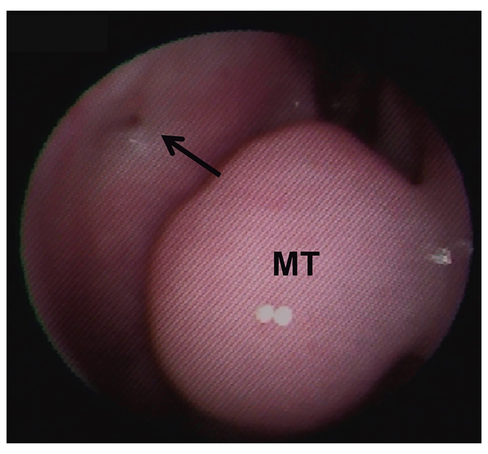
To perform the marsupialization, the wall of the medial lacrimal sac was incised vertically along its entire length and then horizontally in a "cross-like shape". The flaps of the lacrimal sac were exteriorized toward the lateral wall, leaving the lacrimal sac open (Figure 1). The superior and inferior canaliculi were canalized; then, a bicanalicular silicone probe was passed whose ends were knotted inside the nostril (Figure 1). A Gelfoam sponge with a dexamethasone patch was lightly squeezed into the exposed sac.

MT, middle turbinate.
Once the lacrimal sac is exposed, a resection of the medial wall is performed with various surgical instruments, such as rongeurs, and/or Blakesly forceps. There is no intent to preserve the lacrimal sac. The superior and inferior canaliculi were canalized; then, a bicanalicular silicone probe was passed whose ends were knotted inside the nostril.
A follow-up assessment of the patients was conducted. The results were measured subjectively based on improvements in the symptomatology (i.e., the absence of ocular symptoms and lacrimal sac permeability) compared with the preoperative conditions. Objective measures were conducted via endoscopic controls that enabled the observation of an open fistula (Figure 2).
MT, middle turbinate.
The Research Committee at Hospital Civil de Culiacán approved this research (Comité de Investigación del Centro de Investigación y Docencia en Ciencias de la Salud, number: 278). Since this was a retrospective chart review and the clinical images were non-identifying, the ethics committee waived the need for participant consent.
The information was entered into a database using SPSS version 22 for Windows. Frequencies and percentages were calculated for the categorical variables. The numerical variables were evaluated considering the means, confidence intervals, minimums, and maximum. The qualitative variables were measured using frequencies. The continuous variables were compared with Student's t-test, whereas the categorical variables were compared with a chi-square test. A p-value of ≤0.05 was considered significant.
During the study period, 24 endoscopic DCRs were performed on 17 women and 7 men with a mean age of 47.21 years (7–82 years). Of these patients, two patients presented with congenital disease, five suffered from traumatism, and one patient reported a history of eye surgery (Table 1).
A total of seven patients (29.2%) underwent endoscopic DCR with lacrimal sac marsupialization. The remaining 17 patients underwent other endoscopic techniques. The follow-up period was 18 months (4–43 months). One patient received previous dacryointubation and two had previous dacryocystorhinostomies. Electric drilling equipment was used for five patients. Bone removal was performed via a Kerrison clamp for 19 patients. There were three patients lost to follow up.
The efficacy (i.e., the absence of symptoms and patent lacrimal sac) was 71% (n=5/7 patients) for the lacrimal sac marsupialization and 71% (n=10/17 patients) for the other endoscopic techniques. No significant differences were found in the surgical outcomes between the techniques (p=0.686). Raw data are available on figshare7.
A total of seven patients presented with postoperative complications: six with infection and one with granuloma and infection (Table 2).
| Variable | Without marsupialization, n | Marsupialization, n | P value |
|---|---|---|---|
| Surgical technique | 17 | 7 | |
| Complications | |||
| • Infection | 4 | 2 | 0.76 |
| • Granuloma | 1 | 0 | 0.51 |
| Technique failure | 4 | 2 | |
| Resolution achieved | 10 | 5 | 1.00 |
| Lost to follow up | 3 | 0 |
A total of seven patients presented with postoperative complications: six with infection and one with granuloma and infection (Table 2). Patients with infection resolved with topical and oral antibiotics (typically a cephalosporin) and the one patient with granuloma resolved once it was removed the silastic tube. No long-term complications were reported in the study.
Three of our patients were lost to follow up. Unfortunately, six cases reported no improvement, regardless of endoscopic technique.
External DCR became popular because of its high success rates. However, the endoscopic DCR described by McDonogh and Meiring in 19891,4,5 has been used more often because of the simplicity of its innocuous endonasal approach. In addition, it offers advantages over the external approach such as reduced surgical trauma and hemorrhage, the avoidance of facial scars, the maintenance of intact medial canthus structures, and a faster time to return to work3,5,6. Failures of up to 12% of patients have been reported8. The main causes of failure of endoscopic DCR have been attributed to failure to locate the lacrimal sac, insufficient osteotomy, granulation tissue, synechiae, and closure due to premature scarring, fibrosis, and osteogenesis1,5,8.
The additional advantages offered by endoscopic DCR are better visualization of intranasal structures, the avoidance of angular vein damage, the preservation of the pumping function of the nasolacrimal sac, the corroboration of the adequate site for nasolacrimal tube insertion, the better correction of errors, and the identification of surgical failures9.
To avoid or prevent the obstruction of the neoformed ostium, multiple techniques have been tried with several modifications (e.g., complete marsupialization of the lacrimal sac, i.e., the use of mucosal flaps after a wide resection of the bone that surrounds the sac)3,10. Massegur et al. proposed this modification in 2004, with surgical success ranging from 87 to 92%3. The present study used a similar technique, an endoscopic DCR with marsupialization of the mucosal flap sac and resection of the bone using Kerrison's rongeur. A mean follow-up time of 18 months was conducted. The other endoscopic techniques used in the study were partial resection of the lacrimal sac mucosa and maxillary line graft, using Blakesley forceps or Kerrison’s rongeur.
A learning curve of the surgeons could explain similar results in both techniques. The first seven cases of DCR marsupialization are described in this case series.
Yigit et al. (2007) compared the results of external DCR (55 patients) to those of endoscopic DCR (48 patients) in 103 patients with chronic dacryocystitis. The evaluated results were considered as successful if the epiphora decreased, infections were reduced, or reflux from the canaliculus was absent during lacrimal irrigation. The patient management success rate was 69.9% for those undergoing external DCR, and it was 89.7% for those receiving endoscopic DCR. These results were evaluated based on a 1-year follow-up period11.
Likewise, the use of a silastic tube has been a matter of debate. Grigori et al. (2008) examined 46 patients undergoing DCR via a prospective, randomized study: half with silastic tube insertion and the other half without a catheter. Success was defined as the absence of epiphora, decreased conjunctival discharge, and fewer infections. The success rate for the 46 patients was 89%; the success rates with and without the use of a silastic tube were 78% and 100%, respectively, a significant difference (p=0.049). The follow-up period was 6 months. In addition, the controversial use of a silastic catheter was demonstrated12.
A similar efficacy was found between endoscopic DCR with lacrimal sac marsupialization and the other endoscopic techniques in this study. Studies with larger patient samples are needed. Appropriate follow-up and postoperative care are recommended for all cases.
Figshare: Dacryocystorhinostomy dataset 2019, celis-aguilar et al.https://doi.org/10.6084/m9.figshare.7716500.v47.
This dataset includes the following files:
DCRSPSSFEB2016f10002.csv (dataset containing surgical information on all patients)
data coding dacryocystorhinostomy article celis et al.docx (data dictionary)
Data are available under the terms of the Creative Commons Attribution 4.0 International license (CC-BY 4.0).
| Views | Downloads | |
|---|---|---|
| F1000Research | - | - |
|
PubMed Central
Data from PMC are received and updated monthly.
|
- | - |
Provide sufficient details of any financial or non-financial competing interests to enable users to assess whether your comments might lead a reasonable person to question your impartiality. Consider the following examples, but note that this is not an exhaustive list:
Sign up for content alerts and receive a weekly or monthly email with all newly published articles
Already registered? Sign in
The email address should be the one you originally registered with F1000.
You registered with F1000 via Google, so we cannot reset your password.
To sign in, please click here.
If you still need help with your Google account password, please click here.
You registered with F1000 via Facebook, so we cannot reset your password.
To sign in, please click here.
If you still need help with your Facebook account password, please click here.
If your email address is registered with us, we will email you instructions to reset your password.
If you think you should have received this email but it has not arrived, please check your spam filters and/or contact for further assistance.
Comments on this article Comments (0)