Keywords
H1N1, ocular, pandemic, infections
This article is included in the Eye Health gateway.
H1N1, ocular, pandemic, infections
Novel A/H1N1 influenza (previously termed as swine flu) is a common infectious disease in India (12,942 infections causing 954 deaths in 20181) and globally2. The respiratory system is primarily affected by infection and extensive data is available for the interaction of A/H1N1 with the respiratory system3. Systemic viral infections (human immunodeficiency virus (HIV), cytomegalovirus, herpes simplex, herpes zoster, chikungunya and West Nile virus) are commonly associated with ocular lesions, usually posterior uveitis, due to their hematogenous dissemination. HIV infection, additionally, shows a classic microvasculopathy (cotton wool spots or hemorrhages) that reflect direct endothelial infection4. However, reports of ocular lesions (infective or vasculopathic) in A/H1N1 infections are limited. Published reports5,6 present single patient findings but aggregate data from multiple cases are not available. This study was intended to ascertain the presence and types of ocular lesions in novel A/H1N1 infections and reports findings from 14 patients.
Approval for retrospective data collection and publication was granted by the institutional ethics committee of Lilavati Hospital and Research Center (approval dated 09/04/2019) The requirement for individual consent was waived.
A retrospective cross-sectional observational study. We evaluated the records of all patients examined in the medical intensive care unit of a medium-sized (320 beds) tertiary referral hospital (Lilavati Hospital and Research Center) from 2015 until 2018. This was done over a month (February 2019). As part of a standard hospital protocol, patients with pyrexia of unknown origin (PUO), at the specific discretion of the admitting physician, undergo a bedside dilated fundus evaluation. A total of 14 patients (six female and eight males, ages ranging from 36–78 years, mean 54.07 years) met the criteria for inclusion (positive tests for novel A/H1N1 infection and having been ophthalmologically evaluated) and their records were analyzed. Of these, 13 (92.8%) were admitted in the intensive care/isolation unit and one (7.1%) chose to be managed on an outpatient basis.
All patients underwent a detailed evaluation on admission, including a complete blood count (hemoglobin estimation, total and differential white cell counts and platelet counts), blood sugar tests (random on admission, fasting if deemed necessary), liver (serum glutamic-oxaloacetic transaminase (SGOT)/serum glutamic pyruvic transaminase (SGPT)) and renal function tests (serum creatinine and blood urine nitrogen, if needed), urine analysis (routine and microscopy), procalcitonin (PCT) estimation and prothrombin time (PT)/partial thromboplastin time (PTT) studies. Radiological evaluation included chest x-rays and a follow-up chest computed tomogram (CT), at the intensivist’s discretion. An infectious disease evaluation included peripheral blood smears for malarial parasites, rapid NS1 antigen detection for dengue infection and serology for chikungunya (serum IG/IgM) and/or hantavirus infection (serum IgG/IgM estimation). A nasopharyngeal swab was sent for reverse transcriptase-polymerase chain reaction (RT-PCR) for H1N1 infection. Patients who were permitted to receive domiciliary care underwent a slitlamp evaluation, dilated fundus evaluation and intraocular pressure evaluation in the outpatient department.
Data were entered into a spreadsheet (Excel for Mac, Microsoft Corp, WA, version 16.16.9) and analyzed with the inbuilt statistical functions.
The presenting complaints of the 14 patients were persistent fever, cough, sore throat and progressively worsening dyspnea for five to seven days prior to admission7. Demographic information and clinical presentations can be found in Table 1. Significant pre-existing diseases included type 2 diabetes mellitus in three patients (21.4%, ages ranging from 74–78 years with a mean of 75.66 years - cases 3, 5 and 14), with additional chronic liver disease in one of these patients (7.4 %, 74-year-old female - case 5).
All of the patients were febrile, tachypneic and tachycardic. Chest imaging (x-rays and chest scans) revealed extensive bilateral fluffy infiltrates in five patients that were admitted with acute respiratory distress syndrome (35.7% - cases 1, 2, 8, 9 and 11). Other chest imaging findings included unilateral/bilateral patchy infiltrates or consolidation in two patients (21.4 % - cases 4 and 6). Five patients (35.7% - cases 3, 5 and 12–14) had normal chest findings on chest radiology.
The findings of hematological tests including complete blood count (hemoglobin estimation, total and differential white cell counts) and platelet counts were normal. A bleeding diathesis was ruled out by normal PT/PTT values (11 to 13.5 seconds), in nine (64.2%) of these patients. Liver function tests (SGOT /SGPT) revealed marked hepatic dysfunction in one patient (case 6 – 7.1%) and were normal (SGOT 5–40 U/L, SGPT 7–56 U/L) or borderline elevated in the rest. Renal function tests (serum creatinine and blood urea nitrogen) were normal in all cases (creatinine <1.2 mg%, blood urea nitrogen 5–20 mg%) except three (cases 3,5 and 13 – 21.4%). Random blood sugars on admission were grossly abnormal in one patient (case 9 – 317 mg%) and were normal (< 160mg%) in the rest. Bacterial infection was ruled out, where available, by normal PCT values (<0.5 ng/ml). The results of the tests for malaria, chikungunya, hantavirus and dengue were negative in all patients. Nasopharyngeal swabs were tested using RT-PCR and found to be positive for novel influenza A/H1N1 in all cases. In cases 2, 3, 8–10 and 12–14 (57.1%), the microbiology laboratory was able to confirm the strain as pandemic novel A/H1N1/pdm (2009) strain, but confirmatory evidence of the pdm strain was not available for the rest.
Following a dilated fundus evaluation, three patients (21.4 %) showed ocular lesions. These three patients included one male and two females, aged 36–54 years (mean 46.0 years - cases 7, 8 and 9) who demonstrated a unilateral intraretinal hemorrhage. These three patients had platelet counts ranging from 173,000 – 258,000/cu.mm and had no evidence of a bleeding diathesis.
One patient in this group (case 8) had a large disc hemorrhage and an area of retinal whitening consistent with arterial occlusion and retinal ischemia in the macular area. This patient was a 36-year-old female who rapidly deteriorated with the development of a right cerebral bleed and a right arm radial artery thrombosis that required surgical embolectomy.
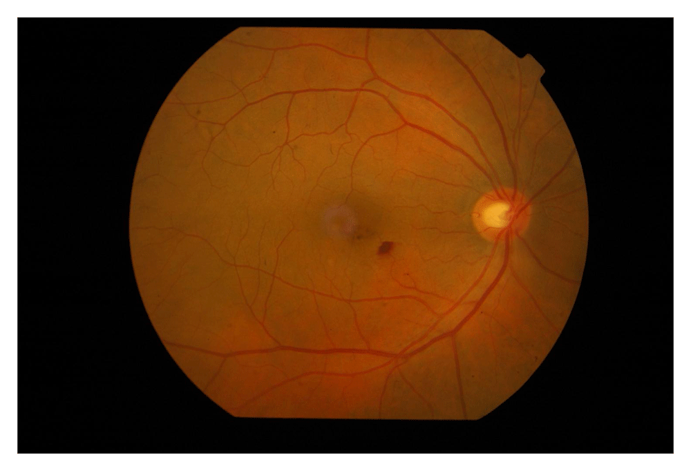
The single patient (case 7) who opted for domiciliary care, underwent a detailed evaluation. This patient’s vision was 6/6, N6 bilaterally with a normal anterior segment examination on slit-lamp evaluation. Dilated fundus evaluation revealed a 1/3-disc diameter (DD) sized retinal hemorrhage in the right eye (Figure 1) and was normal in the left.
We observed ocular lesions in three patients in the form of intraretinal and disc hemorrhages. One patient had disc hemorrhages and macular ischemia, consistent with arterial occlusion/retinal ischemia along with systemic thrombotic events in the form of a cerebral bleed and a radial arterial thrombus. In our patients, thrombocytopenia or a bleeding diathesis did not seem to be the mechanism of these bleeds, thus suggesting an alternate mechanism.
Several authors have postulated the virus has a direct cytopathic effect on retinal, neuronal and vascular tissue. Studies of lung tissue have shown similar cytopathic effects on bronchial and alveolar epithelial cells8, as well as intraluminal fibrin thrombi and partial loss of the endothelium in intrapulmonary blood vessels9. We hypothesize that similar patterns of retinal intravascular events, possibly induced directly by the virus or mediated through an immune reaction, may be responsible for our findings.
Bunce et al. studied 119 patients with H1N1 infections and were able to identify seven patients with systemic thrombotic events10. Among the likely prothrombotic mechanisms, they identified enhanced platelet activation or endothelial dysfunction, suggesting that the etiopathogenesis of the arterial thrombi may involve endovascular injury or endothelial dysfunction with the release of proinflammatory mediators.
Breker et al. reported a case of a 13-year-old girl with a H1N1 infection who developed bilateral retinal and lateral geniculate body infarction5. They noted an ischemic retinal whitening with inner retinal thickening and hyperreflectivity which they hypothesized was an immune mediated complication. Roesel et al. described a 11-year-old girl with a H1N1 infection, who developed vitritis with massive choroidal exudation which was subsequently confirmed on ultrasound and optical coherence tomography (OCT). Following steroid therapy, the effusion significantly improved6.
The weaknesses of our study include a referral bias towards critically ill patients and the fact that referral for ophthalmic evaluation was at the discretion of physicians. Hence, this data does not reflect an accurate prevalence of all admitted patients, with a likely skew to a higher frequency of ocular lesions. Virtually all of these patients were isolated or too ill to be moved for a more detailed evaluation including retinal angiography to assess the vascular integrity and patterns of blood flow.
This small study suggests that hitherto unreported ocular involvement in the form of hemorrhages/retinal ischemia may occur in these patients. Further data is needed to assess their significance, especially as prognostic indicators.
Figshare: H1N1_figshare.xlsx. https://doi.org/10.6084/m9.figshare.80797437
This project contains the following underlying data:
Data are available under the terms of the Creative Commons Attribution 4.0 International license (CC-BY 4.0).
| Views | Downloads | |
|---|---|---|
| F1000Research | - | - |
|
PubMed Central
Data from PMC are received and updated monthly.
|
- | - |
Provide sufficient details of any financial or non-financial competing interests to enable users to assess whether your comments might lead a reasonable person to question your impartiality. Consider the following examples, but note that this is not an exhaustive list:
Sign up for content alerts and receive a weekly or monthly email with all newly published articles
Already registered? Sign in
The email address should be the one you originally registered with F1000.
You registered with F1000 via Google, so we cannot reset your password.
To sign in, please click here.
If you still need help with your Google account password, please click here.
You registered with F1000 via Facebook, so we cannot reset your password.
To sign in, please click here.
If you still need help with your Facebook account password, please click here.
If your email address is registered with us, we will email you instructions to reset your password.
If you think you should have received this email but it has not arrived, please check your spam filters and/or contact for further assistance.
Comments on this article Comments (0)