Keywords
Trichilemmal carcinoma, pilar cyst
Trichilemmal carcinoma, pilar cyst
Trichilemmal carcinoma is a rare tumour derived from the outer root sheath of hair follicles1. It typically occurs in elderly patients on sun-exposed areas of the body1. Such tumours may occur de novo, but more commonly they arise from trichilemmal cysts, which are benign lesions arising from the isthmus of hair follicles, or proliferating trichilemmal tumours2. It is thought that trauma and inflammation can induce the transformation of a benign tumour into a malignant tumour2. The tumour may have a prolonged benign period before cancer develops.
This case report is important as it illustrates that a diagnosis of trichilemmal carcinoma is often delayed due to it mimicking other skin lesions.
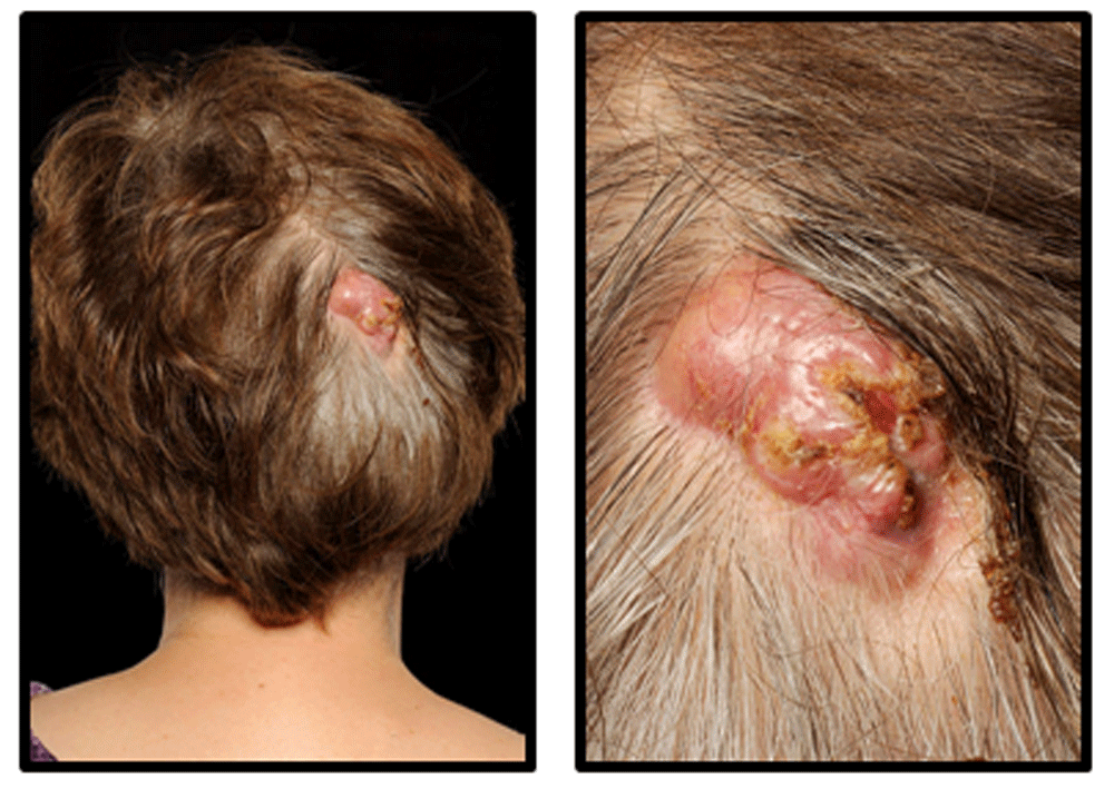
A 62-year-old Caucasian British female presented with a 20-year history of three 1-cm cysts on her scalp. She was previously fit and well and had no significant medical history. Over a seven-month period, the cyst overlying the occiput had become painful and grown in size. During this time, the patient had visited her general practitioner and local emergency department, both of which suspected infection. The lesion was incised, and the patient was treated with three courses of oral flucloxacillin (each course, 500 mg four times per day for 1 week) and one course of oral clarithromycin (250 mg twice per day for 1 week). At the time of surgical excision, the lesion measured 3 x 4 cm. It was raised, indurated, crusted, demonstrated a sparsity of hairs on the surface, had superficial ulceration and exuded serosanguinous fluid when pressed (Figure 1). There was no palpable lymphadenopathy.
Microscopic examination of the lesion identified rounded dermal lobules of squamous epithelium with trichilemmal keratinisation in keeping with a pre-existing pilar cyst (Figure 2A). Areas with nuclear pleomorphism, mitoses and an infiltrative architecture were noted, and they retained trichilemmal keratinisation (Figure 2B–D). The features were of a trichilemmal carcinoma arising in a pilar cyst.
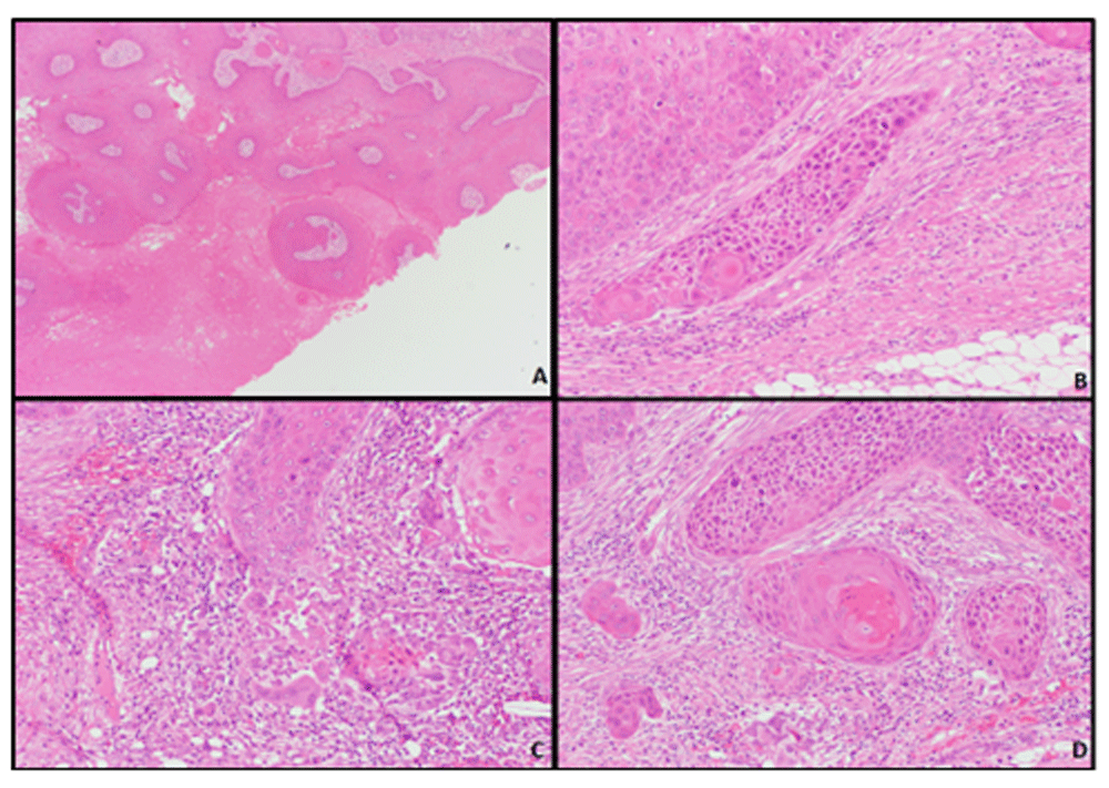
(A) Squamous epithelium with trichilemmal keratinisation (x4 objective). (B–D) Epithelium with nuclear pleomorphism, mitoses and an infiltrative architecture (x20 objective).
She was reviewed three months post-operatively. The wound had healed well and there was no sign of recurrence.
Trichilemmal carcinomas can be difficult to distinguish clinically and histologically from other skin lesions, particularly squamous cell carcinoma (SCC). Microscopically they are characterised by an abrupt transition of nucleated squamous epithelial cells to keratinised cells, without the formation of a granular layer3, and a lobular proliferation of epithelial cells which exhibit nuclear pleomorphism, prominent mitotic activity and infiltration beyond the basement membrane4.
Trichilemmal carcinomas are considered to be a low-grade tumour, but they do have the potential to spread to lymph nodes and to metastasise to distant sites in the body5. There are also reports of death due to the disease6. Therefore, prompt treatment is necessary to reduce morbidity and mortality. Surgical excision with a 1-cm border is the recommended treatment. However, in recent years, Mohs surgery has been used with success.
For recurrent disease, or cases with lymph node or distant metastases, radiotherapy and chemotherapy are sometimes considered, but often there is no standard protocol for trichilemmal carcinoma treatment, and regimens similar to those used for SCC are employed. Following treatment, patients will need to undergo regular follow-up due to the risk of recurrence and/or metastases. Because of the tumour’s rarity, standard treatment and follow up protocols have not been established.
Trichilemmal carcinoma is a rare adnexal tumour. It can mimic common skin lesion such as cysts or squamous cell carcinoma. Diagnosis is dependent on microscopic examination, and the identification of features including the absence of a granular cell layer, a lobular architecture, cellular pleomorphism, mitoses and invasion beyond the basement membrane. The tumour can behave aggressively. Adequate excision and appropriate follow-up are required.
Trichilemmal carcinoma is a rare adnexal tumour.
It can mimic common skin lesion such as cysts or squamous cell carcinoma.
Diagnosis is dependent on microscopic examination, and the identification of features including the absence of a granular cell layer, a lobular architecture, cellular pleomorphism, mitoses and invasion beyond the basement membrane.
The tumour can behave aggressively. Adequate excision and appropriate follow-up are required.
All data underlying the results are available as part of the article and no additional source data are required.
Written informed consent for publication of their clinical details and clinical images was obtained from the patient.
| Views | Downloads | |
|---|---|---|
| F1000Research | - | - |
|
PubMed Central
Data from PMC are received and updated monthly.
|
- | - |
Provide sufficient details of any financial or non-financial competing interests to enable users to assess whether your comments might lead a reasonable person to question your impartiality. Consider the following examples, but note that this is not an exhaustive list:
Sign up for content alerts and receive a weekly or monthly email with all newly published articles
Already registered? Sign in
The email address should be the one you originally registered with F1000.
You registered with F1000 via Google, so we cannot reset your password.
To sign in, please click here.
If you still need help with your Google account password, please click here.
You registered with F1000 via Facebook, so we cannot reset your password.
To sign in, please click here.
If you still need help with your Facebook account password, please click here.
If your email address is registered with us, we will email you instructions to reset your password.
If you think you should have received this email but it has not arrived, please check your spam filters and/or contact for further assistance.
Comments on this article Comments (0)