Keywords
pigmented paravenous retinochoroidal atrophy, Retinochoroidal atrophy
This article is included in the Eye Health gateway.
pigmented paravenous retinochoroidal atrophy, Retinochoroidal atrophy
Pigmented paravenous retinochoroidal atrophy (PPRCA) is a bilateral and symmetrical condition1,2, which is characterized by atrophy of choriocapillaris and retinal pigment epithelium (RPE), and pigmentation along the retinal veins3. Patients are usually asymptomatic and diagnosis is made during routine examination based on typical fundus appearance and non-progressive nature of disease2. Here we present, to the best of our knowledge, the youngest typical case of PPRCA reported to date.
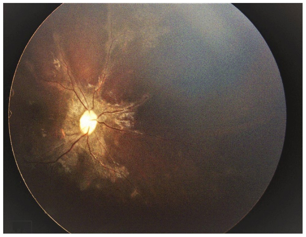
A 27-month-old girl presented with exodeviation in his right eye to retina clinic, at Farabi eye hospital in April 2016. She experienced a normal birth and development, and had unremarkable family history. She used no medications. Visual acuity testing was not feasible due to patient’s young age. Her parents had noticed outward deviation in her right eye. The cyclorefraction of both eyes were +2.25 diopters. The anterior segment and vitreous examination were normal and there were no inflammatory signs. In funduscopy, typical bilateral radial paravenous pigmentary changes and retinochoroidal atrophy were noticed in both eyes (Figure 1). The pigmentations consisted of coarse black pigmentations and fine subretinal yellowish round flecks. They arborized into the peripheral retina along the veins. Unaffected areas between the lesions seemed to be normal.

These pigmentary changes were arborized into the peripheral retina along the veins. Unaffected areas between the lesions appeared normal.
The fundus examination of her parent and newly born brother showed no abnormality. Electroretinogram (ERG) responses showed mild to moderate reduction in both scotopic and photopic responses, equally. Based on retinal examination and ERG findings, PPRCA was diagnosed. At 9 months later, with no treatment, the fundus findings showed no changes and cyclorefraction was +1 diopter. On 16-month follow up, fundoscopy and ERG findings were the same as those at the initial presentation.
To the best of authors’ knowledge, this patient is the youngest with PPRCA ever reported. The patient showed typical characteristics of PPRCA and considerable stability over the course of a 16-month follow up. This presentation is in concordance with the congenital origin hypothesis of PPRCA3,4. In 1937, Brown described this condition for the first time in a 47-year-old man with alopecia areata and named it as retinochoroiditis radiata. The patient had already been under treatment for a disseminated choroiditis at the age of 26 years and had symptoms of tuberculous spondylitis. Considering that his close relatives had died of tuberculosis, the condition was presumed to be a form of tuberculous periphlebitis5,6. The hypothesis of inflammatory origin was further confirmed by other case reports with congenital syphilis, rubeola, measles and Behçet's disease. However, in the years after, a hypothesis of congenital origin was developed and the condition was considered a hereditary disease4,7.
Later in 1962 Franceschetti, changed the name of this condition from retinochoroiditis radiata to a more generally accepted term of PPRCA4,8. So far, there are more than 100 case reports in literature and most of the affected patients are men3,4. In 2003, after lengthy follow ups, Yanagi et al.9, found that this disease is stationary in younger patients while is slowly progressive in older subjects. The progression in older patients may be attributed to wrong diagnosis of PPRCA and these patients may actually have pseudo-PPRCA.
Although the majority of cases have been sporadic, there are reports of familial occurrence2,3. In 1986, Traboulsi and Maumenee10 described a mother and her three sons with PPRCA. Every member had a different chief complaint. The youngest son. who was 4 years old. had poor fixation, nystagmus, and peripheral pigmentary abnormalities. The 10-year-old son had no signs of pigmentary change and the second son had mild pigmentary changes at age of 7 years. To our knowledge, the members of this family were the youngest cases ever been reported to date.
ERG findings are not the same in all cases and have a wide spectrum. While in some cases, ERG shows noticeable involvement, in others it may be normal or show only mild involvement. Reduction of b wave amplitude is the most common finding, followed by a wave amplitude reduction and prolonged latency3,4,7. In some cases, rod responses may be affected more than cone responses, while in others cone response reduction is the dominant feature. In our case both responses were almost equally diminished. This variation may reflect the heterogeneous impairment of various cell types in the retina4.
Our patient presented with exotropia in right eye and bilateral fundus involvement. Previous studies have reported the association of PPRCA with different ocular problems, including anisometropia, amblyopia, esotropia, exotropia, nystagmus, optic disc drusen, and macular changes, such as cystoid macular edema, pigmentary macular degeneration, lamellar macular holes and macular coloboma3,4. Since secondary PPRCA or pseudo-PPRCA has been reported, clinicians should be aware of differential diagnoses, which include chorioretinal degenerations, serpiginous choroidopathy, retinits pigmentosa, tuberculous disseminated choroiditis, helicoid peripapillary chorioretinal atrophy and angioid streaks7,9,11,12.
One of the main advantages of this case report was a relatively long-term follow-up period. This case showed no progression during 16 months of follow up, which may indicate that primary congenital PPRCA with no inflammatory association may be a non-progressive disease. Cases with progressive course or other concomitant findings may be secondary PPRCA and pseudo-PPRCA can be a better term for them.
All data underlying the results are available as part of the article and no additional source data are required.
Written informed consent for publication of their clinical details was obtained from the parents of the patient.
| Views | Downloads | |
|---|---|---|
| F1000Research | - | - |
|
PubMed Central
Data from PMC are received and updated monthly.
|
- | - |
Is the background of the case’s history and progression described in sufficient detail?
Partly
Are enough details provided of any physical examination and diagnostic tests, treatment given and outcomes?
No
Is sufficient discussion included of the importance of the findings and their relevance to future understanding of disease processes, diagnosis or treatment?
No
Is the case presented with sufficient detail to be useful for other practitioners?
Partly
Competing Interests: No competing interests were disclosed.
Is the background of the case’s history and progression described in sufficient detail?
Partly
Are enough details provided of any physical examination and diagnostic tests, treatment given and outcomes?
Partly
Is sufficient discussion included of the importance of the findings and their relevance to future understanding of disease processes, diagnosis or treatment?
Partly
Is the case presented with sufficient detail to be useful for other practitioners?
Partly
Competing Interests: No competing interests were disclosed.
Alongside their report, reviewers assign a status to the article:
| Invited Reviewers | ||
|---|---|---|
| 1 | 2 | |
|
Version 1 04 Jun 19 |
read | read |
Provide sufficient details of any financial or non-financial competing interests to enable users to assess whether your comments might lead a reasonable person to question your impartiality. Consider the following examples, but note that this is not an exhaustive list:
Sign up for content alerts and receive a weekly or monthly email with all newly published articles
Already registered? Sign in
The email address should be the one you originally registered with F1000.
You registered with F1000 via Google, so we cannot reset your password.
To sign in, please click here.
If you still need help with your Google account password, please click here.
You registered with F1000 via Facebook, so we cannot reset your password.
To sign in, please click here.
If you still need help with your Facebook account password, please click here.
If your email address is registered with us, we will email you instructions to reset your password.
If you think you should have received this email but it has not arrived, please check your spam filters and/or contact for further assistance.
Comments on this article Comments (0)