Keywords
Femoral hernia, Incarceration, appendicitis, De Garengeot Hernia
Femoral hernia, Incarceration, appendicitis, De Garengeot Hernia
In this version 2, three figures have been added, we give further description of femoral hernia repair, and discuss imaging and operative management of the case.
See the author's detailed response to the review by Zhamak Khurgami
The presentation of femoral hernia with an incarcerated appendix accounts for 0.5-3.3% of all femoral hernias1; very few cases have been described2. The condition is named after Rene De Garengeot, a French surgeon who first described it in 17313. The condition may be described as the femoral counterpart of the more widely described Amyand hernia, involving appendicitis within the inguinal hernia sac1.
A 48-year-old woman presented to Dalby Hospital (a small rural facility) with a 3-day history of an irreducible right inguinal swelling, which came on while cycling a mountain bike. A timeline of care is given in Table 1. She had initially not presented as she suspected a muscle strain but presented when the pain became worse. She reported 2–3 previous occurrences of a lump in the same location many years ago, which had self-resolved. Her prior medical history was notable: 13 previous pregnancies with 10 natural deliveries and 3 terminations, LLETZ procedure for cervical cancer, no previous abdominal surgeries. She was an active smoker with a 25 pack-year history. She took no regular medications.
The patient was initially examined by a rural general practitioner who was concerned for incarcerated hernia. He discussed the case with the surgical registrar at the treating regional hospital and arranged for interhospital transfer. On transfer that evening to this regional facility the swelling was red and inflamed. The patient was haemodynamically stable, afebrile and was moving her bowels. A right sided, painful swelling could be palpated in the right inguinal region. The registrar examining the patient was suspicious for an incarcerated femoral hernia. In the absence of obstructive symptoms it was suspected that this was incarcerated fat only.
In order to exclude the more serious diagnosis of incarcerated bowel with the hernia and to confirm the diagnosis of femoral hernia, an initial ultrasound was ordered by the treating surgical registrar after discussion with the consultant of the night. In addition a full blood count and electrolytes with liver function tests was ordered. The blood tests were all in the normal range. Meanwhile the ultrasound was unable to exclude incarcerated bowel and femoral from inguinal hernia (Figure 1). The discrepancy of this radiological finding with the clinical findings caused further discussion between the radiology sonographer, consultant and the surgical team; the diagnosis of the type of hernia and its contents mandated the urgency of theatre, approaches and timing. As a result, a CT was ordered to further investigate the anatomy in this case in order to plan operative approach.
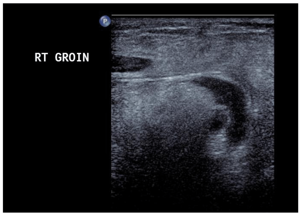
The ultrasound failed to confidently describe the hernia as inguinal or femoral and was unable to exclude the presence of bowel involvement within the hernia.
The initial findings of the CT scan were suggestive of an inflamed inguinal hernia with predominant fat contents and probable bowel involvement (Figure 2 and Figure 3). There was no radiographic evidence of a small or large bowel obstruction. As this patient’s bowels were still moving, it was felt that this was most likely caused by incarcerated, strangulated fat, rather than bowel.
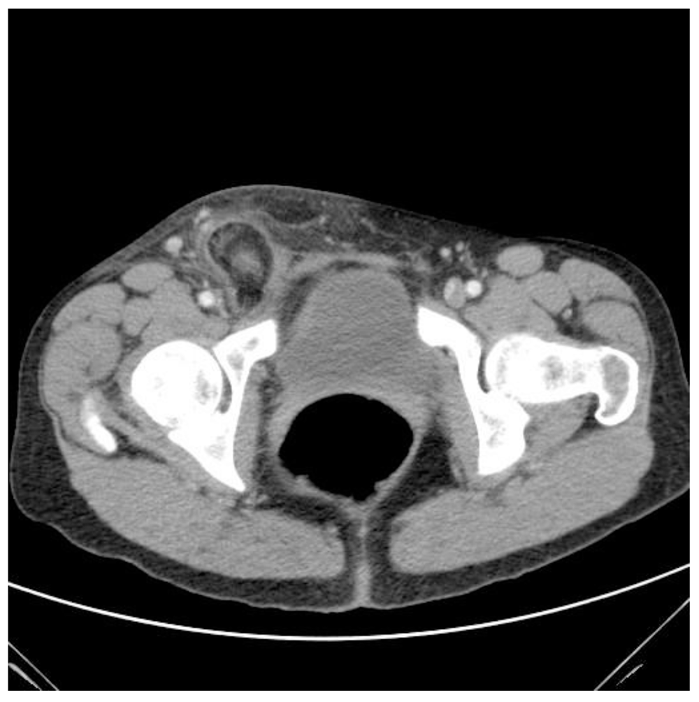
This was reported at the time as an inguinal hernia with bowel involvement but without signs of small bowel obstruction. We were suspicious clinically of a femoral hernia and no bowel involvement

As reported at the time it was reported as probable bowel involvement within an inguinal hernia.
The patient was taken to theatre. Because of the above findings, an incision was made over the inguinal ligament, expecting to find an incarcerated inguinal hernia. Instead, on dissection, a femoral hernia was encountered. The sack was opened and necrotic mucosal content was encountered. It was suspected that this was necrotic bowel requiring resection, so the decision was made a low midline laparotomy to ensure safe resection. On opening, a necrotic appendix was found to be incarcerated in the femoral hernia (Figure 4).
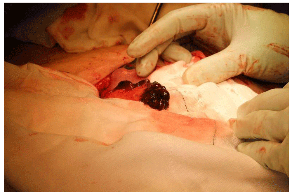
The appendix being reduced, the mesoappendix was clamped, divided and ligated, and the appendix was removed with a purse string suture used to bury residual mucosa. The femoral hernia was repaired primarily with nylon sutures from to the conjoin tendon to the shelf of the inguinal ligament after excision of the sac. A Blake drain was placed and laparotomy wounds were closed with looped Novafil sutures. The patient was placed on Ceftriaxone, 1 g daily and metronidazole 500 mg BD.
The patient recovered swiftly, and was discharged on day 3 following surgery (Table 1). Her antibiotics were ceased after 24 hours and she was treated with simple analgesia only as required. She was subsequently seen in the outpatients’ clinic at 2 weeks after surgery, and had made a full recovery.
This case was notable for its rarity and the clinical and radiological difficulty anticipating the incarceration of the appendix in the femoral canal. This might have mandated a different approach on surgery than might have been undertaken. Accurate diagnosis of the condition would allow for appropriate choosing of incision, or a laparoscopic approach. On review of the literature, this is typical of this rare condition, however. Excepting one Japanese study4, the diagnosis was typically made serendipitously at surgery. This is not unique, to De Garengeot’s hernia; there can often be confusion between femoral hernia and inguinal hernias, particularly upon clinical examination. Littre’s hernia containing Meckel’s diverticulum and a Richter’s hernia are often also diagnosed on the table rather than in the radiologist’s suite.
The rarity of this condition contributes to this. De Garengeot’s is an exceedingly rare condition, accounting for 0.5–3.3% of femoral hernias1, which are rare in and of themselves, accounting for only 3–5% of all presentations of hernia. This can make clinical and radiological suspicion all the more challenging. In this case despite ultrasound and CT scan the operative approach was as for an inguinal hernia without incarcerated bowel. We anticipated using mesh in this case but elected not to in the presence of an infarcted inflamed appendix.
The suspicion of a De Garengeot’s hernia earlier on imaging would have dictated either a laparoscopic approach, which may have also been useful to confirm the diagnosis, or a midline mini-laparotomy. Laparoscopic repair has been described as feasible for treatment of this condition; however, its rarity and difficulty of diagnosis prior to operation would make prospective comparison extremely difficult.
No data are associated with this article.
Written consent for publication of their clinical details and clinical images was obtained from the patient.
| Views | Downloads | |
|---|---|---|
| F1000Research | - | - |
|
PubMed Central
Data from PMC are received and updated monthly.
|
- | - |
Provide sufficient details of any financial or non-financial competing interests to enable users to assess whether your comments might lead a reasonable person to question your impartiality. Consider the following examples, but note that this is not an exhaustive list:
Sign up for content alerts and receive a weekly or monthly email with all newly published articles
Already registered? Sign in
The email address should be the one you originally registered with F1000.
You registered with F1000 via Google, so we cannot reset your password.
To sign in, please click here.
If you still need help with your Google account password, please click here.
You registered with F1000 via Facebook, so we cannot reset your password.
To sign in, please click here.
If you still need help with your Facebook account password, please click here.
If your email address is registered with us, we will email you instructions to reset your password.
If you think you should have received this email but it has not arrived, please check your spam filters and/or contact for further assistance.
Comments on this article Comments (0)