Keywords
Hypomineralization, resin infiltration, microabrasion, bleaching
Hypomineralization, resin infiltration, microabrasion, bleaching
Hypomineralization defects are considered one of the most common reasons patients seek esthetic dental treatment. However, management of such defects is always challenging as they imply proper identification of the cause, nature, severity of the defect, and proper understanding of all available treatment modalities that suites various degrees of the defect, thus achieving satisfactory results.
The probable etiological factors for enamel hypomineralization defects in permanent teeth can be broadly divided into two main categories, according to whether those defects have a localized or generalized distribution1. Localized hypomineralization defects could be caused by trauma, localized infection, and irradiation. While generalized (diffuse) hypomineralization may be caused by a wide range of factors1. Genetic disorders resulting from a single gene defect, as an X-linked, autosomal dominant or autosomal recessive trait are considered one of the factors2,3. Fluoride intoxication is one of the most common types of intoxications that causes enamel hypomineralization. Fluorotic lesions are characterized by opaque white spots or discolorations ranging from yellow to dark brown. The severity of those lesions depends on the duration and amount of fluoride intake during tooth development4. Perinatal and postnatal illnesses that may occur in premature and low birth weight neonates could be also responsible for the occurrence of enamel hypomineralization5. Infectious diseases and fever during early childhood such as chickenpox, measles, and mumps have also been linked to the occurrence of hypomineralization defects6.
A 27 year-old single Egyptian male, working as a clothes vendor, visited the outpatient clinic of Operative Dentistry Department, Faculty of Dentistry, Cairo University in March 2017 requesting to remove the discoloration from his front teeth. The patient was not satisfied with his smile because of the discolored teeth. He was healthy, with no systemic diseases. The patient’s dental history showed an irregular attendance to dental care with history of fillings, root canal fillings, and extraction.
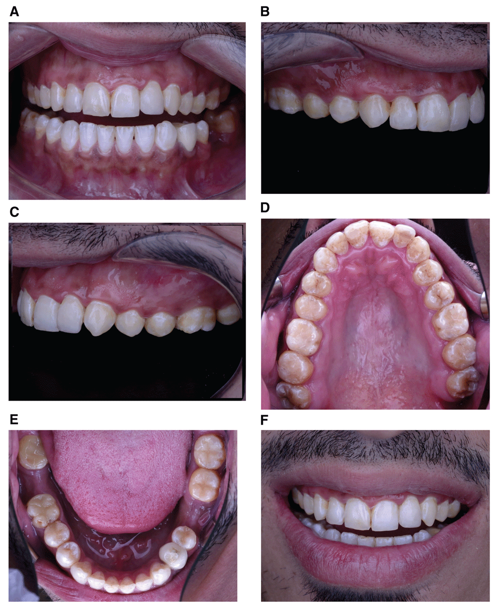
Clinical examination showed a diffuse opaque white spots and streaks, together with subsurface brown discolorations and pitted enamel representing severe hypomineralization. Figure 1 illustrates the pre-operative photos of the case. This clinical appearance was very confusing concerning its cause, as it has long been considered a typical clinical picture of enamel fluorosis. Nevertheless, developmental defects of enamel with similar appearances are not necessarily caused by similar aetiologic factors.
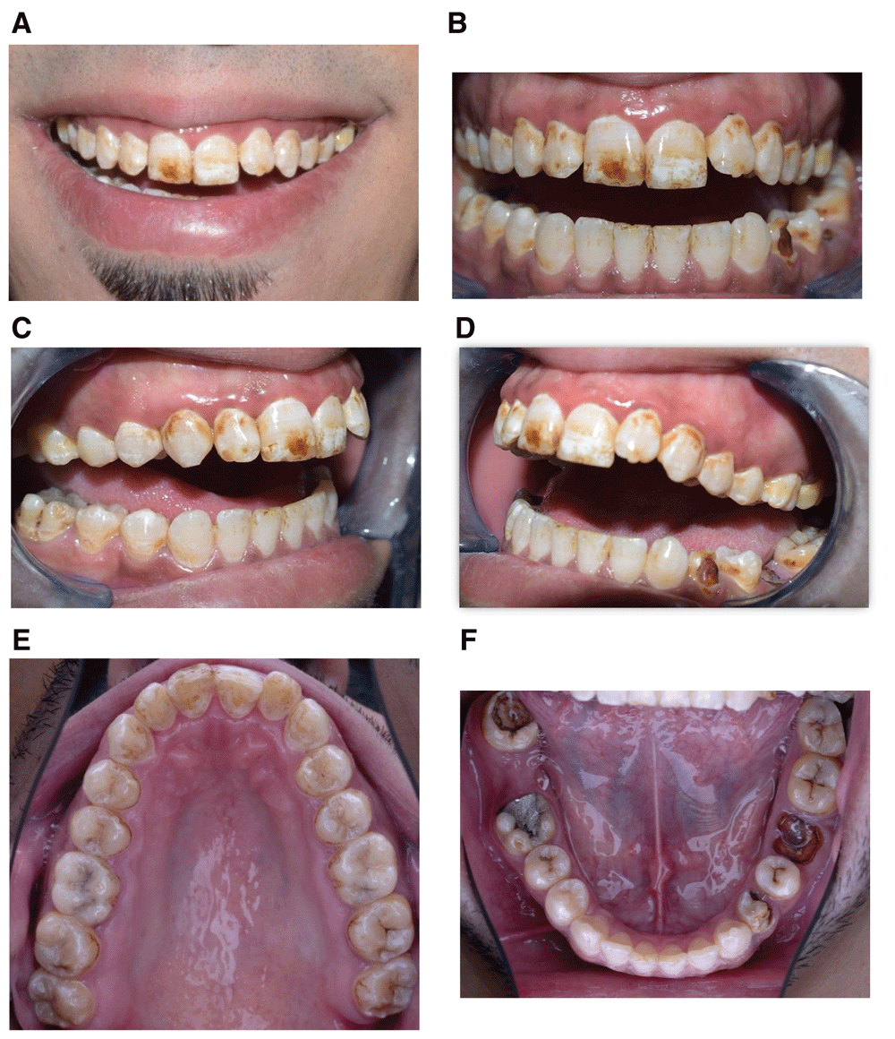
(a) Smile view. (b) Frontal retracted view. (c) Right side retracted view. (d) Left side retracted view. (e) Maxillary occlusal view. (f) Mandibular occlusal view.
Conversely, the same aetiologic factors can produce different defects at different stages of tooth development1. Consequently, not all white or brown hypomineralized enamel are caused by fluorosis7. In addition, the medical history of this patient was noncontributory regarding his exposure to fluoride, as this patient did not report that he had lived in an area where water is fluoridated during his childhood, nor had he taken any fluoride supplements, Thus, based on the patient’s history, fluoride was excluded to be the cause of hypomineralization.
The patient had calculus deposits on the upper left canine and premolar teeth, and bleeding on probing, thus full mouth debridement was performed together with instructions about maintenance of good oral hygiene. Besides, the clinical and diagnostic cast examination revealed a protrusion (malalignment) of tooth #10 relative to the adjacent teeth. In addition, teeth #21 and #32 were badly decayed. Tooth #21 was restored with a root canal treatment, ready- made post, resin composite core, and a definitive full coverage restoration. As for teeth # 32, root canal treatment, and a definitive composite restoration was made.
Periapical radiographs (Figure 2) showed that teeth #19 and #30 had periapical radiolucencies related to their defective root canal fillings. Tooth # 19 was extracted as it was unrestorable. While tooth # 30 was subjected to a re-treatment of its root canals, and a definitive composite restoration.
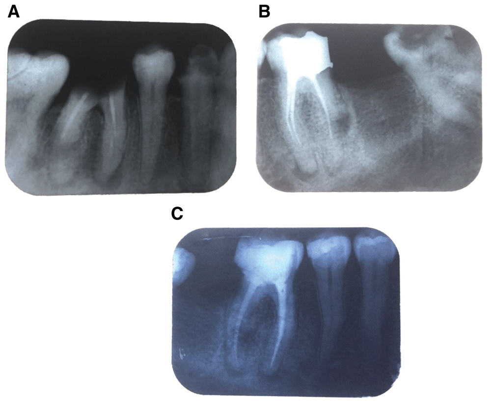
(a) Periapical radiograph of tooth #19. (b) Periapical radiograph of tooth #30 (c) Periapical radiograph of tooth #30 after endodontic re-treatment and a definitive restoration.
The initial treatment plan for the hypomineralization defects was presented to the patient, this included in-office bleaching with 35% hydrogen peroxide (polaoffice in-office Tooth Whitening System, SDI Australia, 7700031) to target the brown discolorations, followed by resin infiltration to mask the white spot areas with Icon (DMG Germany, 220343). The patient was informed that the correction of the protruded tooth #10 would have to be delayed until after completion of the whitening and resin infiltration procedures.
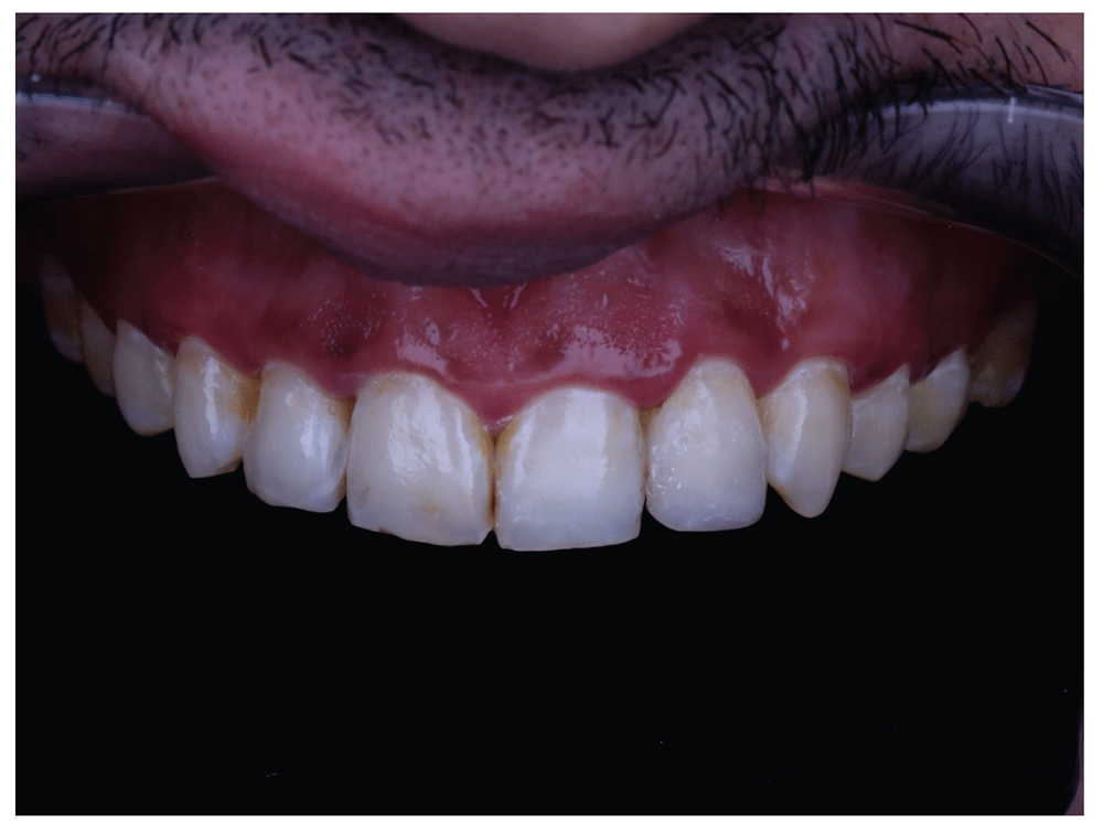
One session of 3 applications of an in-office whitener (polaoffice in-office Tooth Whitening System, SDI Australia, 7700031) was performed for the patient, each of 8 minutes duration. The patient mentioned no post-treatment sensitivity, and no inflammation in his soft tissue was observed. The patient was informed that a period of 3 weeks was necessary between the bleaching sessions and the resin infiltration procedures. Figure 3 illustrates the clinical picture after the in-office bleaching, it was noticed that a residual light brown color still remains on the anterior teeth especially tooth #8. It was decided at this moment that a change in the initial treatment plan would have to occur, and that teeth microabrasion (Opalustre, Ultradent USA, 555) was going to be carried out before resin infiltration to selectively target those resistant stains.
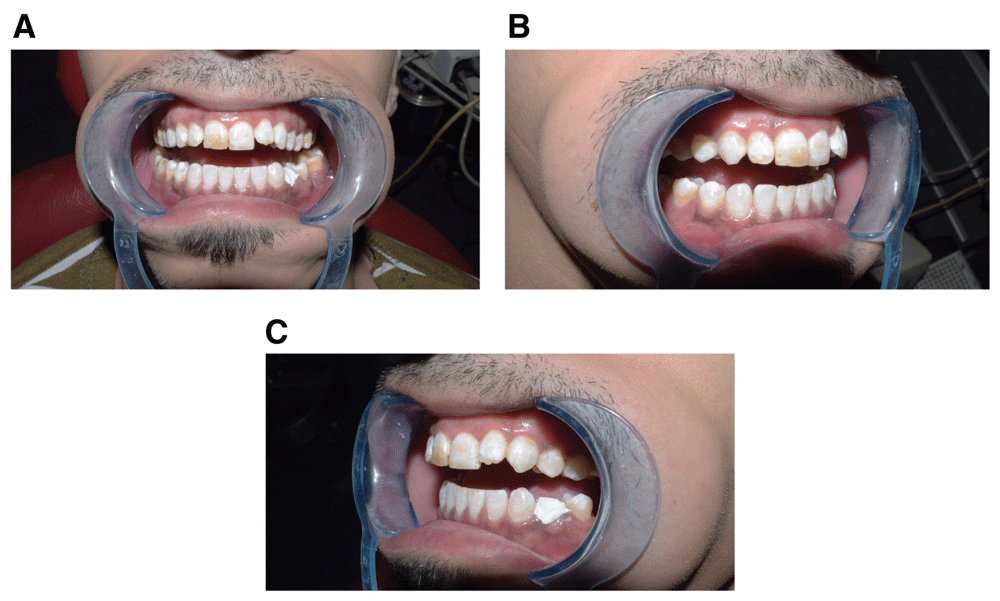
(a) Frontal retracted view. (b) Right side retracted view. (c) Left side retracted view.
The disappearance of those resistant stains was evident immediately after the microabrasion session (Figure 4). The patient returned to the clinic one week later for the resin infiltration procedures (Figure 5 & Figure 6). The patient was now told at this stage of the treatment plan that he would have the correction of his protruded #10 done two weeks after completion of the resin infiltration. All the materials used in this case were applied according to the manufacturer’s instructions. Table 1 illustrates the materials used in this clinical case, their composition, and catalogue number. Table 2 illustrates the timeline of the esthetic rehabilitation of the case.
(a) Application of Opalustre microabrasion gel. (b) After microabrasion, frontal view. Notice the brightening of the brown stain that was present on tooth #8. (c) After microabrasion, right sided view. (d) After microabrasion, left sided view.
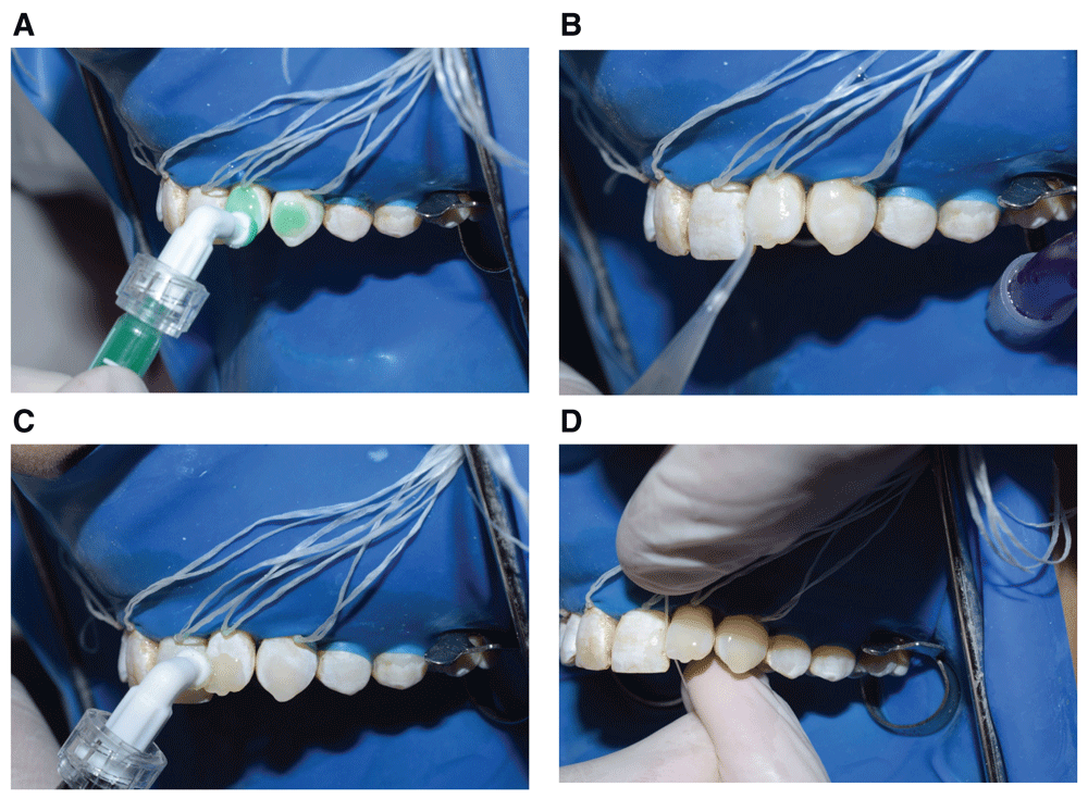
(a) Icon-Etch gel (DMG Germany) was applied to the surface and left undisturbed for two minutes. The gel was thoroughly rinsed with water for 30 seconds. The teeth were then air dried with water- and oil-free air for 15 seconds. (b) Icon-dry (DMG Germany) was applied to the surface and left undisturbed for 30 seconds. The teeth were then air-dried with water- and oil-free air for 15 seconds. (c) Icon-Infiltrant (DMG Germany) was applied and left undisturbed for three minutes. Excess material was gently air-blown to prevent pooling around the incisal edge. (d) Excess resin was removed with dental floss. The resin was then light cured for 40 seconds in each tooth. Notice the difference between the resin infiltrated lateral, canine, and the other teeth.
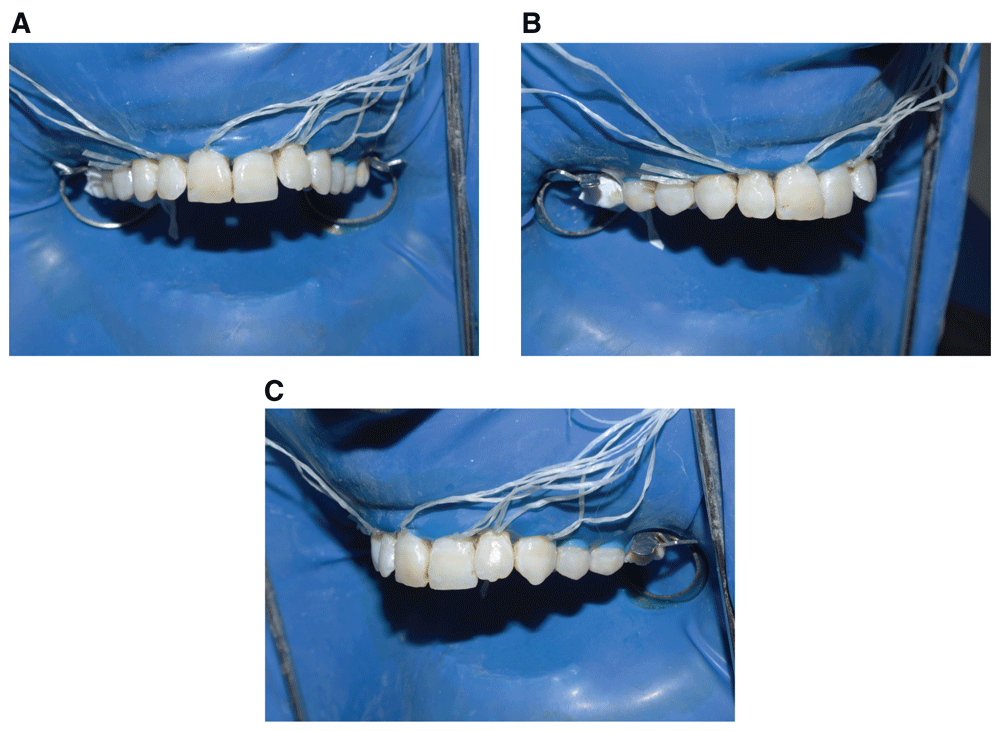
(a) Frontal view. (b) Right sided lateral view. (c) Left sided lateral view.
A waxed up model (Figure 7) was fabricated and shown to the patient to express his opinion. The model included the following: a slight reduction (0.5 mm) of the mesio-incisal aspect of the labial surface of tooth #10, increase of the length of the clinical crown of tooth #10 with white wax, labial realignment of tooth #10 with resin composite, with a similar realignment for tooth #7 to preserve symmetry of the frontal aspect of the teeth.
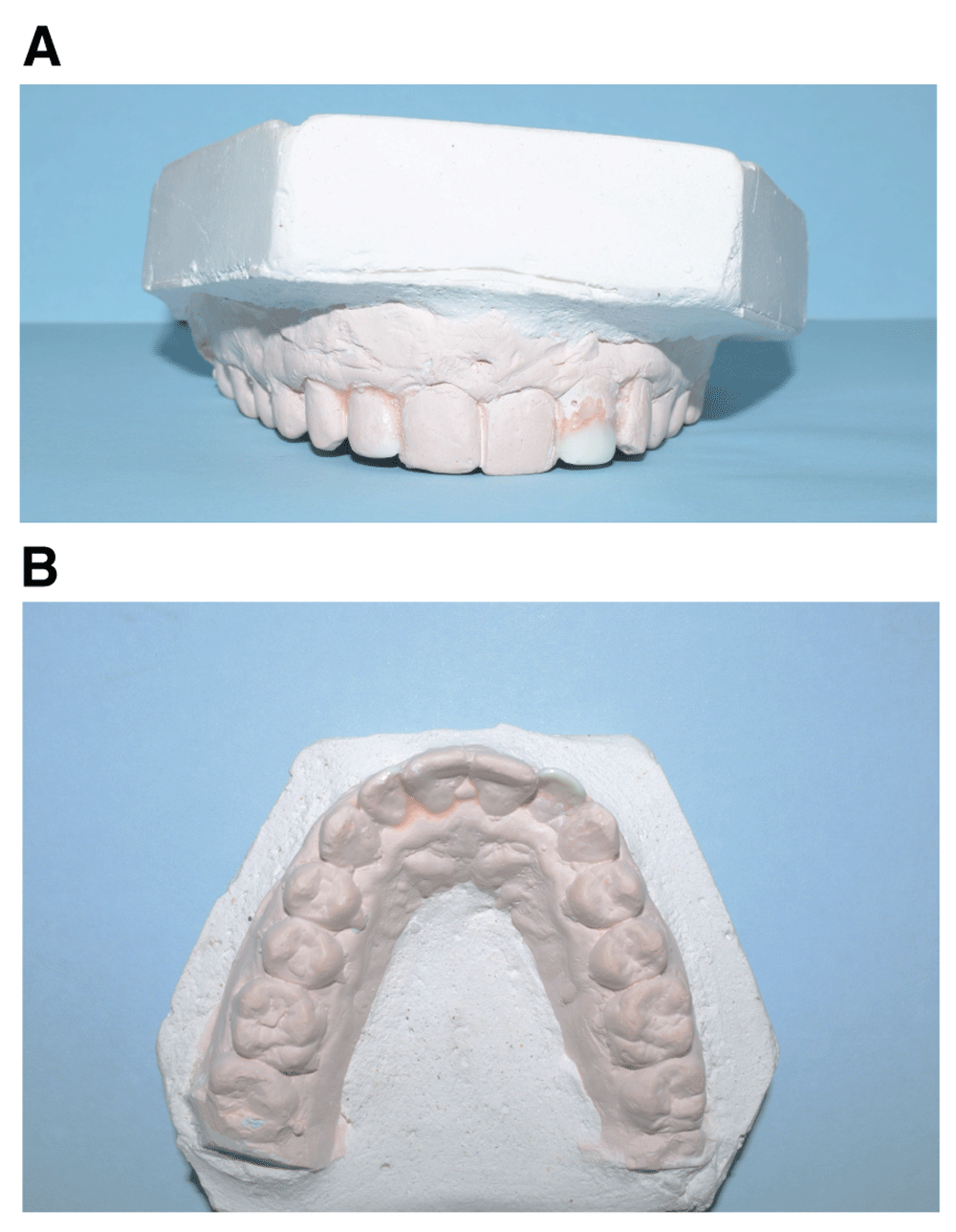
(a) Frontal view. (b) Palatal view.
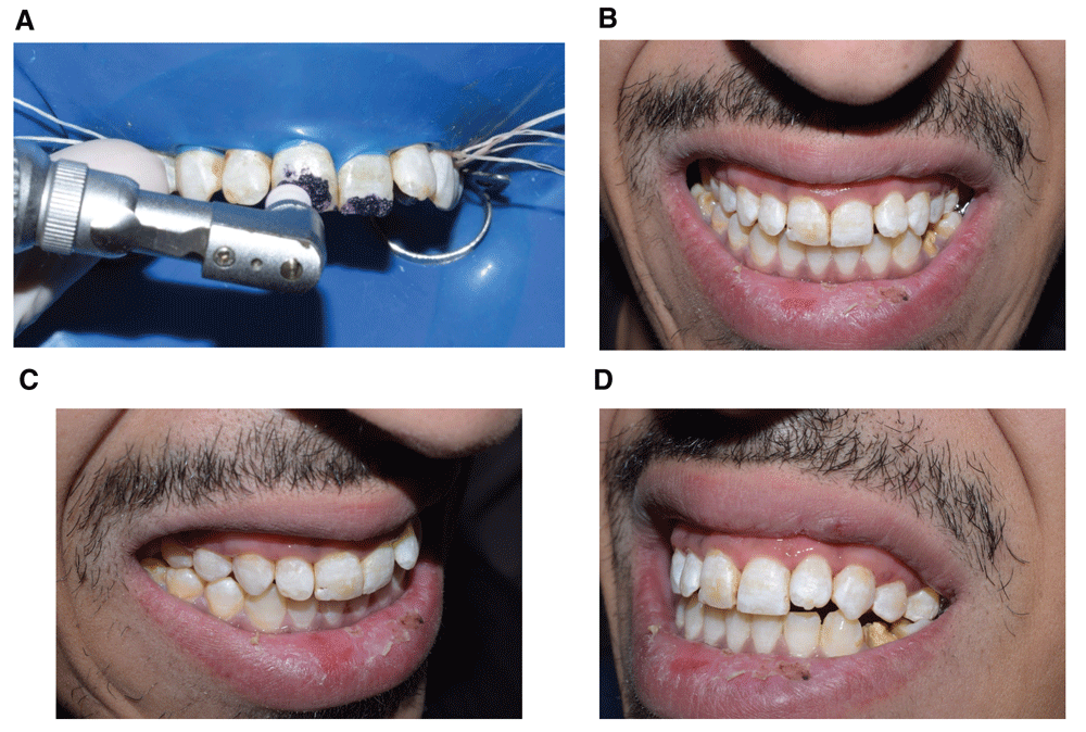
The steps that was done on the model to realign tooth #10 was duplicated inside the patient’s mouth using a putty consistency guide (zetaplus, Zhermack, C 100600) fabricated from the waxed-up model. After the resin composite shade was selected and minimal tooth reduction (0.5 mm) of the mesio-incisal aspect was carried out as previously mentioned, enamel was then etched with 37.5% phosphoric acid gel (Kerr, Italia, 31297) for 15 seconds, rinsed with water for 10 seconds, and dried with air. OptiBond All In One adhesive (Kerr, Italia, 29670) was then applied for 15 seconds in a rubbing motion, and gently air dried followed by light curing (Elipar LED curing light, 3M ESPE) at an intensity of 1200 mw/cm2 for 10 seconds from the facial and palatal aspects. Resin composite was then applied in layers starting with A1 enamel shade (Herculite XRV Ultra, Kerr Italia, 34002) from the palatal aspect, followed by A2 dentin shade (Herculite XRV Ultra, Kerr Italia, 34019) to replace missing dentin and finally A1 enamel shade on the labial aspect. Each layer was light cured for 40 seconds from the both facial and palatal aspects. Finishing was carried out using a tapered flat end finishing carbide bur #7713 (MIDWEST, DENTSPLY 388529), and polishing was performed using fine, extra-fine discs (OptiDisc, Kerr 4182, 4183) and rubber cups (HiLuster Gloss PLUS Polisher, KerrHawe 2653 ). Figure 8 shows the procedure of realignment of tooth #10, and enhancing the final esthetic outcome using resin composite restorations. Figure 9 and Figure 10 show the final photographs of the case.
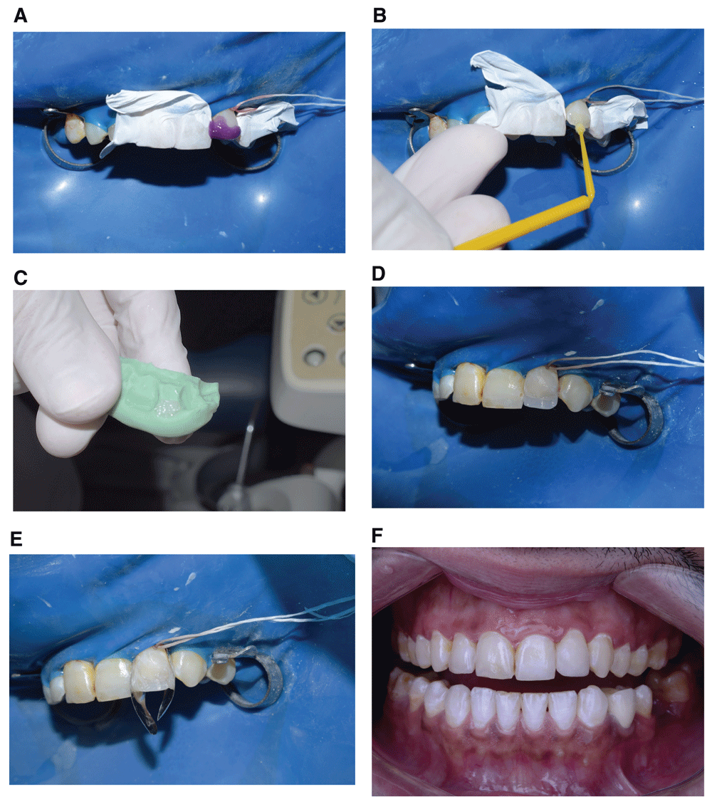
(a) All prepared enamel was etched with 37.5% phosphoric acid gel for 15 seconds. (b) OptiBond All In One adhesive was applied for 15 seconds in a rubbing motion, air dried and then light cured. (c) Trimmed putty consistency index containing A1 enamel shade composite to be adapted on the palatal surface of tooth #10. (d) Tooth #10 having a palatal wall of A1 enamel. (e) Sectional matrix placement. (f) Tooth#10 after re-alignment, finishing and polishing.
Final clinical aspect of the case (a) Frontal retracted view. (b) Right sided retracted view. (c) Left sided retracted view. (d) Maxillary occlusal view. (e) Mandibular occlusal view. (f) Smile view.
The aim of the esthetic rehabilitation in this case was to restore the patient esthetics and self-confidence in the most conservative way. The success of different treatment plans proposed for treating enamel hypomineralization cases depends on the severity of the defect. Most clinical reports aimed at conservative management of those defects have incorporated different interventions such as teeth bleaching, enamel macroabrasion, microabrasion, and resin infiltration in their treatment plans. The main difference between these reports is the sequence with which these interventions were used8–12.
A recent clinical report8 has recommended masking enamel fluorosis stains using at home bleaching with 10% carbamide peroxide in an overnight tray as a first phase of treatment, followed by resin infiltration to mask the residual white spots. They did not recommend the use of in-office bleaching as it would cause post-treatment sensitivity. On the contrary, in-office bleaching was used in our case because the patient compliance to wear a tray was doubtful, additionally no sensitivity was reported after the treatment.
The second phase of the treatment in our clinical case included enamel microabrasion. This was opposite to a previous report8, who recommended resin infiltration as a second phase rather than microabrasion. They stated that resin infiltration would be more conservative as it removes only 40 µm of surface enamel, while microabrasion removes up to 200 µm of enamel corresponding to 10 applications. In our case, only 2 applications of microabrasion, which contains 6.6% HCl and silicon carbide, was carried out, which removed 50 µm of the enamel surface which is nearly equivalent to the amount removed during resin infiltration, and was sufficient in our case to remove those residual brown stains. In addition, the procedure of resin infiltration blocks the enamel from the labial surface because of the thin layer of resin coating the surface, and this would preclude the use of any further intervention from the labial surface (the most important surface). It is for this reason that the previous case report5 had to apply the bleaching gel from the lingual surface to target a residual yellow color after bleaching was carried out. Microabrasion also harmonized the color of the tooth after bleaching, and prepares the surface for resin infiltration.
A previous case report9 has recommended macroabrasion as a first stage in treating hypomineralization defects. They used an ultrafine diamond bur (macroabrasion) followed by eight applications of the microabrasion gel. After one week, the microabrasion procedure was repeated, and finally three sessions of in-office whitening (35% hydrogen peroxide) were carried out. This approach was considered to be aggressive because of the enamel removal by macroabrasion.
In another case report10, two sessions of an in-office bleaching, three applications in each session was the starting protocol for such case. This was followed by 12 applications of the microabrasion material. This was consistent with the strategy adopted in our case, except that the number of applications of the bleaching material and that of the microabrasion material was quite big, so it may be considered also an aggressive treatment.
Realignment of tooth #10 and #7 was carried out in our current case using minimum reduction (0.5 mm) of the mesio-incisal surface, increase in its clinical length, and re-shaping using resin composite. This was considered a perfect alternative for patients unwilling to undergo orthodontic treatment, or when ceramic restorations are not feasible. Resin composite was used to create an optical illusion that the protruded teeth were realigned, conforming to the shape of the arch, and the result was quite acceptable for our patient. Figure 10 illustrates six months postoperative evaluation of the patient and he was satisfied with the result.
The patient was pleased with the provided conservative treatments, he preferred the use of as few clinical procedures as possible. He was satisfied with the outcome and his self- esteem was improved.
The proper management of hypomineralization defects depends on the evaluation of the severity of the defect, and proper evaluation of the outcome after each single step of intervention rather than predetermined interventions based on anticipated outcomes. Thus, we consider in-office teeth bleaching, followed by microabrasion, and resin infiltration an acceptable method in treating hypomineralization discolorations.
Written informed consent for publication of the clinical details and images was obtained from the patient.
| Views | Downloads | |
|---|---|---|
| F1000Research | - | - |
|
PubMed Central
Data from PMC are received and updated monthly.
|
- | - |
Provide sufficient details of any financial or non-financial competing interests to enable users to assess whether your comments might lead a reasonable person to question your impartiality. Consider the following examples, but note that this is not an exhaustive list:
Sign up for content alerts and receive a weekly or monthly email with all newly published articles
Already registered? Sign in
The email address should be the one you originally registered with F1000.
You registered with F1000 via Google, so we cannot reset your password.
To sign in, please click here.
If you still need help with your Google account password, please click here.
You registered with F1000 via Facebook, so we cannot reset your password.
To sign in, please click here.
If you still need help with your Facebook account password, please click here.
If your email address is registered with us, we will email you instructions to reset your password.
If you think you should have received this email but it has not arrived, please check your spam filters and/or contact for further assistance.
Comments on this article Comments (0)