Keywords
Thalamomesecephalic stroke, HIV, Vasculitis
Thalamomesecephalic stroke, HIV, Vasculitis
The main difference is based on:
Establish the criteria of HIV vasculitis in our region and based on the literature reviewed.
Inclusion of the new typical radiological image of vasculitis.
A better description of ischemic stroke in the thalamus and midbrain.
Next time we will deliver a better proposal to review this topic because high experience and skill are necessary to perform a good job.
See the authors' detailed response to the review by Jefferson V. Proano
In 2018, Sato et al. reported a 62-year-old man presenting with a rare eye movement. This patient had a vertical one-and-a-half syndrome caused by unilateral thalamomesencephalic stroke (TMS)1. Other eye movements' abnormalities, such as bilateral vertical gaze palsy, were previously reported due to a unilateral stroke of the rostral midbrain by other authors2,3.
The anatomical circulation of the brain is complex and diverse, and the circle of Willis variations can include absence or fusion of components, incomplete process, fenestrations, fetal branches, and asymmetrical and duplication. From previous studies on autopsy and structural imaging scans, normal anatomical variations were detected in 48–58% of the general population, and even during fetal development4–8.
An investigation done on human cadaveric brains have been demonstrated four major thalamic arterial territories, with notable blood variations. These areas receive blood supply by the polar, paramedian/thalamoperforating arteries, thalamogeniculate, and posterior choroidal arteries9–17. The perforating arteries supply the medial the walls of the third ventricle, hypothalamus, and subthalamic-mesencephalic junctions. These areas include the oculomotor nucleus, red nucleus, subthalamic nucleus, substantia nigra, pretectum, trochlear nucleus, reticular formation of the midbrain, posterior part of the internal capsule, the rhomboid fossa, and the rear part of the thalamus9–20. Because the artery of Percheron occlusion can affect the thalamus and midbrain at the same time, here we have to mention that artery of Percheron is an uncommon vascular variant of the paramedian branches of the posterior cerebral artery, arising from one P1 segment, bifurcates, and bilateral supply to the bilateral paramedian thalami and the rostral midbrain10,11 but not unilaterally. Therefore, occlusion of Percheron arteriole causes an atypical pattern of bilateral infarct of the median thalami with or without mesencephalic damage.
From 2010 and 2017, several authors21–26 also reported a case series of ischemic stroke on the thalamus and different clinical manifestations. Other clinical presentations of TMS include see-saw nystagmus that shows intorsion and elevation of one eye, with synchronous extorsion and depression on the contralateral one, convergence-retraction nystagmus and contraversive ocular tilt reaction probable due to ischemic involvement of the interstitial nucleus of Cajal27, anisocoria. Another author found vertical ocular motor disturbances in the vertical plane and eye movement synkinesis, hypersomnia, and coma as a clinical manifestation of TMS28. Others reported headaches, blurred vision, and diplopia as a particular variant of cerebral lacunae TMS29. In 2012, Benjamin et al. established that HIV infection can cause TMS by opportunistic infections, secondary to a cardioembolic phenomenon, coagulopathy, and vascular diseases such as stenosis, acquired aneurysm, vasculitis, and direct/indirect effect of HIV infection and antiretroviral therapy30. In our region, ischemic stroke due to infectious vasculitis is quite common. In 2017, the first case presenting bi-thalamic infarctions leading to acute vascular dementia associated with HIV infection was reported31.
A 41-year-old female presented with a 5-day history of inability to open the right eye associated with decreased vision of the right eye, which subsequently developed binocular diplopia. The patient also reported a failure to balance herself and could not walk independently. There was no history of trauma, excessive use of NSAIDs, contraceptives, use of vitamin supplements, or complaint of headaches. The patient did not smoke, drink alcohol, or use other recreation or illicit drugs. The patient has a background history of hypertension since her last pregnancy in 2016 and has been on treatment with hydrochlorothiazide (12.5 mg daily) and enalapril (5 mg daily). HIV-reactive with the latest CD4 (01/2020) count of 715 and viral load are lower than the detectable limit on treatment with a combination of tenofovir/emtricitabine/efavirenz TDF/FTC/EFV (300/200/600 mg daily).
On the nervous system examination, the patient was alert and well oriented with no meningeal signs. A cranial nerve exam revealed right cranial nerve 3rd palsy, right complete ptosis, right mydriatic pupil nonresponsive to light, and paralysis of the medial, superior, and inferior rectus plus inferior oblique (Figure 1).
The motor exam showed left hemiparesis (4/5) on upper and lower limbs, bilateral Babinski sign with left hemiataxia despite muscle weakness on the affected side, and no sensory disorder or extrapyramidal signs. The rest of the examination was within normal limits and there were no rashes noted.
The investigations done were as follows: Blood tests (on the day of admission) See Table 1
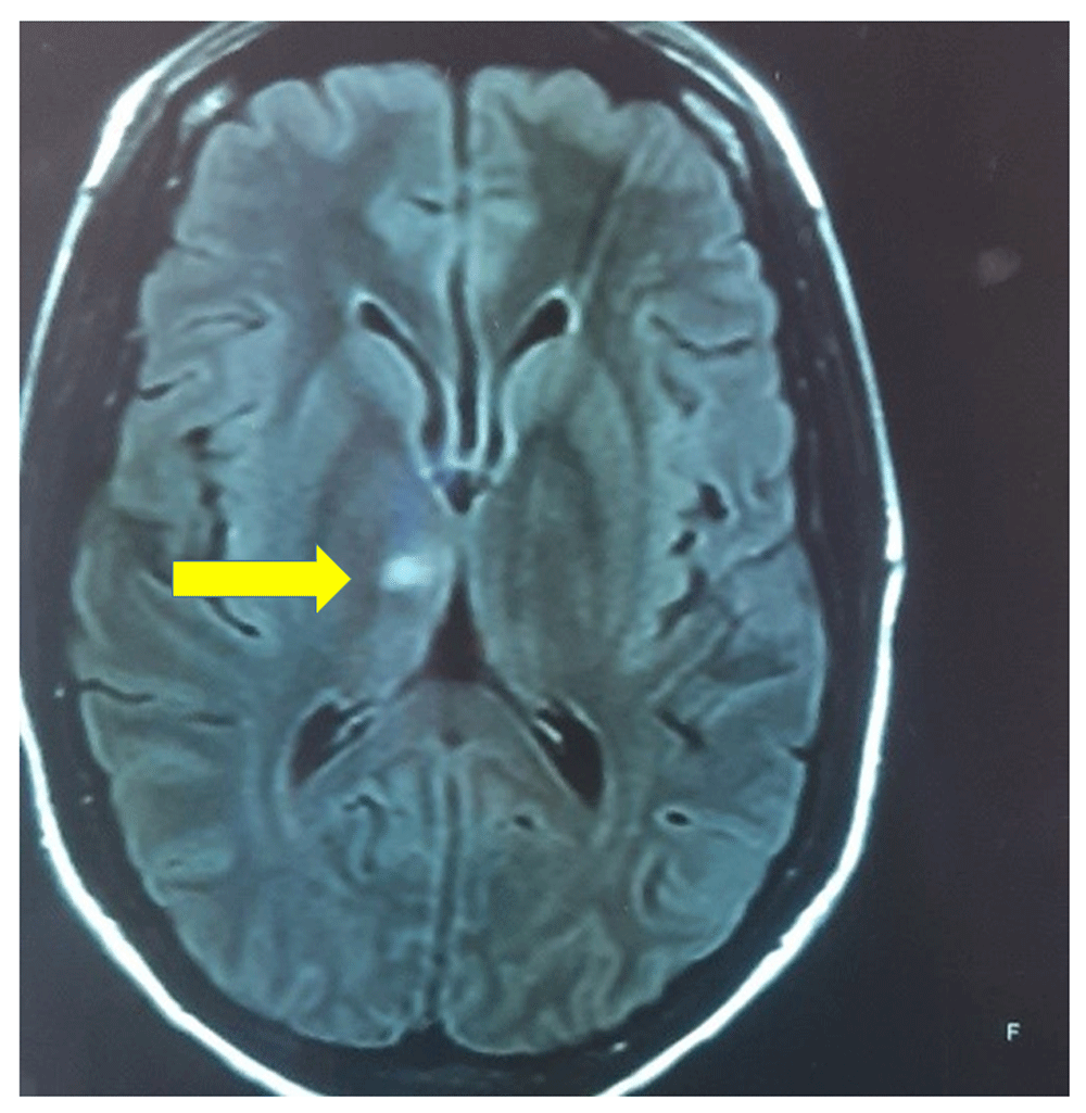
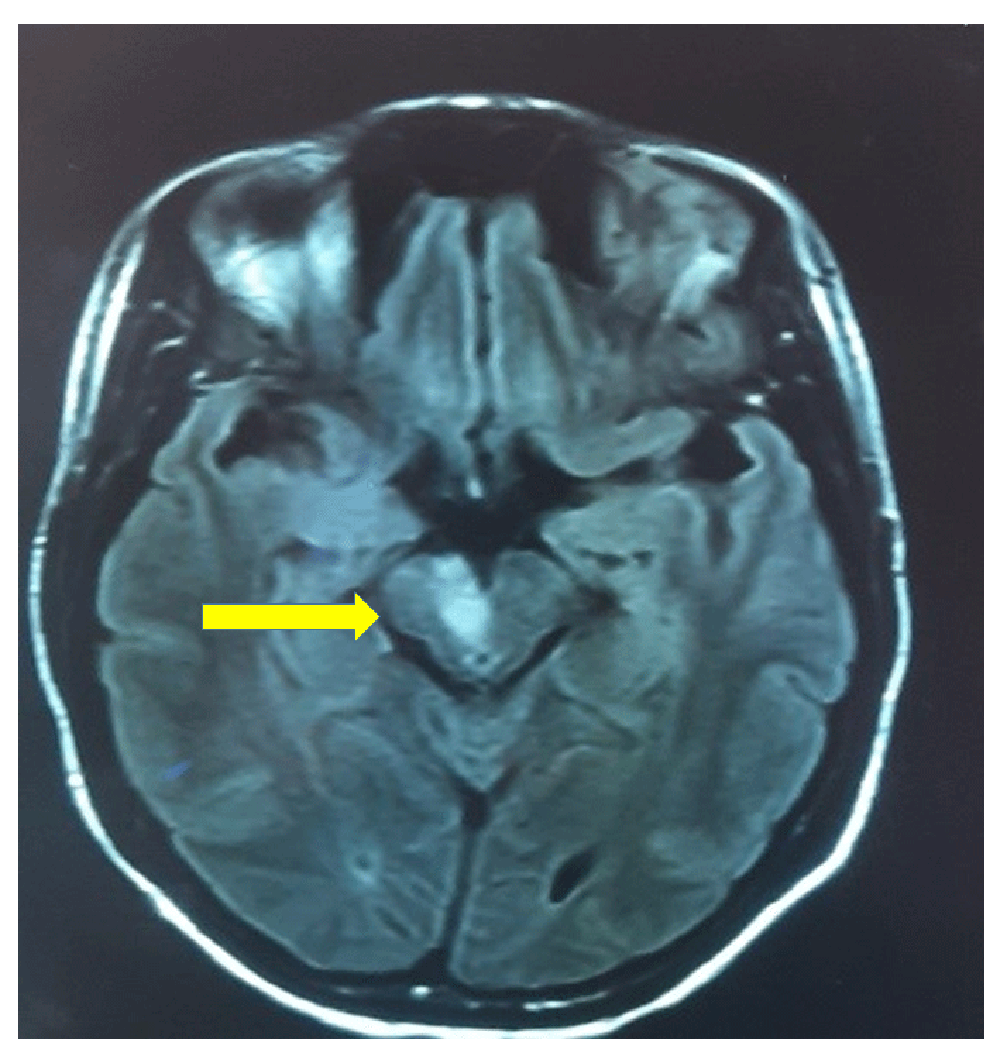
Computed tomography (CT) angiogram and MRI (done two days after admission) showed diffuse vasculitis with parenchymal changes seen in the right thalamus and midbrain and hyperdensity lesion (T2- weighted/FLAIR MRI images) secondary to ischemic infarct in the area supplied by the right paramedian branch of the posterior cerebral artery due to vasculitis (Figure 2–Figure 4).

The axial view shows the right hyperdense lesion at the paramedian thalami caused by ischemic infarct secondary to HIV vasculitis.

The axial view shows a hyperdense lesion on the right midbrain caused by ischemic stroke due to HIV vasculitis.

showing ‘beading’ of the paramedian blood vessels supplying the midbrain. (signs of vasculitis).
The cardiology team requested a cardiac review. A cardiac ultrasound (done the day after admission) showed an ejection fraction of 75%, No signs of valvulopathy or effusion was present. The patient was admitted and started on the following treatment: Vitamin B12 supplementation (1000 µg IM daily for five days in the first week, then weekly for five weeks, aspirin (150 mg daily), enoxaparin (40 mg s/c daily), simvastatin (20 mg daily), pyridoxine (50 mg daily), thiamine (100 mg daily). The patient continued the chronic medication (hydrochlorothiazide 12.5 mg, enalapril 5mg and TDF/FTC/EFV 300/200/600 mg daily). Physiotherapy and occupational therapy are actively working with the patient. [TJ4] The patient received rehabilitation in our ward for two weeks. The right-sided hemiataxia did improve, but the power on the right side was still 4/5. She was referred to her base hospital to continue rehabilitation and a follow-up date with us in 1 month.
Here we report a case of a 41-year-old woman with right-sided TMS. Unilateral TMS is uncommon, and its incidence remains unknown, but one study showed that it comprises about 0.6% to 1% of midbrain ischaemic strokes and often accompanied by other posterior circulation infarcts32.
Baran et al. conducted an observational study in 2018, which showed a male predominance. The study also showed that from an etiological point of view, the most common cause was extensive atherosclerosis, followed by cardio-embolism, apart from small vessel disease33.
The main risk factors associated with extensive atherosclerosis are hypertension, diabetes, hyperlipidemia, smoking, and previous history of stroke. The main risk factors for cardio-embolism in these patients is atrial fibrillation33. Patients who are suffering from peripheral vascular disease and coronary artery disease are at risk34.
HIV is a risk factor for stroke35 and is associated with advanced disease36. There have been numerous mechanisms proposed to explain this. A systematic review done by Addallah et al.37 reported that this could be due to HIV-associated opportunistic infections, HIV-induced coagulopathy, and chronic inflammatory processes that can accelerate atherosclerosis. Another systematic review by Bogorodskaya et al.38 also reported that some antiretrovirals (lopinavir, indinavir, and abacavir) were also associated with an increased risk of stroke.
Lesions of the midbrain can present as distinct syndromes. However, because of the structures' close organization, there can be considerable overlap of these syndromes. The neurological manifestation will depend on which area of the midbrain is affected and whether one half or both halves are involved and whether adjacent structures (thalamus, pons, cerebellum) are also involved. The symptoms may include but are not limited to, low equilibrium, weakness of one or both sides of the body, diplopia, and slurred speech39. The most common examination findings include ataxia, limb weakness, dysarthria, sensory disturbance, oculomotor findings (3rd nerve palsy, internuclear ophthalmoplegia), and dysarthria40. The exact pattern will depend on the area involved and whether surrounding structures are also involved (thalamus, pons, medulla, etc.). There are being midbrain syndromes, which include, among others, Weber syndrome, Claude's syndrome, Nothnagel syndrome, and Benedikt's syndrome. Benedikt's syndrome presents with a contralateral rubral tremor, which she does not have.
Weber syndrome is a result of a lesion involving the ventromedial area of the midbrain. They present with ipsilateral 3rd nerve palsy with contralateral hemiplegia. Our patient has Claude's syndrome, which presents ipsilateral 3rd nerve palsy with contralateral cerebellar ataxia due to the dorsal tegmentum lesion, which involves the 3rd nerve nucleus/fibers and also involving either the red nucleus, superior cerebellar peduncle, or brachium conjunctivum37. Benedikt's syndrome is due to a lesion involving the tegmentum. It presents with ipsilateral 3rd nerve palsy and contralateral ataxia, but there is also the involvement of the fibers of the corticospinal tract and will result in contralateral hemiparesis even41. When assessing these kinds of patients, it is essential to ascertain a good history and physical examination and check the National Institute for Health Stroke Score42. Imaging to confirm the diagnosis is mandatory. CT or MRI angiogram is usually requested to identify the stenosed vessels or identify other possible vascular problems. Blood workup for stroke is compulsory, which includes but is not limited to full blood count, renal function tests, international normalized ratio, lipid profile, HIV ELISA, and if young to include thrombophilia screen, Antinuclear antibodies, and glycosylated haemoglobin. ECG to rule out possible atrial fibrillation and transthoracic or even transesophageal echocardiography to identify cardiac causes.
The management approach depends on the etiology of the stroke. If the infarct is ischaemic, the reviewed literature recommends thrombolysis if posterior circulation strokes meet the established criteria43. Mechanical thrombectomy benefits are not yet well established, but it can be done44. Then after the acute period, it is crucial to managing the risk factors and causes. Then treat the risk factors such as arterial hypertension, diabetes mellitus, hyperlipidemia, and secondary prophylaxis. If there is a cardiac cause, then it should be treated. A multidisciplinary approach is vital for patients presenting TMS. The stroke team should include dieticians, physiotherapy, speech therapy, occupational therapy, and social workers apart from the medical specialists.
Risk factors for developing stroke in our patient were hypertension, HIV, and hyperlipidemia. We had done an extensive workup to rule out other possible contributors to a stroke. The patient also had contralateral hemiparesis and hemiataxia with bilateral Babinski and hyperreflexia on both lower limbs. Her blood workup showed that she was virally suppressed and had hyperlipidemia. The MRI and CT angiograms showed evidence of an infarct involving the ventromedial midbrain and thalamus, and in the absence of other lesions. Cardiovascular investigations ruled out a cardiac source of the infarct. In this patient, hypertension, HIV infection, and hyperlipidemia predisposed her to the stroke. The patient is markedly younger than one would typically expect for a TMS (median age around 64 years)32. Of note in our patient is the presence of a bilateral Babinski sign, which never happens in a patient with Claude's syndrome.
Generally, the strokes involving posterior circulation have a higher mortality rate than those involving anterior circulation unless it involves the smaller blood vessels41, as happened in our case. The present case is unique, among other reasons, owing to the bilateral Babinski sign and the absence of thalamic manifestations without other lesions affecting different segments of the brainstem and the spinal cord. The patient's age (41 years) also makes this case uncommon. The patient's leading risk factor is HIV vasculitis, which has not been implicated for TMS from the literature reviewed. In our setting the commonest causes of infectious vasculitis are HIV/AIDS, tuberculosis, neurocysticercosis, and neurosyphilis that is quite different from other countries. Therefore, HIV-vasculitis should be the first differential diagnosis in our region. However, we could not confirm this aetiology, then we decided to use the terminology: “in patient with HIV” because it was confirmed. We did not find signs of middle longitudinal fascicle (MLF) typical syndrome. MLF syndrome, secondary to ischemic stroke affecting only the mesencephalon, is a rare occurrence45,46. We would highlight that we could not find the cause of the bilateral Babinski signs in this case. This patient did not present thalamic characters despite the ischemic lesion in the right thalamus despite the midbrain's role over the thalamus. The modulation of thalamic neurons is necessary to control adaptive behavior mediated by midbrain cholinergic transmission. The central cholinergic neurons in the mesencephalon are at the pedunculopontine nucleus and the laterodorsal tegmental nucleus, which provide dense innervation of the thamalus. Recently, Huerta-Ocampo et al. confirmed that midbrain cholinergic neurons could innervate all thalamic nucleus. They also found that these axons topographically are well organized and provide a segregated innervation of the thalamic nuclei47. Therefore, we expected some evidence of thalamic dysfunction in this patient. However, these ischemic lesions on the midbrain did not cause abnormal behavior or thalamic manifestations in our patient, which is a novel finding.
To our knowledge, this is the first patient presenting with unilateral TMS secondary to HIV vasculitis with bilateral Babinski signs, and without thalamic manifestations to be reported in the medical literature. In young patients presenting with unilateral TMS, HIV vasculitis is one of the etiological diagnoses to be considered. Still, we recommend an extensive investigation based on a series of cases to support this postulate.
All data underlying the results are available as part of the article, and no additional source data are required.
Written informed consent for publication of their clinical details and clinical images was obtained from the patient.
We wish to thank Dr. M Anwary from the Department of Radiology. Nelson Mandela Academic Central Hospital Mthatha, South Africa, for the investigations done.
| Views | Downloads | |
|---|---|---|
| F1000Research | - | - |
|
PubMed Central
Data from PMC are received and updated monthly.
|
- | - |
Is the background of the case’s history and progression described in sufficient detail?
Partly
Are enough details provided of any physical examination and diagnostic tests, treatment given and outcomes?
No
Is sufficient discussion included of the importance of the findings and their relevance to future understanding of disease processes, diagnosis or treatment?
No
Is the case presented with sufficient detail to be useful for other practitioners?
Partly
References
1. Benjamin LA, Bryer A, Lucas S, Stanley A, et al.: Arterial ischemic stroke in HIV: Defining and classifying etiology for research studies.Neurol Neuroimmunol Neuroinflamm. 2016; 3 (4): e254 PubMed Abstract | Publisher Full TextCompeting Interests: No competing interests were disclosed.
Reviewer Expertise: Clinical neurology, cardiac autonomic dysfunction, neurogenetics
Competing Interests: No competing interests were disclosed.
Reviewer Expertise: Neurology and Infection disease of the nervous system
Competing Interests: No competing interests were disclosed.
Reviewer Expertise: Neurology and Infection disease of the nervous system
Is the background of the case’s history and progression described in sufficient detail?
Partly
Are enough details provided of any physical examination and diagnostic tests, treatment given and outcomes?
Partly
Is sufficient discussion included of the importance of the findings and their relevance to future understanding of disease processes, diagnosis or treatment?
Partly
Is the case presented with sufficient detail to be useful for other practitioners?
Partly
Competing Interests: No competing interests were disclosed.
Reviewer Expertise: Neurology and Infection disease of the nervous system
Alongside their report, reviewers assign a status to the article:
| Invited Reviewers | ||
|---|---|---|
| 1 | 2 | |
|
Version 3 (revision) 30 Mar 21 |
read | read |
|
Version 2 (revision) 01 Dec 20 |
read | |
|
Version 1 16 Oct 20 |
read | |
Provide sufficient details of any financial or non-financial competing interests to enable users to assess whether your comments might lead a reasonable person to question your impartiality. Consider the following examples, but note that this is not an exhaustive list:
Sign up for content alerts and receive a weekly or monthly email with all newly published articles
Already registered? Sign in
The email address should be the one you originally registered with F1000.
You registered with F1000 via Google, so we cannot reset your password.
To sign in, please click here.
If you still need help with your Google account password, please click here.
You registered with F1000 via Facebook, so we cannot reset your password.
To sign in, please click here.
If you still need help with your Facebook account password, please click here.
If your email address is registered with us, we will email you instructions to reset your password.
If you think you should have received this email but it has not arrived, please check your spam filters and/or contact for further assistance.
Comments on this article Comments (0)