Keywords
rheumatoid arthritis, rheumatoid vasculitis, aortitis, immunosuppressive therapy, case report
rheumatoid arthritis, rheumatoid vasculitis, aortitis, immunosuppressive therapy, case report
Rheumatoid arthritis (RA) is a connective tissue disease predominantly affecting the joints. Extra-articular manifestations develop in up to 40% of cases1, of which rheumatoid vasculitis (RV) is the most serious. The widespread vascular involvement, which effects not only the synovia but also other organs such as the skin, eye and nerves, can be life threatening. Mortality can reach up to 40% within five years of disease onset2. Fortunately, RV is a rare complication that occurs in 1-5% of RA patients3. It commonly affects small and medium blood vessels4. Large vessel vasculitis is unusual during RA. Hereby we describe a case of an aortitis revealing RV, which is a rare presentation of a rare complication of RA.
A 54-year-old man, a North African policeman with a personal history of smoking and pulmonary tuberculosis in 1991 was diagnosed in 2009 with seropositive and erosive RA associated with Sjögren’s syndrome. He was being treated with methotrexate (25mg/week). In November 2019, the patient presented to our hospital with fever, fatigue and chest pain that had started one week prior. Physical examination found a high temperature of 38.5°C. Systolic blood pressure was 100mmHg and diastolic blood pressure was 60mmHg in both arms. He had tachycardia, with a heart rate of 115 beats per minute. There were no signs of heart failure and respiratory rate was normal. Mobilization of the wrists, elbows and shoulders was painful, with a swollen right wrist. He had a dislocation of the right ulnar styloid. He had rheumatoid nodules on the outer side of both elbows. The rest of the physical examination was normal.
An electrocardiogram showed atrial fibrillation with a heart rate of 115 beats per minutes associated with diffuse ST-segment elevation with upward concavity.
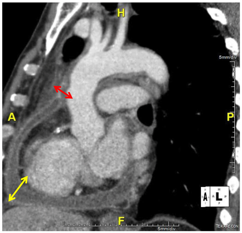
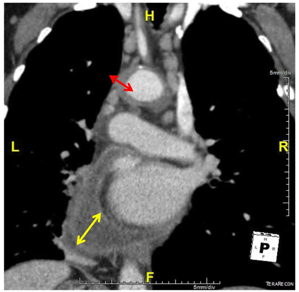
Blood tests showed hyper leukocytosis at 11000/mm3, C-reactive protein (CRP) levels of 117 mg/l, an erythrocyte sedimentation rate (ESR) of 118mm and cholestasis with increased gamma glutamyl transferase and phosphatase alkaline levels of 199UI/L (four times the normal rate) and 348UI/L (five times the normal rate), respectively. Transaminase levels were normal (ALAT level of 35UI/L and ASAT level of 25UI/L). A procalcitonin test was negative (<0.5 μg/L). Blood gas analysis was normal (pH level of 7.40, PaO2 level of 90mmHg, PaCO2 level of 42mmHg, HCO3- level of 25 mmol/l). A chest X-ray showed a flask-shaped enlarged cardiac silhouette. Transthoracic echocardiography confirmed a non-compressive large posterior pericardial effusion. Abdominal ultrasound was normal. A thoraco-abdominal computed tomography (CT) scan showed pericardial effusion with enhancement of the pericardium, compatible with pericarditis, and regular parietal hypodense circumferential thickening of the aortic arch and supra aortic arterial trunk root, confirming aortitis (Figure 1 and Figure 2). There was emphysema in the pulmonary parenchyma but no evidence of active tuberculosis on chest CT.
A biopsy of the cardiac effusion was not possible due to its posterior location. The sputum test for Koch’s bacillus was negative. Viral hepatitis B and C and syphilis serology were negative. Antinuclear antibodies were positive at 1/1200. Anti-LKM1, anti-CCP and rheumatoid factor levels were 1/80, 40 IU/ml and 50 IU/ml, respectively. Immunoglobulin levels were normal (IgG level of 14.3 g/L, IgM level of 2.08 g/L and IgA level of 1.8g/L).
Liver biopsy showed peliosis with no sign of auto-immune hepatitis nor auto-immune biliary cholangitis.
The diagnosis of RV with large vessel and cardiac involvement was retained. Disease activity was evaluated as moderate by the DAS-28 CRP score.
The patient was treated with a high dose of prednisone (60 mg/day) and IV pulses of 1000mg cyclophosphamide every month for six months. Treatment with methotrexate was stopped. Because of a personal history of pulmonary tuberculosis and the prescription of corticosteroids and cyclophosphamide in an epidemic country of tuberculosis, we administered prophylactic antitubercular therapy. The patient was also by treated with 200mg/day of amiodarone for the atrial fibrillation.
The patient was apyretic after day 2 and their heartbeat became normal. Chest pain and articular manifestations decreased and disappeared after one month of treatment. CRP levels decreased to 12mg/L after steroids and cyclophosphamide pulses. ESR became normal after two months of treatment. Cholestasis disappeared after one and a half months of methotrexate withdrawal.
Investigations of the cholestasis highlighted the iatrogenic involvement of methotrexate, confirmed by a report from the National Drug Safety Department in Charles Nicolle’s Hospital of Tunis.
Cardiac echography showed the disappearance of the cardiac effusion at two months. A chest CT scan showed a significant regression of the vasculitis.
The epidemiology of RV is hard to define. Heterogeneous clinical presentation, paucity of specific data to confirm the diagnosis and lack of a unanimous definition of RV are some of the reasons why it is hard to define. Many authors claim that RV can be observed in up to 5% of RA patients5.
Our patient was 54 years old. The mean age of patients at diagnosis of aortitis in a literature review was 56±15.2 years old6.
RV commonly affects small and medium sized vessels. The skin is the most commonly damaged organ (90% of patients). The association of aortitis with RV is not widely recognized. However, many authors reported an association of RA with aortitis6. Our patient had a lesion typical of vasculitis of the aorta on his chest CT scan. Differential diagnoses were ruled out such as syphilis, tuberculosis, systemic erythematous lupus, Takayasu’s arteritis and Horton’s disease.
Aortitis was the only manifestation of RV in our patient. Other similar cases have been reported. In half of the cases, aortitis was isolated, with no other features of vasculitis6.
In our patient, RV was revealed after 11 years of RA onset while the patient was treated with methotrexate and a low dose of corticosteroids. In a review of the literature, RV appeared after a mean disease duration of six years. Rheumatoid nodules were observed in up to half of patients with RV6.
Our patient had poor articular manifestations with disease activity evaluated as moderate by the DAS-28 score. The concept of ‘burnt out’ disease is described by many authors, consisting of the contrast between benign articular presentation and severe life-threatening RV4. This leads us to insist on actively screening this rare but fatal complication.
RV typically occurs in long-standing seropositive and erosive RA, especially in males, smokers and patients with rheumatoid nodules or rheumatoid pericarditis1,5. Our patient had all these conditions.
There are no randomized controlled studies to guide the management of RV. However, treatment must be guided by the severity of organ involvement. High doses of corticosteroids and cyclophosphamide have been known to be the treatment of severe forms of RV such as aortitis1,7,6–9. Our patient had a good response to high doses of prednisolone and cyclophosphamide. Biotherapy such as TNF inhibitors, rituximab, abatacept and anakinra could be a good alternative4,10.
Another particularity of our patient is his liver injury. The patient had increased gamma glutamyl transferase and phosphatase alkaline levels with normal transaminases. This cholestasis is mostly due to methotrexate. This was confirmed by the complete normalization of liver enzyme levels after methotrexate withdrawal and the report from the drug safety department. However, our patient had autoimmune hepatitis (AIH) antibodies without any histological pattern of AIH and with normal levels of transaminase and immunoglobulins. There was not enough evidence to retain the diagnosis of AIH. Furthermore, surveillance of liver biology is recommended to assess the risk of developing AIH11–13.
In the past years, the incidence of RV has decreased. Early diagnosis of RA, treat-to-target treatment strategies and the large use of methotrexate and biological molecules has improved the quality of life of RA patients5. Better management of the disease has led to a diminishing incidence of RV. However, clinical presentation remains unchanged. The mortality rate remains high, making RV a life-threatening condition that must be screened and treated early and aggressively. In addition, liver injury in RA patients varies from infectious (hepatitis B or C), toxic (paracetamol, methotrexate) and autoimmune.
All data underlying the results are available as part of the article and no additional source data are required.
Written informed consent for publication of their clinical details and images was obtained from the patient.
| Views | Downloads | |
|---|---|---|
| F1000Research | - | - |
|
PubMed Central
Data from PMC are received and updated monthly.
|
- | - |
Provide sufficient details of any financial or non-financial competing interests to enable users to assess whether your comments might lead a reasonable person to question your impartiality. Consider the following examples, but note that this is not an exhaustive list:
Sign up for content alerts and receive a weekly or monthly email with all newly published articles
Already registered? Sign in
The email address should be the one you originally registered with F1000.
You registered with F1000 via Google, so we cannot reset your password.
To sign in, please click here.
If you still need help with your Google account password, please click here.
You registered with F1000 via Facebook, so we cannot reset your password.
To sign in, please click here.
If you still need help with your Facebook account password, please click here.
If your email address is registered with us, we will email you instructions to reset your password.
If you think you should have received this email but it has not arrived, please check your spam filters and/or contact for further assistance.
Comments on this article Comments (0)