Keywords
Lansium domesticum Corr., cytotoxic, T47D cell line, Lamesticumin A
Lansium domesticum Corr., cytotoxic, T47D cell line, Lamesticumin A
In this version, we present cytotoxic data on normal cells (Vero cell line) in Table 2 and Figure 3 has been revised as recommended by the reviewer for major point revision. Besides, we also present the minor points revision such as grammar and typological errors.
See the authors' detailed response to the review by Ratana Banjerdpongchai
The most frequent cancer in women and that which causes the highest mortality is breast cancer. In Indonesia, it was reported that approximately 21% of cancer deaths among women were due to breast cancer1. Therefore, new medicines to eradicate this type of cancer is required. Duku (Lansium domesticum Correa) widely grows in Indonesia. Traditionally, L. domesticum bark and seeds have been used to treat dysentery and fever2. Based on previous studies, chloroform and methanol extracts of L. domesticum displayed cytotoxic activity on murine melanoma (B16F10) and colon cancer (HT29) cells3. In addition, it has been shown that ethanol and ethyl acetate fractions of the peel have a deterrent activity on DNA damage in lymphoblast cells induced by H2O2 exposure4. Onoceranoid-type of triterpenoids have been isolated from twigs and leaves of L. domesticum, and these compounds showed antibacterial and antimutagenic activities5,6. In this study, the cytotoxic effects of compound extracted from the peels of L. domesticum are assayed against breast cancer T47D cells.
The fruits of L. domesticum were collected on March 2018 from Bantul, Yogyakarta (GPS : -7.871098, 110.394854) and identified at the Department of Pharmaceutical Biology, Faculty of Pharmacy, Universitas Gadjah Mada.
Organic solvents (methanol, ethyl acetate, chloroform, n-hexane) used were pro analytical grades obtained from Merck. Silica gel F254, Silica gel PF254, (Merck), RPMI 1640, Fetal Bovine Serum, Penicillin-Streptomycin, Fungizon, Sodium bicarbonate (Gibco), HEPES (Invitrogen), Phosphate Buffered Saline, MTT (Sigma Aldrich cat. M5655), Doxorubicin (Sigma Aldrich). Infrared (KBr) spectrum was obtained from spectrophotometer (Shimadzu) using a method previously described by Ashokkumar and Ramaswamy7. Ultraviolet spectrum (CHCl3) was obtained from UV spectrophotometer (Hitachi UH 5300). Sample (1 mg) were diluted in 1 mL CHCl3 and was run between 200–400 nm. Spectra of 1H- and 13C- NMR in CDCl3 solvent were measured using JEOL JNM-ECZ 500R/S1 at 500 MHz.
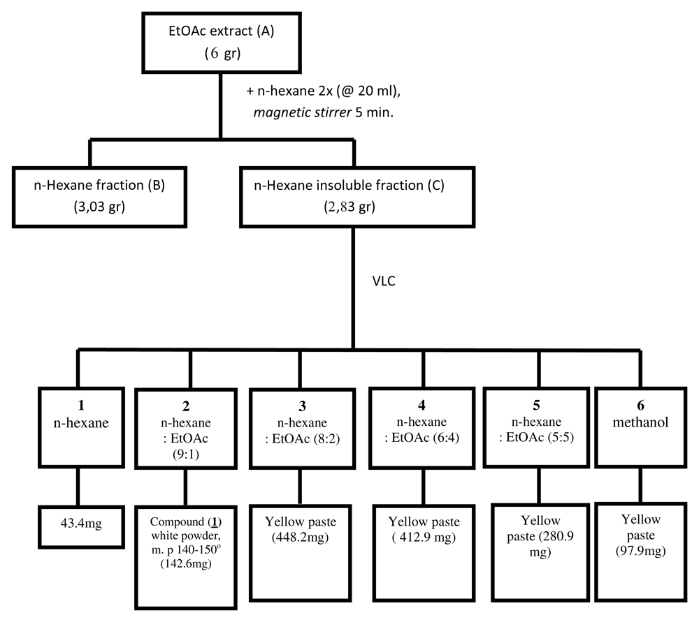
The peel was separated from the fruit, dried in oven 50°C for 24 hours and powdered using a blender. Powdered L. domesticum fruit peel (200g) was macerated with ethyl acetate (EtOAc; 2 L) overnight. This solution was filtrated (0.45μm) and the filtrate was evaporated to dryness with a rotary evaporator set at 50°C, to give dried EtOAc extract (A, 50.13 g). In order to separate the extract into non-polar and polar fractions, 6 g of extract A was diluted 5 times using n-hexane (20 mL) to give soluble n-hexane fraction B (supernatant; 3.03 g) and insoluble n-hexane fraction C (residue; 2.83 g).
Fraction C was the most active among other fractions (see Results), and was therefore further fractionated using vacuum liquid chromatography as described by Mae Sri Hartati et al.8. In brief, using silica gel preparation grade (15g) as stationary phase this was eluted with n-hexane and increasing amounts of ethyl acetate. Six subfractions were obtained and subfraction 2 contained a major compound which appeared as white crystals (referred to as compound 1). Compound 1 was obtained as a single compound from subfraction 2, while the other subfractions still contained various compounds. Compound 1 (142.6 mg), had a melting point at 140–150°C (Figure 1). Compound 1 was identified using spectroscopy data such as ultraviolet (UV), infrared (IR), 13C-NMR and 1H-NMR (see section Chemicals and equipment).
The bioassay followed the methodology described by Bahuguna et al.9 with modifications. In brief, 100 μl T47D and Vero cell line (in RPMI mediaFaculty of Medicine, Universitas Gadjah Mada) were placed in each well of a 96 well micr oplates, resulting in 1 × 10⁴ cells/well. The cells were incubated for 24 hours at 37°C in a CO2 incubator.
Extract A, fractions B and C and compound 1 (5mg) were dissolved in DMSO (50 μL). Serial concentrations of extract and fractions (50, 25, 12.5, 6.25, 3.125 μg/mL), compound 1 (25, 12.5, 6.25, 3.125 μg/mL) and doxorubicin (positive control; 0.5, 0.25, 0.125, 0.0625, 0.0312 μg/mL) were obtained. Cells were treated with the dose dependent samples and incubated for 24 hours at 37°C. The culture medium was removed by pipette, and MTT solution (100μL) was added to each well and incubated for 4 hours at 37°C. After incubation, stop solution (10% SDS, 100 μL) was added to each well and let stand at room temperature for 24 hours.
Absorbance was measured by microplate reader (Bio Rad) at 595 nm. positive control The data generated were used to plot a dose-response curve and IC50 of the samples was determined.
Identified as Lamesticumin A.
White crystal. IR (KBr) vmax cm-1: 3074, 2960, 1712; UV (MeOH)λmax 236,5; 1H,13C-NMR: see Table 1; m/z 502; (Calculated for C31H50O5)
The infrared spectroscopy (KBr) spectrum of 1 showed a broad band at 3400–2800 cm-1, which indicated the presence of –OH group, specifically –COOH due to intermolecular bonding. This data is supported by the appearance of –C=O at 1712 cm-1. Compound 1 displayed UV absorption at 236,5 nm. The 13C-NMR spectrum (500 MHz, CDCl3) of compound 1 showed 30 carbons (Table 1). There were two down field carbon signals (δ, 147.6 and 148.1 ppm) identified as C=O signal carbons. Two characteristic terminals =CH2 signals (δ, 107.4 and 114.2 ppm) were observed, and this identity was confirmed by 2D (Het-Cor) NMR technique. Based on 13C-NMR and 1H-NMR data, compound 1 (Figure 2) was identified as Lamesticumin A (C31H50O5, m/z, 502) which was previously isolated from L. domesticum twigs5.

The cytotoxicity of extract A, fractions B and C, compound 1 and doxorubicin (positive control) is shown in Table 2. Fraction C was the most cytotoxic (IC50 25.57 μg/mL) compared with extract A (29.41 μg/mL) and fraction B (43.51 μg/mL). The IC50 of the isolated compound from fraction C, compound 1/Lamesticumin A was 15.68 μg/mL. All samples inhibited T47D cell growth in a dose dependent behavior (Figure 3).
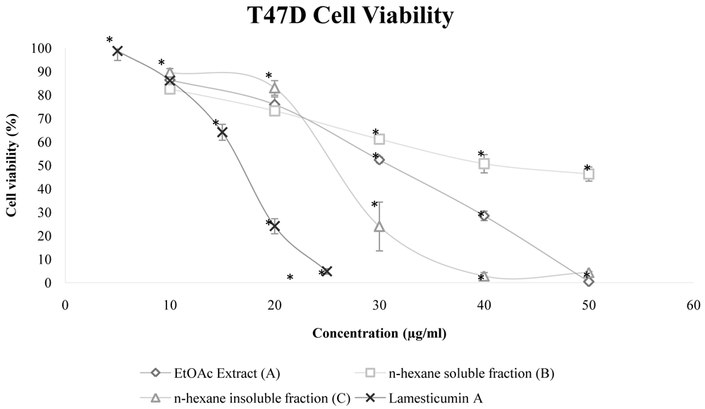
Error bar shows standar deviation (SD, n=3). symbol (*) indicates statistical significance (p<0.05)
Error bars shows standard deviation.
In this study, the cytotoxic activity of Lamesticumin A, derived from the peel of L. domesticum, was demonstrated in the T47D cell line with IC50 15.68 (μg/ml). The T47D cell line is an epithelial breast cancer cell subtype luminal A cell line that express estrogen and progesterone receptors10. Based on National Cancer Institute guidelines, a natural compound has potent anticancer activity if it has IC50 <4 μg/ml or 10 μM11.
Many triterpenoid compounds have been previously isolated from L. domesticum. Most of these compounds are UV inactive or have no strong UV absorbance because triterpenoid’s lack of a conjugated functional group12. Lansiosida A and Dukunolida A has been isolated from n-hexane extract of L. domesticum fruit peel13,14. Lamesticumin A is an onoceranoid-type triterpenoid, isolated previously from L. domesticum twigs, that has antibacterial activity against Staphylococcus aureus, Staphylococcus epidermidis, Micrococcus luteus, Bacillus subtilis, Micrococcus pyogenes and Bacillus cereus with minimum inhibitory concentration of <15 μg/ml5. Another onoceranoid-type triterpenoid Lansium acid I-IX were isolated from L. domesticum leaves, which was reported to have antimutagenic activity6.
Based on several in vitro tests, some terpenoid compounds had anticancer activity. Sesquiterpene lactone compounds are known to inhibit Nf-kB, thereby inducing apoptosis15. Celastrol has anticancer properties by regulating various transcription factors, angiogenesis processes, cell cycle arrest and induction of apoptosis16. Betulinic acid can induce apoptosis in HT-29 colon cancer cells and acts as a chemosensitizer for chemotherapeutic agents in wildtype adenocarcinoma cancer cells (SNU-C5/WT)17. Clematangoticosides D and F from Clematis tangutica are known to have cytotoxic activity against human gastric cancer cell line (SGC-7901) with IC50 24.22 and 21.35 μM, respectively18. Cycloartane-type and oleanane-type triterpenoids from Ligularia przewalskii show cytotoxicity in Hela, HEPG2, SGC7901, MDA-MB-231, HL-60, and Lewis cell lines with IC50 8.40–24.39 μM19.
It has been reported that natural compounds combined with low doses of antineoplastics can increase effectiveness and reduce toxic effects20. Betulinic acid can induce apoptosis when combined with 5-fluorouracil, irinotecan and oxaliplatin4. Ursolic acid (UA), a pentacyclic triterpenoid, is known to have anticancer activity through interfering with multiple signaling pathways. Furthermore, UA has been shown to act as a chemosensitizing agent to increase the effect of conventional anticancer drugs21, and to increase the effect of doxorubicin by increasing the cellular amount of the drug in the MCF-7 cell line22. Further study is needed to investigate the possibility of Lamesticumin A to be combined with doxorubicin for its potential to have synergistic effect.
Extract, fractions and Lamesticumin A derived from the peel of L. domesticum showed cytotoxic activity against the T47D breast cancer cell line. Further research is needed to investigate the potential of the natural compound Lamesticumin A derived from L. domesticum fruit peel as an anticancer therapy.
Zenodo: A bioactive compound isolated from Duku (Lansium domesticum Corr) fruit peels exhibits cytotoxicity against T47D cell line, http://doi.org/10.5281/zenodo.353967023.
This project contains the following underlying data:
- UV, infrared, 13C-NMR and 1H-NMR spectra of compound 1.
- Cell viability and IC50 values of extract A, fractions B and C, compound 1 and doxorubicin in T47D cell line.
Data are available under the terms of the Creative Commons Attribution 4.0 International license (CC-BY 4.0).
| Views | Downloads | |
|---|---|---|
| F1000Research | - | - |
|
PubMed Central
Data from PMC are received and updated monthly.
|
- | - |
Provide sufficient details of any financial or non-financial competing interests to enable users to assess whether your comments might lead a reasonable person to question your impartiality. Consider the following examples, but note that this is not an exhaustive list:
Sign up for content alerts and receive a weekly or monthly email with all newly published articles
Already registered? Sign in
The email address should be the one you originally registered with F1000.
You registered with F1000 via Google, so we cannot reset your password.
To sign in, please click here.
If you still need help with your Google account password, please click here.
You registered with F1000 via Facebook, so we cannot reset your password.
To sign in, please click here.
If you still need help with your Facebook account password, please click here.
If your email address is registered with us, we will email you instructions to reset your password.
If you think you should have received this email but it has not arrived, please check your spam filters and/or contact for further assistance.
Comments on this article Comments (0)