Keywords
Emergency surgery, Breast cancer, Cholecystitis
This article is included in the Oncology gateway.
Emergency surgery, Breast cancer, Cholecystitis
Cholecystitis is one of the leading causes of emergency surgical interventions. The diagnosis of acute cholecystitis is usually based on physical examination, laboratory tests and abdominal ultrasound. The surgical options for cholecystitis are either open and laparoscopic cholecystectomy; the latter is nowadays considered the gold standard of treatment. Surgical specimens must be sent for histopathological examination to rule out cancer1.
The occurrence of metastases to the gallbladder is rare and has only been reported in the literature exceptionally2. Primary tumors can metastasize to the gallbladder either by proximity, such as hepatocellular carcinoma and pancreatic carcinoma, or by blood diffusion3.
Chan reported, in a series of 7910 cholecystectomy specimens, that 36 cases of metastatic carcinoma were found, more often secondary to the stomach, lower gastrointestinal tract, liver, kidney or skin (malignant melanoma) cancer4. Another more recent study shows that metastasis to the gallbladder accounted for 7/225 (3.1%) of the incidental gallbladder malignancies5. Metastasis from breast cancer to the gallbladder is even less common; in fact, breast cancer usually metastasizes to bone, lung, lymph nodes, liver and brain.
We describe here the case of a patient who underwent cholecystectomy for acute cholecystitis with gallbladder metastasis from breast cancer. Subsequently, we present the results of a literature search concerning this disease.
We report the case of an 83-year-old female patient with a previous history of breast surgery with axillary dissection in 1997, followed by adjuvant chemotherapy due to invasive ductal carcinoma of the left breast. The family history was negative for neoplastic diseases, both mammary and belonging to the gastrointestinal tract. Oncological follow-up was negative, and the patient considered disease-free for almost 15 years. During 2012, an X-ray of the spine, performed for the appearance of lumbar pain, revealed the presence of vertebral metastases. The patient was treated with radiotherapy and spinal stabilization. In addition to this, a deep venous thrombosis episode was reported in 2017, and treated with anticoagulant therapy. In the same year, multiple myeloma associated with mild chronic kidney disease was diagnosed. Neither myeloma nor kidney disease had requested specific treatments.
In July 2018, the patient was admitted to the emergency department for sepsis and an episode of acute kidney failure, anuria and fever. Right-upper quadrant abdominal pain triggered by food intake and abdominal tenderness was also present, placing the diagnostic suspicion of biliary sepsis due to acute cholecystitis.
This condition was conservatively treated with intravenous antibiotic therapy with renal adjusted dose of piperacillin-tazobactam and hemodialysis for two weeks. Subsequently, kidney function improved, diuresis had an increasing glomerular filtration rate and sepsis was cured. Abdominal CT-scan performed during this hospitalization had shown a diffuse thickening of the gallbladder’s wall associated with stones as well as pericholecystic fluid (Figure 1). The CT-scan didn’t highlight pathological findings on the liver, such as enlarged regional nodes. A dilated common bile duct with the presence, in its proximal portion, of tenuously hyperdense material was described.

Endoscopic ultrasound was performed, and it confirmed the presence of both gallbladder and common duct stones, the largest was 7 millimetres, and biliary sludge with lack of dilatation of the intrahepatic biliary tract. Several stones were removed via endoscopic retrograde cholangiopancreatography, and a nasobiliary tube was left behind. Subsequent cholangiography demonstrated the regular calibre and morphology of the cystic duct, the principal biliary tract, and the intrahepatic biliary tree. However, the gallbladder appeared distended with several little stones inside.
The patient, after 6 days from the admission, finally underwent laparoscopic cholecystectomy. Intraoperative findings showed the gallbladder with thickened walls and densely fused with the liver but without other pathological findings. No intraoperative complications occurred. Histological examination of the surgical specimen highlighted the presence of metastasis from an infiltrating ductal breast carcinoma with positive hormone receptors: Estrogen Receptors (MoAb SP1) 98%, Progesterone Receptors (MoAb 1E2) 95%, Cytoprolferative Activity (MoAb MIB-1) 10%, c-erbB2 (MoAb 4B5) score: 0. The cystic lymph node showed no evidence of metastasis. The postoperative course was regular, and the patient was transferred to a rehabilitation ward five days after surgery.
After completion of the rehabilitation program, the patient was discharged, and hormone therapy (letrozole 2.5 mg once a day) was started. The patient died 15 months later due to peritoneal and bone progression of the disease.
We conducted a systematic review in which all articles describing cases of gallbladder metastasis from breast cancer were considered eligible for inclusion. Abstracts, conference papers and studies concerning animals were excluded. No restrictions were applied to publication date or languages, if there was an English version of the article available.
A systematic search for articles published up to February 2020 using PubMed, Scopus, Google Scholar and Web of Science databases was performed, and references of articles that were retrieved in the full text were also searched. The search strategy utilized in all databases included the combination of the keywords: “gallbladder metastasis”, “breast cancer”, “acute cholecystitis”, “biliary colic”, “cholelithiasis”. A minimum number of two search keywords were utilized, one of which was always “breast cancer”.
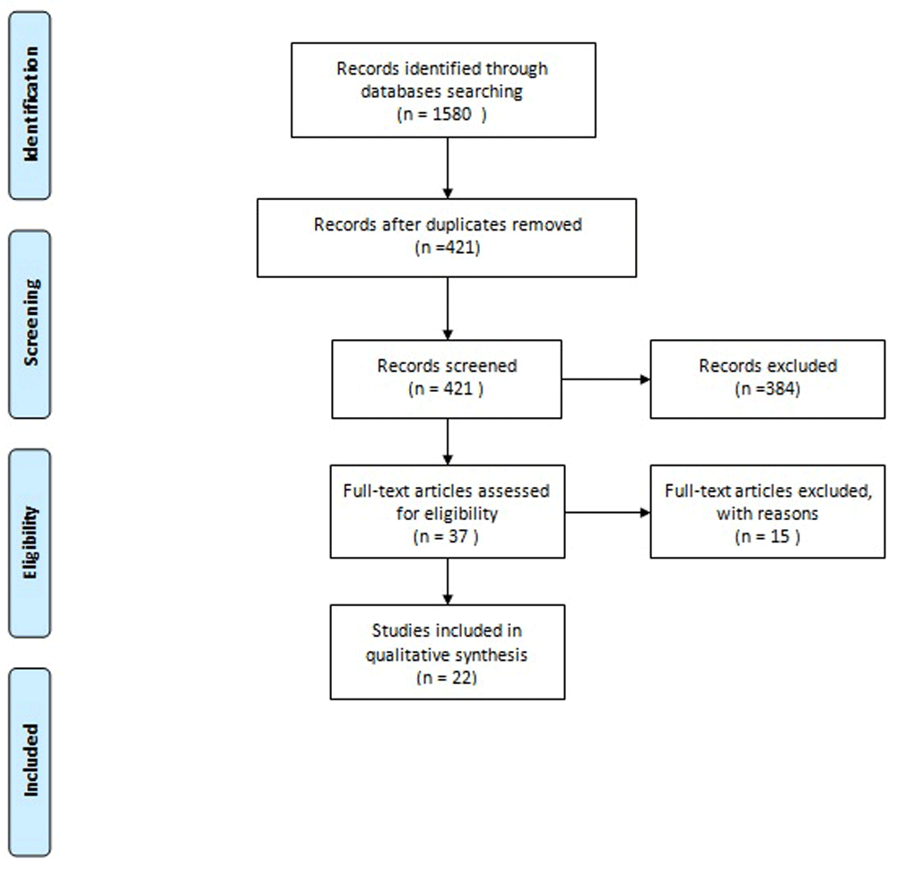
A total of 848 potentially relevant articles were retrieved in Google Scholar, 427 in Scopus, 182 in Web Of Science and 123 in PubMed. Among these 22 studies were identified to be strictly matched with our research (Figure 2). Our case was also included in the review.
In consequence of advances in medical chemotherapy and endocrine therapy in the last years, the outcomes for breast cancer are improved. Disease recurrence is more common within five years of surgery while late recurrences after more than 10 years are very uncommon. The literature outlines risk factors for late recurrence as lymph node metastases, ER + status and HER-2 negative status6,7. Breast cancer metastases occur through contiguous, lymphatic and hematogenous spread. It usually metastasizes to bone, lung, lymph nodes, liver and brain. Less frequently invaded are the endocrine organs, pericardium, abdominal cavity and eyes. Metastasis in the extrahepatic digestive system are infrequent and characteristically appear after a long latent period, which takes from three to up to 20 years5.
Concerning gallbladder metastases by breast cancer, autopsy findings have shown that secondary hematogenous metastases (also from other primary organs) to the gallbladder initially generate small flat nodules below the mucosal layer. They grow as a pedunculated tumor, rarely reaching higher than several millimetres in size. The growth pattern clarifies why gallbladder metastases rarely result in clinical symptoms and that they are not diagnosed during patients’ lives. Metastatic gallbladder tumors rarely show signs; acute cholecystitis is the most frequent clinical presentation8. Obstructive jaundice, haemobilia, even bile peritonitis due to perforation, are seldom described. When a gallbladder metastasis is identified after surgery, the primary tumor can be not easily defined. Distinguishing between primitive gallbladder carcinoma and metastases from breast cancer is crucial for proper post-surgery therapy; in this way, immunohistochemical evaluation is necessary. The most reliable markers are gross cystic disease fluid protein such as 15 (GCDEP -15), plus cytokeratin 7, cytokeratin 20, and estrogen and progesterone receptors. Usually, their positivity is present in metastatic breast cancer, but not in all cases9.
At microscopic pathological examination, metastases are often represented by small clusters and chains of neoplastic cells, commonly of the signet-ring histotype. Pathological diagnosis of metastases from lobular breast cancer can be difficult because signet-ring cells could be present in tumors originating from different organs, such as the stomach10.
Our review of the literature conducted on secondary lesions of the gallbladder from breast cancer has confirmed the rarity of this disease (see Table 1 for a summary of the cases). Gallbladder metastasis is only described in 23 patients, including our case: 11 from infiltrating lobular, 7 ductal origins, 3 mixed ductal and lobular infiltration, and 3 not specified. This analysis reveals how, in most cases (12), the diagnosis of metastatic lesions was made after surgery was performed for acute cholecystitis. There was evidence of gallstones in 8 cases; 9 cases were patients who often suffer from abdominal pain and/or vomiting (symptoms of biliary colic), and so they underwent an elective cholecystectomy. Only in 2 cases, the main symptom was obstructive jaundice or bile peritonitis for necrotic gallbladder.
| Author (year) | Age of patients (years) | Symptoms and signs | Timing of biliary symptoms after breast surgery | Gallstones | Type of breast cancer | Histology | Immunophenotype | Recurrence (months) | Exitus |
|---|---|---|---|---|---|---|---|---|---|
| Di Vita 201111 | 48 | Abdominal pain in the last 3 months. | 3 weeks after surgery diagnosis of chronic cholecystitis at the ultrasound | No | Mixed ductal- lobular k (G3, pT2 N3 M0) | Isolated neoplastic epithelial cells in the muscular layer of the gallbladder | CK 7+, EMA +, ER+, PR+ | 12 SNC mets | Died 14 months after surgery |
| Beaver 198612 | 73 | Abdominal pain and vomiting (cholecystitis), also 10 months before | 3 years after surgery | Yes | Not specified | Small cell tumour growing in an indian file pattern | N/A | N/A | N/A |
| Shah 200013 | 78 | Bile peritonitis for necrotic gallbladder | 11 years after | Yes | Not specified | Focus of poorly differentiated adenocarcinoma characterized by gland formation and cells with eccentric cytoplasm | N/A | N/A | Died 5 days after surgery |
| Rubin 198914 | 55 | Biliary colic for 12 months | Synchronous | Yes | Lobular carcinoma | Carcinoma cells infiltrating singly an in file, mostly in the fibrous tissue deep to the muscular layer focally extended up to the mucosa | N/A | N/A | N/A |
| Manouras 20089 | 46 | Cholecystitis | 2 years after surgery | Yes | Ductal | Glandular poorly differentiated metastases invading the muscular and serosa layers; scattered signet-ring cells infiltrating the mucosa | Lactalbumin +; CKT 7+; CKT 20 -; ER -; PR - | N/A | Died 1 year after surgery |
| Hashimoto 201615 | 59 | Abdominal pain (Cholecystitis) | 12 years after surgery | No | Ductal (pT1c, pN0) | Poorly differentiated carcinoma full- thickness in the cystic duct and gallbladder neck | ER+; PR+; CKT 7+; her 2 -; CKT 20-; GCDEP 15 - | N/A | Died 5 years after surgery |
| Coletta 201416 | 56 | Obstructive jaundice | 13 years after surgery | No | Ductal | Solid honeycombs of malignant epithelial cells localized only in the external side of the biliary duct wall; mucosa free | ER+; PR+; CK 7+; her 2 -; CK 20 - | N/A | Alive 1 year after surgery |
| Nair 201217 | 54 | Symptomatic gallstones | 5 years after surgery | Yes | Lobular (T3 pN1, pMx) | The wall infiltrated by very small regular cells arranged in Indian file | N/A | N/A | Died 2 years after surgery |
| Al-Rawi 201218 | 61 | Cholecystitis | Synchronous | Yes | Lobular | Serosa and adjacent fat showed focal infiltrates of cells with rounded nuclei and small cytoplasmic vacuoles. The cells | Cytokeratins +; Epithelial Membrane antigen +; CK 7 +; ER +; CK 20 - | N/A | Died 5 years after surgery |
| Ebrahim 201519 | 65 | Asymptomatic cholelithiasis at the diagnosis of the tumour; after 2 months of chemo cholecystitis | After 2 months of therapy | Yes | Inflammatory ductal breast cancer | 6–7 mm module with a pale yellow-white solid cut surface in the gallbladder wall | ER + PgR + | N/A | N/A |
| Molina-Barea 201420 | 62 | Biliary colic | After 5 years from surgery | Yes | Lobular | Infiltrated | CK 7 +; ER + | N/A | Died 12 months after surgery |
| Muszynska 20192 | 71 | Biliary colic | Few months before the diagnoses of k | Not specified | Bilateral ductal and lobular | N/A | N/A | N/A | N/A |
| Murguia 20065 | 62 | Symptomatic cholelithiasis | 10 years after surgery | Yes | Ductal | Focal broad- based lesion on the mesenteric face of the body with poorly differentiated adenocarcinoma infiltration, without mucosa involvement | CK 7 +; CK 20 –; ER +0; PgR + | N/A | Died 2 years after surgery for myocardial infarction (2 months before she had done PET and CA 15.3, normal) |
| Mouchli 201921 | 52 | Acute cholecystitis | 1 year after surgery | No | Ductal | N/A | N/A | N/A | Died several days after the surgery |
| Riaz 201222 | 42 | Asymptomatic (finding of a focal area of thickening in gallbladder’s body during the US for staging) | Synchronous | No | Lobular | Cords and nests of malignant cells showing moderate amount of eosinophilic cytoplasm containing irregular hyperchromatic nuclei; indian file pattern is present | Cytoplasmic mucin +; CK 7 +; CK 20 –; E-cadherin –; ER +; PgR+ | N/A | Stable disease until her last follow up |
| Markelov 201123 | 67 | Nausea + weight loss (gallbladder dyskinesia) | 6 years after surgery | Not specified | Lobular with some foci of in situ ductal | Foci of tumour with a single file arrangement present outside the muscularis propria and some tumour cells within the muscolaris propria | ER +; PgR +; Ki67 +; HER 2 - | N/A | N/A |
| Zagouri 200724 | 59 | Acute cholecystitis | 20th month after surgery | Yes | Bilateral synchronous lobular + ductal | The muscular layer and adventitia of the body of gallbladder was infiltrated | ER +; PgR –; CK AE1/AE3 + | N/A | Alive 1 year after surgery |
| Abdelilah 201425 | 45 | Acute cholecystitis | 3 months after surgery | Yes | Lobular (T3 N1 M0) | 1.5 cm palpable mass | ER + PgR+ | N/A | N/A |
| Zamkowski 201726 | 64 | Acute cholecystitis | Synchronous | No | Lobular bilateral | Not described | ER +; PgR – ; HER2 –; Ki67 + | N/A | Alive at the moment of the drafting of the article |
| Fleres 201427 | 83 | Biliary colic with gallstones also in VBP | Synchronous | Yes | Lobular | Parietal infiltration | Ck AE1/AE3 +; CK 7 +; CK 8 +; ER +; PgR - | N/A | Alive 3 years after surgery |
| Herrera 201028 | 46 | Acute cholecystitis | 10 years after surgery | Yes | Lobular | Not specified | N/A | N/A | N/A |
| Machida 200729 | 53 | Acute cholecystitis | 18 years after surgery | No | Lobular | Necrotic change was seen until the muscular layer; white nodules were detected in the submucosal layer of the neck | N/A | N/A | N/A |
| Our experience | 86 | Acute cholecystitis | 21 years after surgery | Yes | Ductal | Parietal infiltration | ER +; PgR +; Mib 1 10%; HER2 0 | 13 months peritoneal and bone | Died 15 months after surgery |
Instrumental diagnostics are useless as they do not show significant data on gallbladder walls that are suspicious for malignancy; the identification of the neoplastic disease is possible only after surgery during histological examination of the specimen, as was shown in our case. From the analysis of the cases described in the literature, it follows that the most frequent tumor histology associated with gallbladder metastasis by breast cancer is infiltrating lobular carcinoma.
This review shows how the detection of gallbladder metastasis usually occurs any time after the surgery for the primary tumor. In essence, we would highlight that in 6 cases, it happened after more than 10 years from primary surgery, in 7 cases between 1 and 6 years, and 3 cases within the first year. Only in 6 cases was the detection of breast cancer and gallbladder metastasis synchronous.
This report emphasizes the importance of long-term follow up in patients with a history of breast cancer.
Our experience and data from the literature suggest carefully evaluating every anomaly observed during routine staging examinations, even when apparently due to benign, mild disease. Metastatic disease always should be included in the differential diagnosis of a patient with a history of invasive breast cancer and new onset of abdominal pain. Conventional methods of documenting gallbladder disease are nonspecific concerning the malignant disease. This may pose a diagnostic challenge in patients with abdominal symptoms after resection of malignancies, also because they need to be aggressively treated as it can improve the poor prognosis of these cases. From our case and literature review, we recommend the following:
Written informed consent for publication of clinical details and clinical images was obtained from the patient on admission to hospital prior to the patient’s death.
No data is associated with this article.
| Views | Downloads | |
|---|---|---|
| F1000Research | - | - |
|
PubMed Central
Data from PMC are received and updated monthly.
|
- | - |
Provide sufficient details of any financial or non-financial competing interests to enable users to assess whether your comments might lead a reasonable person to question your impartiality. Consider the following examples, but note that this is not an exhaustive list:
Sign up for content alerts and receive a weekly or monthly email with all newly published articles
Already registered? Sign in
The email address should be the one you originally registered with F1000.
You registered with F1000 via Google, so we cannot reset your password.
To sign in, please click here.
If you still need help with your Google account password, please click here.
You registered with F1000 via Facebook, so we cannot reset your password.
To sign in, please click here.
If you still need help with your Facebook account password, please click here.
If your email address is registered with us, we will email you instructions to reset your password.
If you think you should have received this email but it has not arrived, please check your spam filters and/or contact for further assistance.
Comments on this article Comments (0)