Keywords
NIFTP, children, multifocality
NIFTP, children, multifocality
More details have been added to the discussion section
See the authors' detailed response to the review by Osama Sharaf Eldin
Generally, the diagnosis of papillary thyroid carcinoma (PTC) has increased over the past several decades1, partly due to increased recognition of the follicular variant of PTC2. The subjectivity in diagnosis of this variant and the indolent behaviour of encapsulated or non-invasive forms, led to revision and follow-up of a large number of these cases by international multidisciplinary collaborative group3,4. Consequently, the encapsulated variant of PTC was reclassified as non-invasive follicular thyroid neoplasm with papillary-like nuclear features (NIFTP), which had strict inclusion and exclusion criteria for this diagnosis. The term NIFTP was then introduced as a separate entity by the World Health Organization in 2017, with a category of follicular tumour of uncertain malignant potential and well-differentiated tumour of uncertain malignant potential5. The majority of NIFTP reports have been in adults. Here, we present a classic case of NIFTP affecting a 10-year old female child.
A female patient of 10 years presented to our department with an enlarged thyroid that had been observed by her mother. No previous relevant family history was recorded.
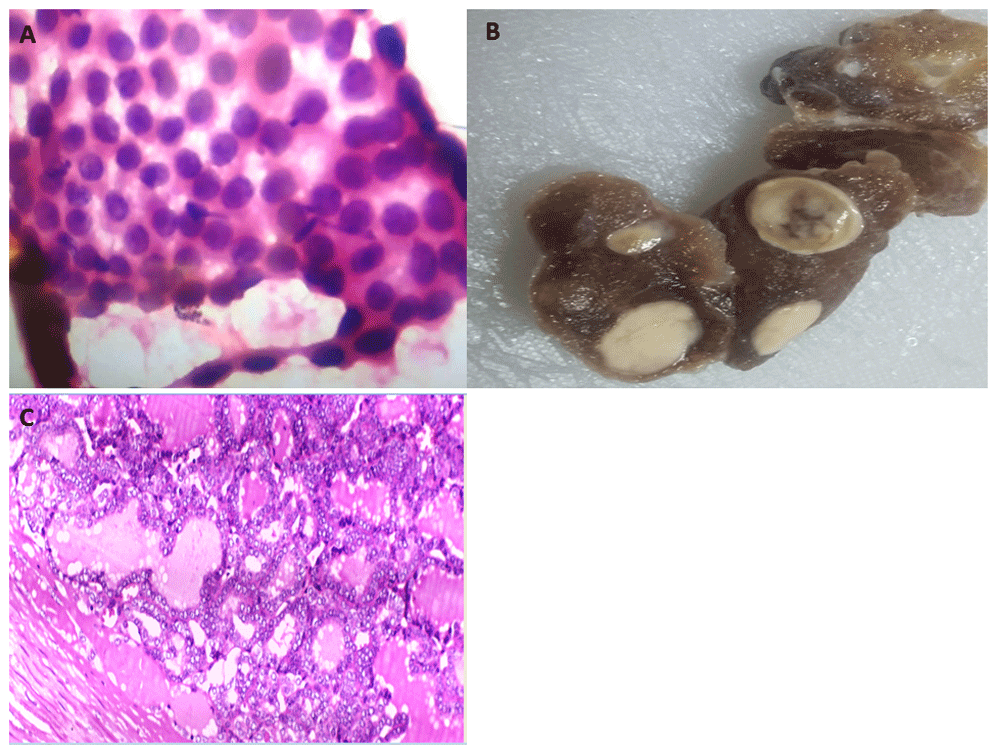
Ultrasound revealed two suspicious nodules on the right side of the thyroid lobe. No pathological lymph node enlargement was reported. Ultrasound guided fine needle aspiration cytology was performed and the results showed sheets of follicular epithelial cells, some were elongated with occasional nuclear grooves and inclusions (Figure 1A). This was diagnosed as atypical thyroid lesion indefinite for malignancy (THY3a).

(A) Cytologic features of fine needle aspiration cytology showing cohesive sheet of follicular epithelial cells, including some which were rounded and others that were elongated with occasional grooved nuclei (hematoxylin and eosin, mag. ×600). (B) Gross picture of affected right lobe after total thyroidectomy showing two well circumscribed whitish nodules. (C) Histopathological examination of nodule of resected thyroid revealing a capsulated nodule formed of microfollicles lined by follicular epithelial cells, which had enlarged pale crowded nuclei together with nuclear grooves and inclusions (nuclear features of papillary thyroid carcinoma)(hematoxylin and eosin, mag. ×400).
The patient was submitted for total thyroidectomy within one month from her first presentation. On resection, the right thyroid lobe measured 5.5 × 3.5 × 3 cm with two well-defined, firm, grayish white nodules. One nodule measured 2 × 1.5 cm and the other measured 1.5 × 1.5 cm (Figure 1B). The left lobe and isthmus measured 4.5 × 3 cm and 1 × 0.5 cm, respectively.
Histological examination of the two nodules resected from the right thyroid lobe revealed well-circumscribed capsulated nodules formed of microfollicles, lined by follicular epithelial cells with wide-spread nuclear features of papillary thyroid carcinoma (Figure 1C). There was no evidence of capsular or vascular invasion, true papillae, trabeculae or solid arrangement. The patient did not receive any specific medications before surgery and she was followed up for 12 months with no evidence of recurrence or nodal involvement.
Most NIFTP cases have been previously reported in adults and data concerning this diagnosis in children is scarce; only 21 cases in children have been reported in the English literature within the last two years (Table 1)6–10. Preoperative diagnosis of our case was based on ultrasound data and the cytology was not obviously malignant. The cytologic smears of NIFTP were usually hypercellular showing follicular epithelial cells arranged in microfollicles without papillae formation and they showed subtle features of papillary thyroid carcinoma but with infrequent or absent nuclear inclusions. NIFTP cytology was commonly interpreted as follicular lesion of undetermined significance in 30% (categories III and IV according to Bethesda system), follicular neoplasm in 21%, suspicious for malignancy in 24%, malignant in 8%, bnign in 10% and non-diagnostic in 3%11,12. Although the above findings would suggest lobectomy, our patient was submitted for total thyroidectomy and as has been done in previously reported cases6,7,9,10.
| Age (years) | Gender F:M | Size (cm) | Focality | Recurrence | Metastasis | Operation | Follow up (months) | |
|---|---|---|---|---|---|---|---|---|
| Wang et al., 2019 (3 cases)6 | 16–17 | 2:1 | 0.4–3.1 | Single | No | No | Total thyroidectomy | 15–138 |
| Rosario and Mourão, 2018 (4 cases)7 | 9-15 | 3:1 | 1.7-2.4 | Single | No | No | Total thyroidectomy | 24-108 |
| Rossi et al., 2018 (2cases)8 | <19 | 1:1 | <2 > 2 | Single | No | No | NA | 84 |
| Mariani et al., 2018 (10 cases)9 | 14.4 | 3.5:1 | 2.1 | 7 cases single 3 cases multifocal | No | 2 cases with lymph node metastases | Total thyroidectomy | NA |
| Samuels et al., 2018 (2 cases)10 | 14 | 2:1 | 1.1-4.5 | NA | No | No | Total thyroidectomy | NA |
| The current case | 10 | Female | 1.5-2 | Multifocal | No | No | Total thyroidectomy | 12 |
On a molecular level, NIFTP shares follicular neoplasm in RAS mutations but it lacks BRAFV600E mutations, which is a common event in papillary thyroid carcinoma13. Immunohistochemistry for BRAFV600E mutations is available on paraffin blocks. Nuclear pseudinclusions are important diagnostic criteria for PTC, which could be highlighted by CK19 immunostaining in comparison to routine hematoxylin and eosin14. The latter authors demonstrated absence of CK19 positive nuclear pseudoinclusions in the investigated 7 cases of NIFTP.
The current report demonstrated a classic case of NIFTP affecting a young female child, agreeing with previous reports that there are more cases in women than men (Table 1). Although not common, multifocality has been reported previously for NIFTP in adults15 and in children9. The size of NIFTP lesion is usually small, rarely exceeding 2 cm in diameter (Table 1).
More aggressive therapy is recommended for PTC in childhood and adolescence16 but the indolent behaviour reported for NIFTP necessitates less aggressive management in children, as well as adults. Therefore, completion lobectomy is not recommended for postoperative cases diagnosed as NIFTP8. NIFTP in children has a similar outcome as cases reported in adults, suggesting that paediatric NIFTP behaves indolently, as evidenced by the absence of local recurrence and nodal metastasis6.
The present report adds a new case of NIFTP in the paediatric age group characterized by multifocality, absence of nodal invasion and indolent course - until last follow-up, recommending less aggressive management of this disease.
Written informed consent was obtained from the patient's father for the publication of this case report and any associated images.
All data underlying the results are available as part of the article and no additional source data are required.
| Views | Downloads | |
|---|---|---|
| F1000Research | - | - |
|
PubMed Central
Data from PMC are received and updated monthly.
|
- | - |
Is the background of the case’s history and progression described in sufficient detail?
Yes
Are enough details provided of any physical examination and diagnostic tests, treatment given and outcomes?
Yes
Is sufficient discussion included of the importance of the findings and their relevance to future understanding of disease processes, diagnosis or treatment?
Yes
Is the case presented with sufficient detail to be useful for other practitioners?
Yes
Competing Interests: No competing interests were disclosed.
Reviewer Expertise: Molecular Pathology, Digital Pathology and experimental pathology
Is the background of the case’s history and progression described in sufficient detail?
Yes
Are enough details provided of any physical examination and diagnostic tests, treatment given and outcomes?
Yes
Is sufficient discussion included of the importance of the findings and their relevance to future understanding of disease processes, diagnosis or treatment?
Partly
Is the case presented with sufficient detail to be useful for other practitioners?
Yes
Competing Interests: No competing interests were disclosed.
Reviewer Expertise: histopathology
Alongside their report, reviewers assign a status to the article:
| Invited Reviewers | ||
|---|---|---|
| 1 | 2 | |
|
Version 2 (revision) 21 Sep 20 |
read | |
|
Version 1 25 Jun 20 |
read | |
Provide sufficient details of any financial or non-financial competing interests to enable users to assess whether your comments might lead a reasonable person to question your impartiality. Consider the following examples, but note that this is not an exhaustive list:
Sign up for content alerts and receive a weekly or monthly email with all newly published articles
Already registered? Sign in
The email address should be the one you originally registered with F1000.
You registered with F1000 via Google, so we cannot reset your password.
To sign in, please click here.
If you still need help with your Google account password, please click here.
You registered with F1000 via Facebook, so we cannot reset your password.
To sign in, please click here.
If you still need help with your Facebook account password, please click here.
If your email address is registered with us, we will email you instructions to reset your password.
If you think you should have received this email but it has not arrived, please check your spam filters and/or contact for further assistance.
Comments on this article Comments (0)