Keywords
Sarcoidosis, Malignancy, Liver mass, Azygous vein
Sarcoidosis, Malignancy, Liver mass, Azygous vein
Sarcoidosis is systemic inflammatory condition of unknown etiology that is characterized histologically by the presence of noncaseating granulomas in the affected organs. It is called the ‘great mimicker’ because it can present in various ways similar to other diseases. It can affect multiple organ systems, but most commonly involves the lungs and the lymph nodes. Other less commonly involved organs include the eyes, skin, liver, spleen, bone marrow, heart and brain1,2. The age adjusted annual incidence of sarcoidosis in the united states is 35.5 per 100,000 for African Americans compared with 10.9 per 100,000 for Caucasians, according to a five-year cohort study3. We present a case presented with asymptomatic cervical lymph node enlargement, multiple liver lesions and azygos vein enlargement, which were suggestive of malignant or infectious etiologies. However, investigations resulted in the diagnosis of the “great mimicker disease’ sarcoidosis.
A previously healthy 53-year-old Caucasian man on no regular medication presented to his primary care provider complaining of a painless slowly growing cervical lump that he noticed one month prior to presentation. Review of systems were negative for fever, weight loss, night sweat, fatigue, loss of appetite, cough, chest pain, hemoptysis, or shortness of breath. Family history was significant for lymphoma. The patient never smoked, and was not exposed to any chemicals at work. Physical examination showed normal vital signs and enlarged right cervical lymph nodes. The rest of the physical examination were unremarkable. Initial laboratory workup showed normal complete blood cell count and basic metabolic panel. (Table 1). A decision to proceed with fine needle aspiration was taken, which showed a non-necrotizing inflammatory reaction. Therefore, a core biopsy was recommended to rule out lymphoma and he was referred to the rheumatology clinic for possible sarcoidosis.
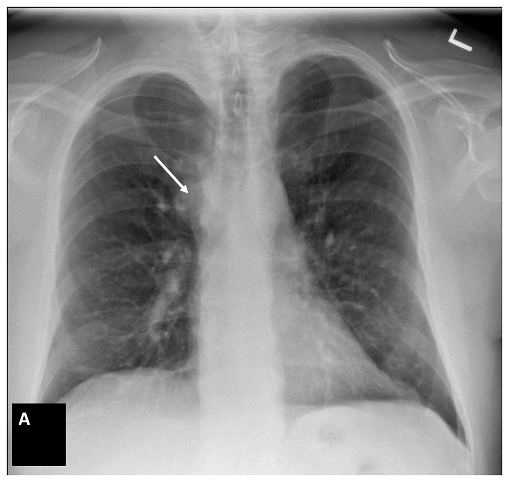
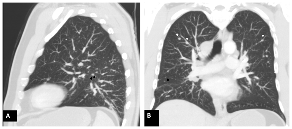
Three months after the initial presentation, he was seen in the rheumatology clinic; further workup was ordered, including an angiotensin-converting enzyme level which was normal, and chest radiograph (CXR), which showed azygous vein enlargement and thus a high suspicion of malignancy (Figure 1). Next, computed tomography (CT) scan of the chest was done and it showed pathologic lymph node enlargement within the thoracic inlet and mediastinum, innumerable bilateral small pulmonary nodules, and multiple liver cystic lesions concerning for metastatic disease. (Figure 2–Figure 4). Further metastatic workup with CT abdomen/pelvis showed multiple hepatic cysts without any other organ involvement (Figure 5). At the same time, histoplasma antigen/antibody were negative, and his erythrocyte sedimentation rate was within normal limit. (Table 2)

Possibly Azygous vein enlargement.

Sagittal (A) and coronal (B) images in lung window showing multiple nodules in centrilobular (white arrowheads) and subpleural (black arrowheads) location consistent with peri lymphatic distribution that can be seen in cases of sarcoidosis.
Incidental liver cysts are also noted.
| Component (reference, units) | 3 months after first presentation |
|---|---|
| Angiotensin converting enzyme (14–82 U/l) | 30 |
| Sedimentation rate (0–15 mm/h) | 8 |
| Histoplasma antibody (negative) | Negative |
One month later, the patient was referred to the pulmonary clinic and underwent general surgery for an excisional biopsy on the most accessible lymph node, which showed non-caseating granulomas without evidence of malignancy, fungal or mycobacterial infection, and confirmed the diagnosis of sarcoidosis. The patient was subsequently started on tapered prednisone (40 mg for two weeks then 20 mg for two weeks then 10 mg for one month). Three months later, he had repeated imaging which showed the same lesions in the lungs and the liver that are of the same size. Therefore, as he was completely asymptomatic, a decision to discontinue prednisone with 1-year radiology imaging follow-up was taken.
The clinical manifestations of sarcoidosis vary and can range from asymptomatic disease detected incidentally on imaging studies to the presence of constitutional symptoms and symptoms attributed to the organ system involved, to multiorgan failure1,2. The diagnosis of sarcoidosis requires fulfillment of certain criteria, which include existence of typical clinical and radiological findings, histopathological evidence of noncaseating granulomas, and exclusion of other causes of granulomatous inflammation4.
The azygos vein is formed by the union of the ascending lumbar vein and the right subcostal vein. It is usually too small to be seen on chest x-ray. The enlargement of the azygous vein on chest x-ray can be seen in multiple diseases, such as congestive heart failure, inferior vena cava thrombosis, and intrathoracic malignancy5.
In this report, we present a patient with asymptomatic cervical lymph node and azygos vein enlargements, with innumerable lung bilateral nodules, hilar, para-esophageal and mediastinal lymph node enlargement and multiple liver nodules which were highly suspicious for malignancy or fungal infection. However, histological evaluation was consistent with sarcoidosis. To our knowledge, this is the first reported case of sarcoidosis and azygous vein enlargement. Hence, we conducted a systematic review of the literature for studies published from 1960 to October 2019 in PubMed, Scopus, Web of Science, and Cochrane Central databases. The following search terms were used: “sarcoidosis”, “azygous vein enlargement’ and “malignancy”. Our search was limited to individuals aged 18 years and older. Our search revealed a total of three patients. None of the cases showed azygous vein enlargement.
Our findings were similar to previous cases reported by Oketani et al.6 who were among the first to report a case of sarcoidosis mimicking metastatic cancer. They reported a case of a 49-year-old female who was found to have mediastinal and intraabdominal lymphadenopathy on radiologic imaging, in addition to presence of space occupying lesions in the liver and the spleen, which raised suspicion for metastatic hepatocellular cancer. However, histologic examination of liver biopsy specimen showed evidence of noncaseating epithelioid granulomas suggestive of sarcoidosis, which responded to treatment with steroids. Giovinale et al.7 reported another case of a 55-year-old female who presented with weight loss and abnormal liver function tests. Imaging revealed the presence of abdominal lymphadenopathy, as well as hepatic and splenic lesions, which were thought to be related to metastatic cancer, as the patient has family history of colorectal cancer. However, histological examination of specimens obtained during exploratory laparotomy showed chronic granulomatous inflammation, and the work up for neoplasia was negative7. Finally, Jafari et al.8 reported a case of a 39-year-old male who was found to have hilar lymphadenopathy and a hepatic nodule on radiologic imaging, which were initially thought to be related to metastatic cancer; however, biopsy confirmed chronic granulomatous inflammation and a diagnosis of sarcoidosis was made after ruling out tuberculosis.
Azygous vein enlargement is a commonly missed finding on chest radiograph which can be a sign of underling malignancy. However, it can also be seen in sarcoidosis. Further appropriate tests are required to confirm the underlying etiology.
All data underlying the results are available as part of the article and no additional source data are required.
Written informed consent for publication of their clinical details and/or clinical images was obtained from the patient/parent/guardian/relative of the patient.
| Views | Downloads | |
|---|---|---|
| F1000Research | - | - |
|
PubMed Central
Data from PMC are received and updated monthly.
|
- | - |
Is the background of the case’s history and progression described in sufficient detail?
Yes
Are enough details provided of any physical examination and diagnostic tests, treatment given and outcomes?
Partly
Is sufficient discussion included of the importance of the findings and their relevance to future understanding of disease processes, diagnosis or treatment?
Yes
Is the case presented with sufficient detail to be useful for other practitioners?
Yes
Competing Interests: No competing interests were disclosed.
Reviewer Expertise: Rheumatology
Is the background of the case’s history and progression described in sufficient detail?
Yes
Are enough details provided of any physical examination and diagnostic tests, treatment given and outcomes?
Yes
Is sufficient discussion included of the importance of the findings and their relevance to future understanding of disease processes, diagnosis or treatment?
Yes
Is the case presented with sufficient detail to be useful for other practitioners?
Yes
Competing Interests: No competing interests were disclosed.
Reviewer Expertise: Medicine and Clinical Epidemiology
Alongside their report, reviewers assign a status to the article:
| Invited Reviewers | ||
|---|---|---|
| 1 | 2 | |
|
Version 1 30 Jun 20 |
read | read |
Provide sufficient details of any financial or non-financial competing interests to enable users to assess whether your comments might lead a reasonable person to question your impartiality. Consider the following examples, but note that this is not an exhaustive list:
Sign up for content alerts and receive a weekly or monthly email with all newly published articles
Already registered? Sign in
The email address should be the one you originally registered with F1000.
You registered with F1000 via Google, so we cannot reset your password.
To sign in, please click here.
If you still need help with your Google account password, please click here.
You registered with F1000 via Facebook, so we cannot reset your password.
To sign in, please click here.
If you still need help with your Facebook account password, please click here.
If your email address is registered with us, we will email you instructions to reset your password.
If you think you should have received this email but it has not arrived, please check your spam filters and/or contact for further assistance.
Comments on this article Comments (0)