Keywords
COVID-19, Acute limb ischemia, Arterial thrombosis, Misan
This article is included in the Emerging Diseases and Outbreaks gateway.
This article is included in the Coronavirus (COVID-19) collection.
COVID-19, Acute limb ischemia, Arterial thrombosis, Misan
Coronavirus disease 2019 (COVID-19) is a global pandemic disease caused by the SARS-COV-2 virus surface spike protein binding to the human angiotensin-converting enzyme 2 (ACE2) receptor, which is expressed in the lung (type 2 alveolar cells), heart, intestinal epithelium, vascular endothelium, and kidneys, providing a mechanism for multi-organ dysfunction. The median incubation period time is 4 to 5 days and 97.5% of patients will exert symptoms within 11.5 days1.
To the best of our knowledge, there is currently no research available in Iraq showing that ischemia of the lower limb caused by thrombosis is a rare presenting clinical feature of COVID-19. Here, we report a rare presentation of lower limb ischemia in a patient one day before the development of the classic symptoms for COVID-19, such as fever and dyspnea.
A 49-year-old female patient admitted to the emergency unit with severe agonizing left lower limb pain of one-day duration. No drug, medical, surgical, and smoking history was reported, but the patient did report contact with COVID-19 patients. On examination, the patient was a middle-aged woman, who seemed to be in discomfort. The left leg was cyanosed and discolored up-to the mid-leg, cold, with no detected dorsalis pedis or posterior tibial artery pulsations. There was popliteal artery pulsation, and no detected sensation in the whole left foot and distal third of leg without movement (Figure 1).
Neurological, cardiac, abdominal, and locomotor system examinations were normal with mild crackles in the lung bases. Blood pressure on admission was 130/70 mmHg, heart rate was 123 beats/minute and the patient had a regular body temperature of 37.3°C. The patient’s respiratory rate was 18 breaths/minute and SPO2 was 75% on room oxygen. The patient underwent a blood test, electrocardiogram (ECG), Doppler study for lower limb arterial examination, and chest radiograph. The blood test results showed hemoglobin of 10.2 g/dl (normal range adult females, 12–16 g/dl), white blood cell count of 17200/ul (normal range, 4000–10000/ul), platelets of 847000/ul (normal range, 165000–415000/ul), lymphocytes of 2800/ul (normal range, 800–2600/ul), blood urea of 8 mg/dl (normal range, 15–45 mg/dl) and D-dimer of 2 (normal range, <0.5). Other investigations such as prothrombin time (PT), activated partial thromboplastin time (aPTT), international normalised ratio (INR) and liver enzymes were not available. The Doppler study showed normal flow in the left common femoral artery, superficial femoral artery, popliteal artery, and both proximal thirds of anterior tibial artery (ATA) and posterior tibial artery (PTA) with abrupt interruption of blood flow in the distal two-thirds of ATA and PTA. Chest X-ray showed large bilateral multiple lung shadows (Figure 2), and the ECG showed sinus tachycardia (Figure 3).
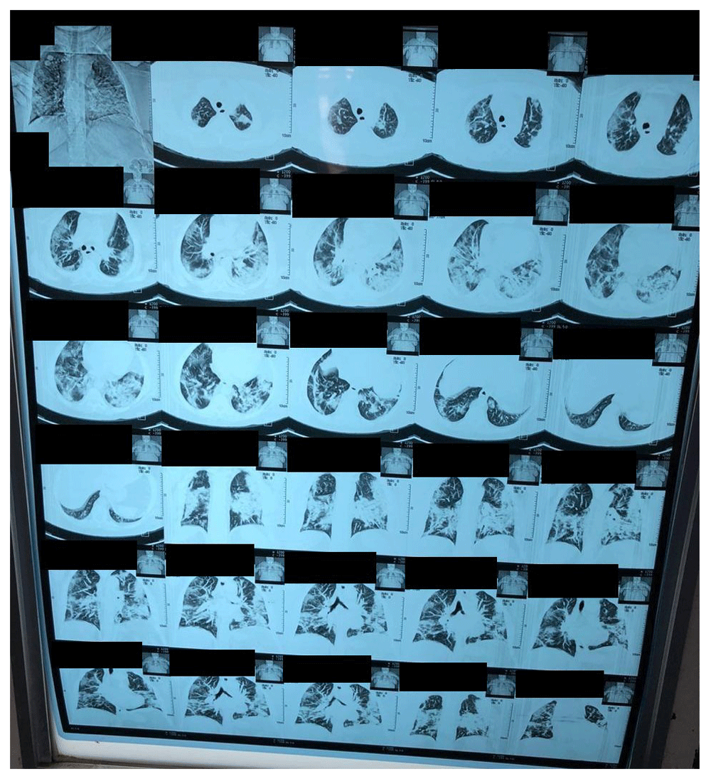
The patient was initiated with an anticoagulant – unfractionated heparin (therapeutic dose of 10,000 Units, in 100mL of 5% Dextrose intravenously (IV) every 6 hours) – fluid therapy (5% Dextrose 250 ml IV every 8 hours; 0.9% Sodium Chloride 250ml IV every 8 hours), oxygen therapy, pain killers (paracetamol injection; (1 g (10mg/100mL) IV every 8 hours), and ceftriaxone injection (1 g IV every 12 hours). The patient was then sent for a CT scan of the chest. This showed bilateral multiple opacities of the lung, suggesting a suspicion of COVID-19 infection (Figure 4). Consequently, the patient was sent to a COVID-19 isolation ward, and nasal and throat PCR test for COVID-19 was taken. Three days post-admission, the COVID-19 test returned a positive result.

On the second day post-admission, the patient developed clear dyspnea and tachypnea. Her saturation dropped from 93% to 60% (normal pulse oximeter readings range from 95–100%, values under 90% are considered low); therefore, the patient was intubated. The left lower limb was non-viable up to the mid-leg, meaning that the patient was a good candidate for amputation. However, the patient was still unstable and unfit for such a surgery. On the fourth day post-admission, the patient developed sudden cardiac arrest due to persistent hypoxia and unfortunately died.
In severe cases, COVID-19 infection can develop disseminated intravascular coagulopathy with fulminant activation of coagulation leading to widespread microvascular thrombosis2. This may be reflected by high D-dimer, prolongation of PT, aPTT, INR and decreased fibrinogen levels, which are not available in our center3. Our case shows that one should keep in mind that acute limb ischemia may be a clinical feature of COVID-19 infection as an isolated early symptom, in addition to other features, or may develop as a complication of disease during the admission period due to intravascular thrombosis2. In our patient, as in many cases of acute lower limb ischemia, the diagnosis was early as it depended on clinical history and examination with Doppler study.
Our case differs from two case reports by Kaur et al. In the first, the patient was a 43-year-old man with a history of hypertension and diabetes mellitus, who presented with acute lower limb ischemia and later developed a fever and exertional dyspnea3. The second reported a 71-year-old Hispanic male with a history of diabetes mellitus who presented with fever, dry cough and exertional dyspnea followed by development of sudden onset severe upper right arm pain diagnosed as intraluminal thrombosis of the axillary artery4. Our patient had no comorbidities and presented with acute lower limb ischemia as an early feature to COVID-19 infection before the development of fever or other respiratory features. In addition, our patient did not undergo surgery due to her instability and metabolic derangement, while in the second report by Kaur et al, the patient underwent thromboembolectomy of the axillary artery4. In a study by Bellosta et al, revascularization was done in 17 out of 20 patients, but this was successful in 12 patients only2. Unfortunately, our patient died due to cardiac arrest by persistent hypoxia.
We present an unusual presentation of COVID-19 infection as acute lower limb ischemia in a female patient without comorbidities. Doctors should have a high suspicion of COVID-19 infection in such a case especially during the pandemic period.
Written informed consent for the publication of the article and any associated images was obtained from the patient before she died. Permission to publish was also sought from the patient’s husband after the patient died.
All data underlying the results are available as part of the article and no additional source data are required.
| Views | Downloads | |
|---|---|---|
| F1000Research | - | - |
|
PubMed Central
Data from PMC are received and updated monthly.
|
- | - |
Is the background of the case’s history and progression described in sufficient detail?
Yes
Are enough details provided of any physical examination and diagnostic tests, treatment given and outcomes?
Yes
Is sufficient discussion included of the importance of the findings and their relevance to future understanding of disease processes, diagnosis or treatment?
Partly
Is the case presented with sufficient detail to be useful for other practitioners?
Partly
Competing Interests: No competing interests were disclosed.
Is the background of the case’s history and progression described in sufficient detail?
Partly
Are enough details provided of any physical examination and diagnostic tests, treatment given and outcomes?
Partly
Is sufficient discussion included of the importance of the findings and their relevance to future understanding of disease processes, diagnosis or treatment?
Yes
Is the case presented with sufficient detail to be useful for other practitioners?
Yes
Competing Interests: No competing interests were disclosed.
Reviewer Expertise: health outcomes research, health care quality and safety, served as judge for clinical vignettes for American College of Physicians and Society of Hospital Medicine.
Is the background of the case’s history and progression described in sufficient detail?
Yes
Are enough details provided of any physical examination and diagnostic tests, treatment given and outcomes?
Yes
Is sufficient discussion included of the importance of the findings and their relevance to future understanding of disease processes, diagnosis or treatment?
Partly
Is the case presented with sufficient detail to be useful for other practitioners?
Yes
Competing Interests: No competing interests were disclosed.
Reviewer Expertise: Internal Medicine / Cardiology.
Alongside their report, reviewers assign a status to the article:
| Invited Reviewers | |||
|---|---|---|---|
| 1 | 2 | 3 | |
|
Version 1 28 Jul 20 |
read | read | read |
Provide sufficient details of any financial or non-financial competing interests to enable users to assess whether your comments might lead a reasonable person to question your impartiality. Consider the following examples, but note that this is not an exhaustive list:
Sign up for content alerts and receive a weekly or monthly email with all newly published articles
Already registered? Sign in
The email address should be the one you originally registered with F1000.
You registered with F1000 via Google, so we cannot reset your password.
To sign in, please click here.
If you still need help with your Google account password, please click here.
You registered with F1000 via Facebook, so we cannot reset your password.
To sign in, please click here.
If you still need help with your Facebook account password, please click here.
If your email address is registered with us, we will email you instructions to reset your password.
If you think you should have received this email but it has not arrived, please check your spam filters and/or contact for further assistance.
Comments on this article Comments (0)