Keywords
Intra-oral meningioma, Benign tumor, Ectopic meningioma, Palatal lesion
Intra-oral meningioma, Benign tumor, Ectopic meningioma, Palatal lesion
We have corrected grammar and language mistakes.
We have added a new reference and changed the numbering of citations and added the data to text.
We have added some lesions to the differential diagnosis list and discussed them.
See the authors' detailed response to the review by Maha M. Abdelsalam
See the authors' detailed response to the review by Eman Abdelzaher
Meningioma is a benign neoplasm of meningothelial cells1. Meningioma may develop as a direct extension of a primary intra-cranial meningioma or as a true primary extra-cranial meningioma2.
Extra-cranial (ectopic) tumors are mostly seen in the head and neck region with no connection intra-cranially3. The most common extra-cranial site is the orbits. Meningioma arising in the oral cavity is extremely rare4. To the best of our knowledge, 19 cases have currently been reported in the oral cavity2,4–20 and we are reporting the first case in the hard palate.
A 59-year-old female patient presented to the outpatient clinic in the Oral and Maxillofacial Surgery Department, Cairo University in January 2019 complaining of a large painless swelling in the hard palate (Figure 1). The patient reported that the swelling had been present in her oral cavity for 22 years. The patient’s medical and familial histories were unremarkable. As well as there was not a history of exposure to radiation. Upon clinical examination on the day of admission, a large hard palatal swelling (3 cm × 3 cm) was evident on the right side of the hard palate. The swelling was covered by normal mucosa and showed a slight bluish tinge. A provisional diagnosis of a benign peripheral nerve neoplasm and a minor salivary gland benign neoplasm were made. CT scan was performed with no evidence of bone involvement.

An incisional biopsy of the lesion was performed. Hematoxylin and eosin stained sections revealed meningothelial cells arranged in lobules. The cells exhibited round to oval nuclei (Figure 2). Psammoma bodies were also present (Figure 3). No mitotic activity and no cellular atypia were found. Immunohistochemical staining for tumor-associated markers was performed to confirm the diagnosis of meningioma and to exclude other mimic tumors as metastiatic carcinomas, schwannoma, neurofibroma, paraganglioma and perineurioma. Cells were positively stained using primary antibodies for epithelial membrane antigen (EMA) and vimentin (Figure 4a, b), but were not stained when using primary antibodies for S100, pancytokeratin, p63, chromogranin and renal cell carcinoma glycoprotein (Figure 5a–e).

Indistinct cell membranes with uniform nuclei and no mitotic figures (inset; magnification, ×200).
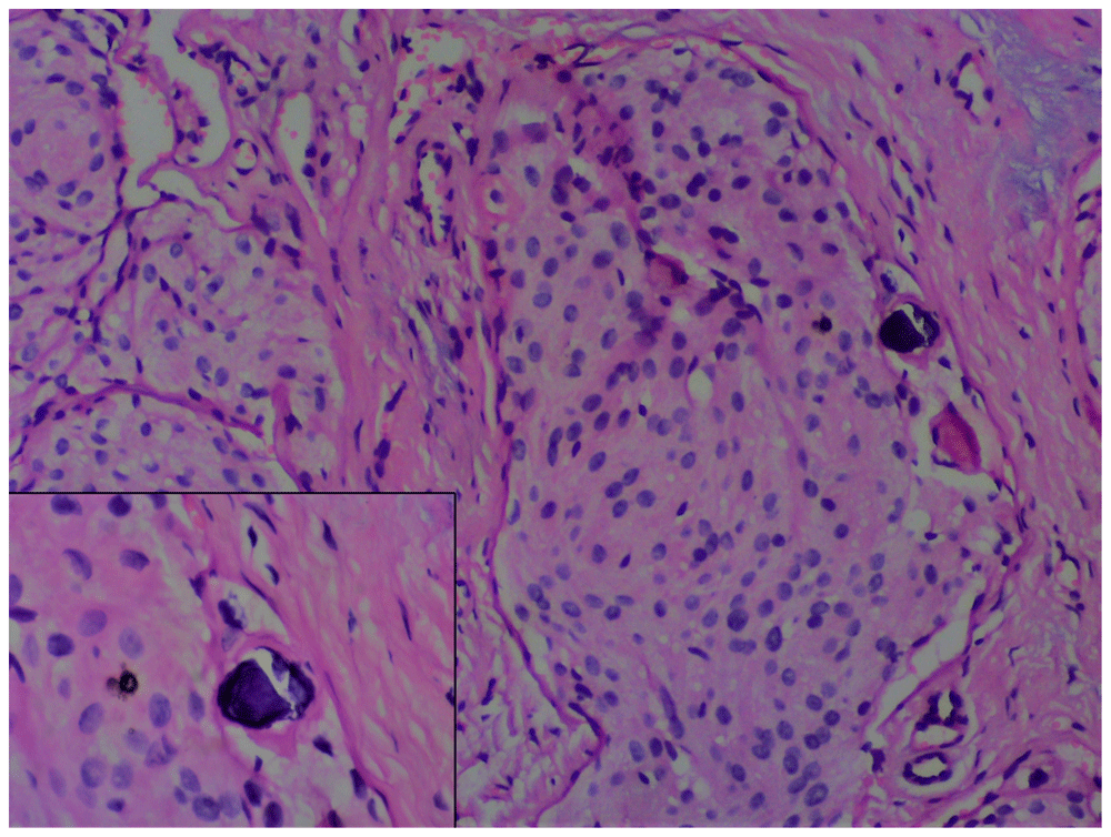
Psammoma bodies seen between meningiothelial cells (inset), (×200).
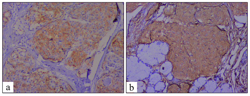
Meningioma tumor cells showing a positive cytoplasmic immunohistochemical reaction for (a) epithelial membrane antigen and (b) Vimentin (magnification, ×200).
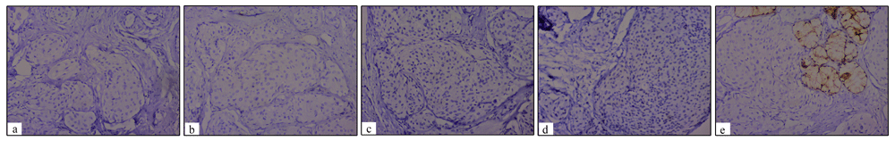
Meningioma tumor cells react negatively following immunohistochemical staining for (a) renal cell carcinoma glycoprotein, (b) S100, (c) chromoginin, (d) p63, (e) PanCK (magnification, ×100).
No therapy was administered to the patient during her admission. Unfortunately, the patient did not show up for surgical excision and follow-up.
| Study | Age, years | Gender | Site | Tumor size | Radiographic findings | Treatment | Follow-up |
|---|---|---|---|---|---|---|---|
| Brown et al.5 | 69 | M | Maxilla | NA | ML RL | Not completed | 8 years |
| Simpson and Sneddon6 | 63 | F | Maxillary alveolus | 4.5 × 2.7 × 2.7 cm | Well-defined mixed RL RO | Surgical excision. | Under review |
| Landini and Kitano7 | 48 | F | Mandible | NA | Well-defined RL | Block resection | 2 years |
| Reddi et al.2 | 26 | F | Maxilla | 3 cm | Ill-defined RL | Surgical excision | 2 years |
| Kishore et al.8 | 44 | F | Soft palate | 3 × 2 cm | NS | Excisional Biopsy | 4 years |
| Pfeifer et al.9 | 77 | F | Maxilla (temporal fossa ) | NA | Dense soft tissue mass | Surgical resection | NS |
| Jones and Freedman10 | 41 | F | Mandible | 4 × 2 cm | Well defined RL | Excisional biopsy | NS |
| Jones and Freedman10 | 74 | F | Mandible | 4 × 3 cm | Well-defined RL | Excisional biopsy | NS |
| Kubotaa et al.11 | 10 | M | Mandible | NA | Well-defined RL | Enucleated | 4 years |
| Mussak et al.12 | 62 | M | Mandible | 7 × 3 cm | Well-defined RL | Segmental mandibulectomy | NS |
| Lell et al.13 | 40 | F | Mandible | NA | Well-defined RL | NS | NS |
| Mosquede-Taylor et al.14 | 53 | F | Mandible | 4 cm | Ill-defined mixed RO RL | Surgical excision | 6 months |
| Rushing et al.15 | NA | Mandible | NA | ||||
| Simsek and Komerik4 | 51 | F | Maxilla | 2 × 2 cm | Ill-defined mixed RL-RO | Surgical excision | 5 years |
| Pinting et al.16 | 59 | M | Maxilla | NA | Well-defined RL | Surgical excision and radiotherapy | NS |
| Maeng et al.17 | 66 | F | Buccal mucosa | 2 cm | Heterogenously enhanced mass | Surgical excision | Year and half |
| Nair et al.18 | 60 | F | Buccal mucosa | 4 × 3 cm | Mass of heterogeneous density | Surgical resection | One year |
| Rege et al.19 | 35 | M | Mandible | NA | Ill-defined ML RL | Partial resection | 5 years |
| Rommel et al.20 | 20 | F | Mandible | 2 × 1.8 cm | Well defined RL | No surgical intervention. | One year |
Primary extra-cranial meningioma is an unusual tumor, especially in the oral cavity4. The first intraoral meningioma reported was by Brown et al. in 1976, which presented as a periapical radiolucency in the anterior maxillary region5.
To the best of our knowledge, 19 cases of primary meningioma in the oral cavity have been reported. Of these, 13 were in female patients, which is also true of the present case. However, the age range was wide in the reported cases – between 10 and 77 years old2,4–20; in the present case, the patient was 59 years old. Regarding the reported cases of intraoral primary meningioma, 6 of the 18 were in the maxilla2,4–6,9,16, 10 were in the mandible7,10–15,19,20, 2 in the buccal mucosa17,18 and one in soft palate8. To the best of our knowledge, we report the first case in the hard palate.
The histopathological criteria of extracranial meningiomas are similar to those of their intracranial counterparts. All documented cases shared the same characteristics: whorls of spindle cells or epithelioid cell proliferation and psammoma bodies. In our case, diagnosis was challenging because of the tumor’s similarity with other tumor entities of peripheral nerve origin, as well as the uncommon location of the tumor. An immunohistochemical panel of tumor-associated markers was used to confirm the diagnosis and to avoid unnecessary aggressive treatment. Most of the 19 cases reported in the literature were diagnosis using immunohistochemical markers. All reported cases that used immunohistochemistry techniques to diagnose meningioma4,9–11,13,14,16,17,19,20 observed that the tumor cells stained positive for monoclonal antibodies against EMA and vimentin, with no immunoreactivity for S-100 protein, which was similar to our findings. However, EMA and vimentin are not useful to differentiate between meningioma and perineuroma as they both express positivity for EMA and vimentin but perineuroma the cells are spindle and elongated however, in our case they are rounded and polyhederal (meningiothelial pattern).
Unfortunately, our patient did not show up for surgical excision and follow-up was not done because of the loss of contact with the patient. However, most of the documented cases were treated successfully without recurrence by surgical excision. Some of the studies, such as that by Rommel et al.20, preferred only to follow-up with the patient rather than conduct surgical intervention. However, others preferred to perform aggressive treatment, such as as segmental mandibulectomy or segmental resection7,12
In conclusion, meningioma is a rare intraoral benign neoplasm. Immunohistochemical markers are an important tool to achieve a final diagnosis, especially for the differentiation from histological mimic entities of peripheral nerve origin, such as perineurioma and neurothekeoma and to avoid unnecessary aggressive treatment. Vimentin and EMA are the two important markers to confirm extra-cranial meningioma diagnosis.
All data underlying the results are available as part of the article and no additional source data are required.
Written informed consent for publication of their clinical details and clinical images was obtained from the patient.
| Views | Downloads | |
|---|---|---|
| F1000Research | - | - |
|
PubMed Central
Data from PMC are received and updated monthly.
|
- | - |
Provide sufficient details of any financial or non-financial competing interests to enable users to assess whether your comments might lead a reasonable person to question your impartiality. Consider the following examples, but note that this is not an exhaustive list:
Sign up for content alerts and receive a weekly or monthly email with all newly published articles
Already registered? Sign in
The email address should be the one you originally registered with F1000.
You registered with F1000 via Google, so we cannot reset your password.
To sign in, please click here.
If you still need help with your Google account password, please click here.
You registered with F1000 via Facebook, so we cannot reset your password.
To sign in, please click here.
If you still need help with your Facebook account password, please click here.
If your email address is registered with us, we will email you instructions to reset your password.
If you think you should have received this email but it has not arrived, please check your spam filters and/or contact for further assistance.
Comments on this article Comments (0)