Keywords
Oral ulcer, Areca nut, Chrysanthemum, TNF-α, Collagen
Oral ulcer, Areca nut, Chrysanthemum, TNF-α, Collagen
Oral ulceration is the most common presentation in the oral cavity caused by many etiologic factors.1 Most of the etiologies of ulcerative lesions on the oral mucosa are divided into four categories; namely, infectious (bacterial and virus infection), immune-related (autoimmune and allergic), traumatic (mechanical trauma), and neoplastic (oral squamous cell carcinoma).2 Traumatic ulcers are injuries to the oral mucosa caused by mechanical or physical trauma such as sharp food, accidental biting during mastication, biting while speaking, punctured by sharp objects, fractured, malformed, carious, or malposed teeth on the superficial epithelial layer or underlying connective tissue or may involve both.3
Currently, increasing the resolution of wound healing is one of the main priorities in the medical field to accelerate the healing of chronic wounds and traumatic injuries.4 The process of tissue regeneration and repair occurs immediately after lesion onset. The linear tissue repair and regeneration involves growth factors that induce cell proliferation, especially parenchymal cells, followed by dynamic changes in soluble mediators, blood cells, and the extracellular matrix.5 The unique oral cavity environment shows advantages in accelerating wound healing compared with skin repair. This is due to differences in the response to inflammation, differentiation and proliferative programs, modulation of stem cells, collagen synthesis, the role of macrophages, and epithelial remodeling.6,7 Collagen synthesis and tumor necrosis factor-α (TNF-α) are important components during acute inflammation and responsible for a diverse range of signaling events within cells in the wound-healing process. TNF-α is one of inflammatory cytokine and regulates the immune system.8
Areca nut is the seed of the areca palm (Areca catechu Linn.), which grows thrive in tropical Pacific region, South and South East Asia, including Indonesia where it is known as “Pinang”. It has been consumed by people worldwide as part of ancient tradition, custom, or ritual for a long time. Several studies have been conducted to prove the healing ability of areca nut, including that the alkaloid and polyphenols content in areca nut could enhance the healing of burn wounds, leg ulcers, and skin graft surgery.9,10 Chrysanthemum or chrysanth (Dendrathema grandiflora) is a flowering plant originating from East Asia and dominantly grows in China. The flowers are generally consumed in herbal teas and as a supplement. Previous studies reported the effect of increasing keratinocyte proliferation and skin regeneration derived from Chrysanthemum boreale.11 Chrysanthemums are usually used for treating allergies, anxiety, hypertension, inflammation, headache, cold, sore throat, and tinnitus by certain communities.12
However, scientific research has not been conducted related to the healing process on the oral mucosa by oral application of these herbal plants. This study aimed to evaluate the potential of the combination of areca nut and chrysanthemum oral gel on oral mucosa using the Sprague–Dawley rat model.
This study was approved by the Ethics Committee for Animal Research of Tropical Biopharmaceutical Research Center (Trop BRC), Bogor Agricultural University, West Java, Indonesia, with number 042-2020-KEH TROP BRC.
Areca nuts were obtained from the Pinang plant from Aceh Besar, Indonesia. 2 kg of areca nuts (gross weight) were cleansed of dirt and dried in the open air and sunlight. Further drying was done using an oven set at a temperature of 50°C. The nuts were crushed using a blender and then strained with a 20-mesh sieve. The maceration process was conducted using 96% ethanol diluent solvent for 7 days before being subsequently filtered and evaporated using a vacuum rotary evaporator at 30–40°C and then reconcentrated using a water bath until a solid dry powder extract was obtained. Chrysanthemum polyethylene (P.E.) (Product No. 1237X17911) was provided by Javaplant Company, Indonesia.
The extract was formulated into four gel dosage forms with various concentrations of the extract combination, namely F1 (20% of areca nut:80% of chrysanthemum), F2 (50% of areca nut:50% of chrysanthemum), F3 (80% of areca nut:20% of chrysanthemum), and base gel (gel without areca nut and chrysanthemum). Preparation of the extract oral gel began with making two mixtures. The first mixture consisted of a carbamoyl and extract combination of areca nut and chrysanthemum and mixed with 10 mL of water at 70°C. The second mixture consisted of methylparaben dissolved in a little water and then mixed with a mixture of glycerin and propylene glycol. This mixture was then combined and stirred with water so that it is mixed homogeneously.
A total of 25 adult male Sprague–Dawley rats weighing 200–240 g were provided by the animal laboratory at Tropical Biopharmaceutical Research Center (Trop BRC), Bogor Agricultural University. The rats were kept for adaptation at 25°C in the well-ventilated laboratory for 14 days under a 12/12 h light/dark cycle and fed with a standard pellet diet and tap water ad libitum before being entered into the experiment. All the animals were given an initial examination for systemic health conditions and stored in boxes with sawdust.
Ulcer induction was performed after the animals were anesthetized by intraperitoneal injection of 33 mg/kg of ketamine and 13 mg/kg of xylazine (2%). The left buccal mucosa was smeared with 10% povidone–iodine on cotton pellets. An abrasion was made with 5 mm diameter, 1 mm depth, and was limited to the mucosa without muscular involvement. The ulceration was performed by using a number 15 scalpel blade. The standardization area was marked with an 8-mm diameter demarcator. The surgical and technique was standardized for animals and performed by the same operator.13 The ulceration was not performed in the normal group. The formation of the ulcers could be observed after 24 hours.
The animals were randomly divided into five groups with 12 animals each: negative control group (rats with base gel treatment), positive control group (rats treated with triamcinolone acetonide), F1 (treatment with 20% areca nut:80% chrysanthemum), F2 (treatment with 50% areca nut:50% chrysanthemum), and F3 (treatment with 80% areca nut:20% chrysanthemum). A normal group (without ulcer and treatment) was added for the microscopic evaluation. All the groups were treated every 12 hours for seven days with topical application. The application of the oral gel was performed by using the individual sterile and disposable dental micro brush (SDent, USA). Each group was sacrificed gradually through the end of the seventh-day study period. After the animals were sacrificed, a section of buccal mucosa containing the ulcer of each rat was collected. Histopathology, immunohistochemistry, and collagenases evaluation were analyzed in Primate Research Center, Bogor Agricultural University, Indonesia.
The animal body weights and diameter of the ulcers were measured before and after treatment on the second and seventh days. The diameter of the ulcers were measured with the naked eyes using a digital stainless vernier caliper which is capable of measuring diameter in 0.05 mm increments.14,15 All measurements were performed by the same operator.
The collected fragments of ulcers were identified and immersed in 10% formol for 24 hours. After fixation in formol, the specimens were macroscopically analyzed, subjected to dehydration in crescent alcoholic series, diaphanized in xylol, impregnated in paraffin, and melted at 60°C. The fragments were packed in paraffin-forming blocks at room temperature. The fragments were sectioned to 5 μm in thickness through the use of microtome and histology using routine coloration by hematoxylin-eosin (HE) was performed. The histopathological parameters were determined and scored from 0 to 4 according to previously published criteria (Table 1).16
The selected tissue sections of the ulcers (2.5 μm) were deparaffinized and rehydrated with distilled water. Blocking endogen peroxidase activity was done using 3% hydrogen peroxide solution for 15 minutes, followed by washing with phosphate-buffered saline (PBS) three times every 5 minutes. After protein blocking, the dripping of Biocare’s Background Sniper was performed and the specimens were incubated for 15 minutes at 37°C. The dripping of normal serum was performed and incubated for 60 minutes at 37°C. The specimens were washed with PBS and anti-TNF-α antibody (TNFA/1172) (ab220210Abcam), was diluted at 1:100, and then dripped and incubated at 4°C for two days. After washing with PBS, the secondary antibody goat anti-rabbit IgG H&L horseradish peroxidase (HRP) (RRID: AB_955447; ab6721-Abcam) was given for 30 minutes at 25°C and followed by Betazoid DAB chromogen solution (BDB2004H-Biocare Medicare LLC). It was used for increasing stability and staining intensity for HRP detection in the specimens. The specimens were washed with distilled water and checked with the microscope. After counterstaining with hematoxylin for 30 seconds, the specimens were rinsed in tap water, dehydrated, purified, and mounted. The percentage of nuclear and cytoplasmic expression in the connective tissue was divided into four scores, namely: 0: no positive cells; 1 (mild): 1–33% of positive cells; 2 (moderate): 34–66% of positive cells; 3 (intense): 67–100% of positive cells. Two observers analyzed the same scores until they were considered as the final scores.17
The same fragments were sectioned to a thickness of 3 μm, de-wax, hydrate paraffin section, and stained using picrosirius red (solution A) for one hour. After washing in acidified water (solution B), the specimens were dehydrated and mounted for observation under polarized light microscopy. The collagen bundles shown in red in the image were then calculated using Adobe Photoshop CC 2017 (RRID:SCR_014199; GNU Image Manipulation Program (RRID:SCR_003182) is an open-access alternative) and ImageJ (v1.50i) (RRID:SCR_003070) software. The percentage of collagen was derived from collagen area pixel divided by tissue pixel and multiply by 100%. The mean of three percentages was used as a sample unit.18
All experiments were carried out in triplicate and the data were expressed as mean ± standard deviation (SD). Statistical analysis was performed by using the SPSS 20 software (SPSS Inc., Chicago, USA) (RRID:SCR_019096); JASP (RRID:SCR_015823) is an open-access alternative. Clinical evaluation in changes of the area of ulcer and body weight of rats analyzed by the dependent t-test. Differences among samples in histopathological and immunohistochemistry analysis were evaluated by using the Kruskal–Wallis test. Collagenesis analysis was evaluated by using analysis of variance (ANOVA/Tukey test). A significant difference was assumed at p < 0.05.
The change comparison of ulcer size analysis in the buccal mucosa of rats showed a significant decrease in ulcer size in F1, F2, F3, and positive control groups (Figure 1A–J, Table 2). This observation was carried out on the second day after the ulcer was formed and at the end of the experiment. The most significant decrease in ulcer area was seen in positive control (triamcinolone acetonide orabase) and F2 treatment groups. Notably, the average ulcer size in positive control and F2 treatment on the second day was 10.10 ± 0.17 and 10.08 ± 0.16 mm2. On the seventh day, it was seen that the size of the ulcer had almost closed, namely 0.03 ± 0.01 and 0.11 ± 0.07 mm2. These data analyses showed that treatment with areca nut and chrysanthemum oral gel caused significantly (p < 0.05) ulcer healing in a dose-dependent manner. Analysis of changes in body weight showed a significant increase in the F2 and positive control groups, while a significant decrease in body weight occurred in the F1 and F3 treatment groups (Table 2). Meanwhile, in the negative control group, there was a decrease in body weight, but it was not significant.
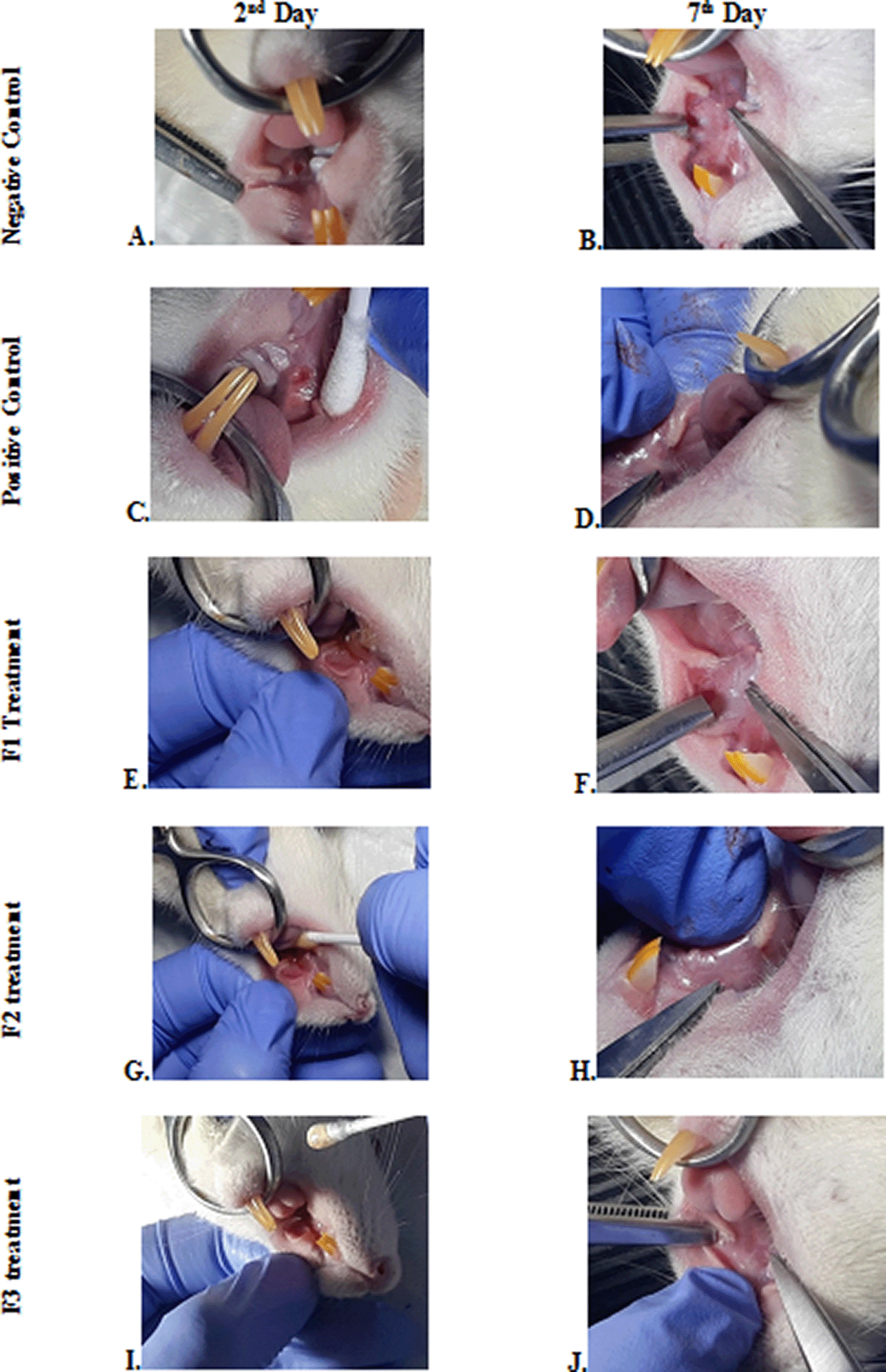
Negative control: base gel treatment group (A and B); Positive control: triamcinolone acetonide treatment (C and D); F1 treatment: 20% areca nut:80% chrysanthemum (E and F); F2 treatment: 50% areca nut:50% chrysanthemum (G and H); F3 treatment: 80% areca nut:20% chrysanthemum (I and J).
To verify the mechanism of ulcer healing by areca nut and chrysanthemum oral gel, the ulcers were subjected to histological examination. As seen in Table 3, the mean of histological scores on the seventh day showed that positive control, F1, and F3 groups indicated that there were no ulcers in the epithelial layer, presence of fibrosis, and slight chronic inflammation in the conjunctive tissue. The score 0 indicating no ulcers and remodeling in the conjunctive tissue was seen in the negative control, F2, and normal groups, but significant differences between groups were not detected in histology examination. Analysis of photomicrograph of ulceration showed loss of ulcer with remodeled connective tissue and accompanied by a slight inflammatory infiltrate in Figure 2B, 2C, and 2E (score 1). There were no visible inflammations in Figure 2A, 2D, and 2F (score 0).
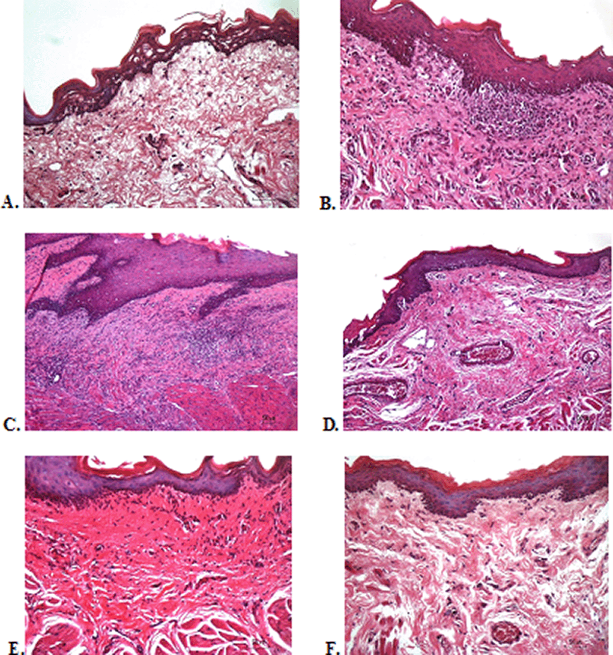
(A) Negative control: base gel; (B) Positive control: triamcinolone acetonide in orabase; (C) F1: 20% areca nut:80% chrysanthemum; (D) F2: 50% areca nut:50% chrysanthemum; (E) F3: 80% areca nut:20% chrysanthemum; (F) Normal group: without ulcer and treatment (Hematoxylin and eosin, 40×).
The immunostaining for TNF-α aims to show changes in TNF-α expression released by connective tissue cells in the cytoplasm during the healing process (Figure 3). The increase in the expressions with a mean score of 2 (moderate) was showed by positive control (2.67 ± 0.58) and F2 groups (2.33 ± 0.58) (Table 3). The lower scores (1-mild) were seen in the negative control, F1, F3, and normal groups. This study showed significant differences between the six groups with changes in TNF-α expression. These results were in line with the pictures that revealed brown diffuse granular-like staining pattern of the connective tissue and epidermis in the Figure 3B and 3D, while the other groups did not show dominant TNF-α expressions except in the F3 group, the immunostaining of TNF-α was prominently found in the basal cell layer (Figure 3E).
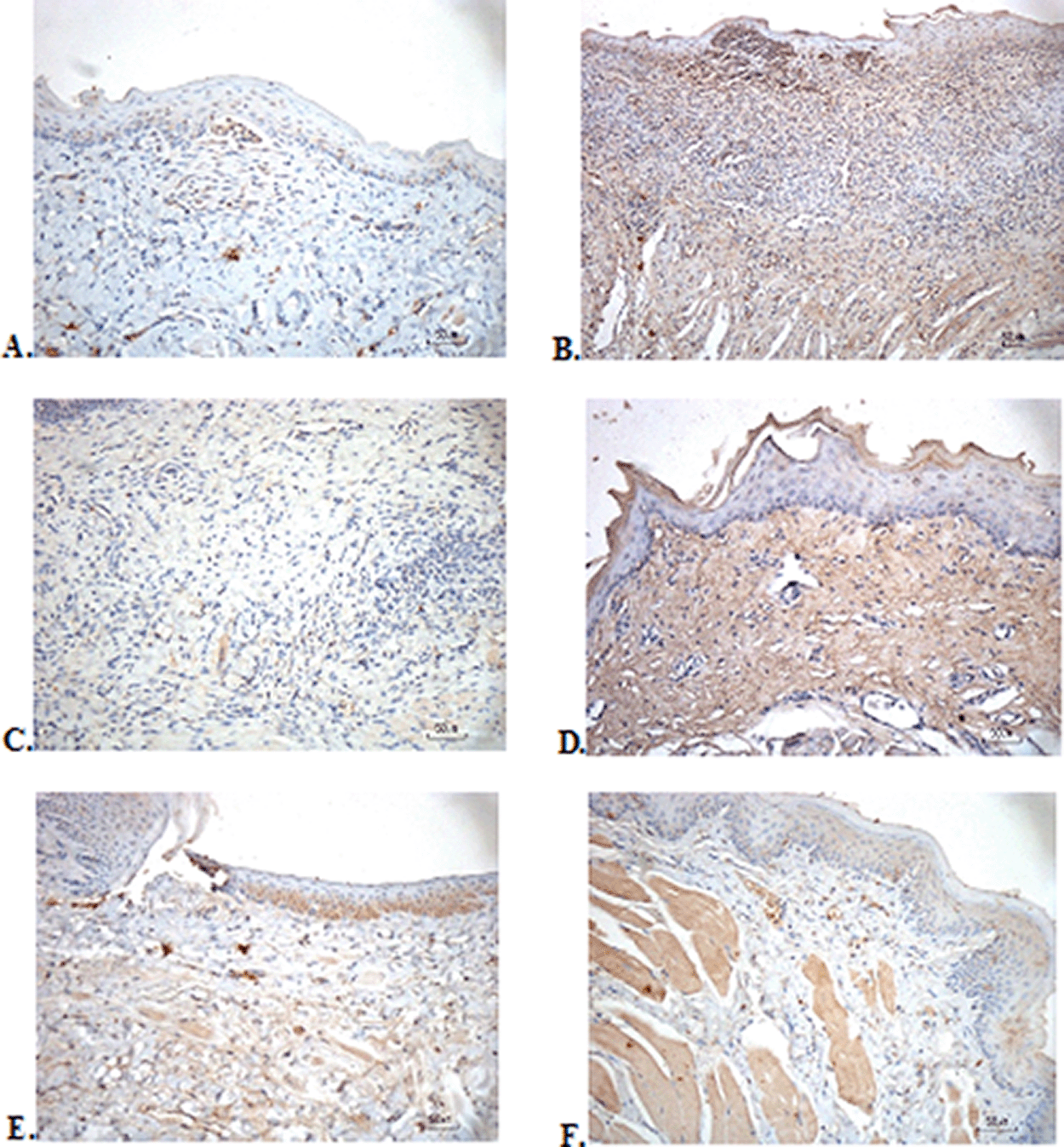
(A) Negative control: base gel; (B) Positive control: triamcinolone acetonide in orabase; (C) F1: 20% areca nut:80% and chrysanthemum; (D) F2: 50% areca nut:50% chrysanthemum; (E) F3: 80% areca nut:20% chrysanthemum; (F) Normal group: without ulcer and treatment (TNF-α immunostaining, 40×)
The results of red picrosirius staining on the last experimental day showed the highest collagen deposition in the F2 group with a mean of 25.4 ± 2.12, while the lowest collagen deposition while collagen deposition in low level was seen in the F3 group (5.63 ± 2.28) (Table 3). Figure 4B (Positive control) and 4D (F2 group) showed regular, dense, and thick collagen, whereas, in the negative control (Figure 4F), collagen deposition showed thin, irregular, and many empty gaps. The ANOVA analysis showed significant differences (p < 0.05) in collagen expression between groups (Table 3). The Tukey test demonstrated significant differences between F2 and F3 groups. Other significant differences were evaluated between F1, F2, F3, and negative control groups.
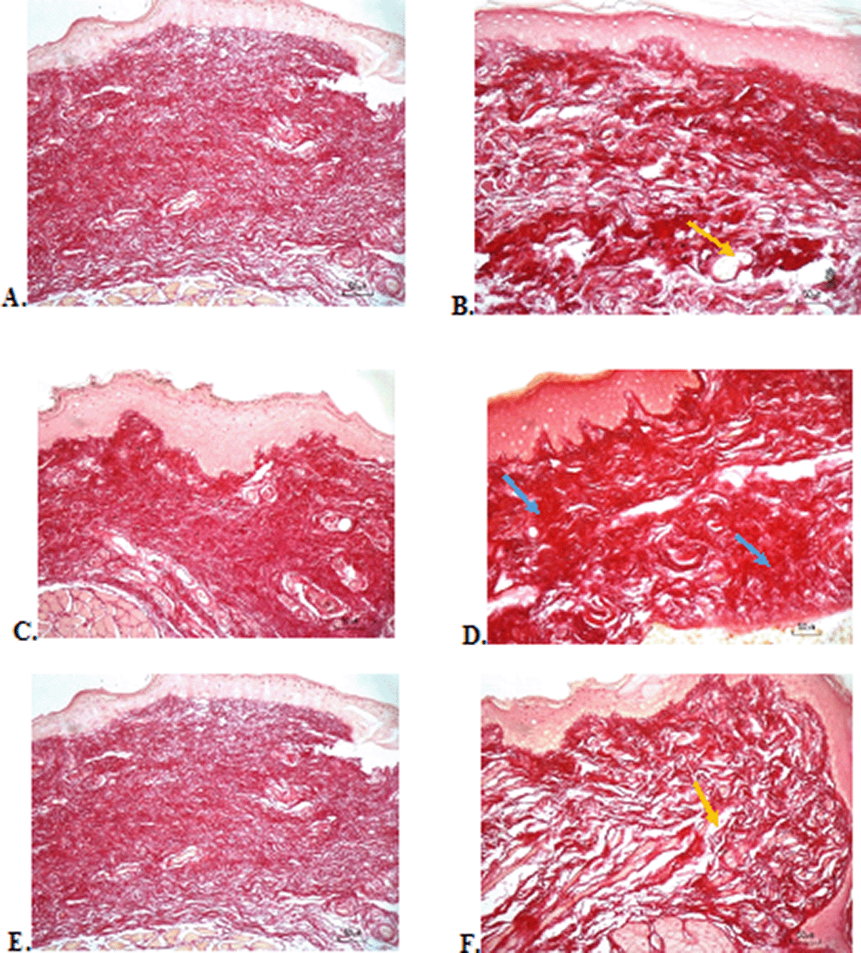
(A) Negative control: base gel; (B) Positive control: triamcinolone acetonide in orabase; (C) F1: 20% areca nut:80% chrysanthemum; (D) F2: 50% areca nut:50% chrysanthemum; (E) F3: 80% areca nut:20% chrysanthemum; (F) Normal group: without ulcer and treatment (Picosirius red staining, 40×). Yellow arrow: Disintact collagen bundles. Blue arrow: Intact collagen bundles.
In the present study, we demonstrated that application of the oral gel to ulcerated lesions for 6 days showed significant lesion healing in the composition of the oral gel containing balanced areca nut and chrysanthemum (50%:50%). This significant wound healing was based on the clinical appearance such as ulcer size reduction, reepithelization over the ulcer base, and formulation of granulation tissue around the ulceration. This result has the same clinical appearance as the positive control group that using triamcinolone acetonide. Areca nut, as one of the main ingredients, contains phenolic and flavonoids which function as antioxidants.19 Catechin is a component of flavonoids present in areca nut that acts as an antioxidant by chelating the ions and scavenging the free radicals particularly, superoxide (O2–), peroxyl, hydroxyl radicals (·OH), and hence inhibit both DNA damage and lipid peroxidation, which can cause membrane damage.20 Catechin can reduce the expression of interleukin-6 (IL-6) and IL-8 which function to overcome inflammation and increase the wound healing process by chemotactic for fibroblasts, accelerates their migration and stimulates deposition of tenascin, fibronectin, and collagen I during wound healing in vivo.21–24 The success of using areca nut in oral gel to heal the ulcers is in line with a previous study using ointment with 2% ethanolic extract on burns on the skin.10 This study also used a combination with chrysanthemum. Previous studies have shown that chrysanthemum has antioxidant, antiproliferative, antimicrobial, acetylcholine esterase inhibition, antitimelanogenic, and antiviral activities, especially Chrysanthemum morifolium, Chrysanthemum indicum, and Chrysanthemum coronarium. The active phytochemistry content of chrysanthemum is apigenin-7-O-glucoside.25
This present study observed the body weight of rats before and after application. There are similarities between ulcer repair and weight gain. A significant increase in body weight in the F2 and positive control showed faster healing of ulcers, making it easier for rats to consume more food. The use of healthy rats aims to evaluate the effect of ulcers in the oral cavity purely based on the ability of rats to gain healing process without being affected by systemic diseases that can affect rat’s body weight. Areca nut is capable of repressing prostaglandin E2 (PGE2) and arachidonic acid-induced inflammation, thereby helping to accelerate ulcer healing.26 The analgesic or antinociceptive activity of the areca nut is originated by inhibition of prostaglandin synthesis. The oral gel form was chosen to facilitate absorption in the oral mucosa and it has a rapid cool soothing effect, although the drawback is that the gel is easily soluble in saliva. Application twice a day in the morning and evening regularly showed a fairly good cicatrization process in both groups (F2 and positive control). The increase in body weight in the rat showed that in the last few days the diet was not disturbed by the size of the ulcer.
Histological examination with HE staining on day seven showed no significant difference between all treatment groups, normal, positive, and negative control groups. The oral gel containing a balanced composition of areca nut and chrysanthemum, negative control, and normal groups did not show any ulcers and the epithelial tissue had been completely remodeled. Meanwhile, the other group still showed fibrosis and inflammation without ulcers. Fibrosis, persistent neutrophil infiltration, and poor angiogenesis in the ulcer base are factors involved in the underlying mechanism of delayed healing ulcers.27 The ulcer healing mechanism begins with ulcer base contraction which includes contraction of the ulcerated area, re-epithelization over the ulcer base, and formation of granulation tissue. The next step is epithelial regeneration and mucus secretion. This epidermal repair is regulated by a key growth factor (KGF) that is expressed at very high levels within 24 h of injury on dermal skin.28 The buccal mucosa is a non-keratinocyte layer, so it is probably the most involved in the re-epithelization process in the oral mucosa is salivary mucin. Mucin is the primary gel-forming component of mucus that provide a critical layer of protection on the wet epithelial surface including gastrointestinal, female genital, and respiratory tracts.29 Angiogenesis is the next step of ulcer healing. It allows the supply of nutrients and oxygen to the ulcerated area thereby giving the collagen in connective tissue and epithelium an opportunity to recover. Matrix formation is the last step of ulcer repair. This remodeling formation is regulated by the temporospatial expression of the fibrillar and basement membrane collagen (types I, III, IV), and matrix metalloproteinase-2 (MMP-2). Fibronectin is important for cell proliferation, adhesion, differentiation, and matrix formation.
In the present study, the analysis of TNF-α showed that the increase in the moderate expressions was showed by positive control and F2 groups. This is in line with research by Kobayashi et al. which showed that the water-soluble component in dried chrysanthemum has antioxidant ability to increase TNF-α. This activity was observed within 3 hours after ulcer formation.8 TNF-α is a cytokine produced primarily by immune cells such as monocytes/macrophages, but several cell types, such as T and B lymphocytes, natural killer cells, neutrophils, fibroblasts, and osteoclasts can also secrete TNF-α in smaller quantities. 30 TNF-α levels which had the same score between F2 and positive control on the seventh day showed that the amount of this cytokine was still high detected in the local connective tissue. Prolonged or remaining elevated expression of TNF-α may occur for up to the third to the fifth day following the traumatic injury.31 Prolonged increase in TNF-α especially in delayed healing reflects their augmented blood TNF-α, suggesting an association between impaired healing and hard work TNF-α in the inflammatory response so that wound healing is achieved.32 In this study, we did not examine the level of TNF-α in the blood so that the role of areca nut-chrysanthemum oral gel to accelerate wound healing systemically cannot be explored.
Triamcinolone acetonide was chosen as a positive control because this preparation has efficacy as a synthetic corticosteroid that has anti-inflammatory, antipruritic, and anti-allergic effects. This preparation has the advantage of being an emollient dental paste that can attach the drug to the oral mucosal tissue so that it is not easily soluble in saliva. An important role of steroids in triamcinolone acetonide is to alter the localization of high junction proteins thereby increasing the adhesion density between epithelial cells.33 A tight epithelial protective barrier can be achieved through tight cell adhesion. Contacts between adjacent cells are made up of tight junctions, adherens junctions, and desmosomes which help in preserving tissue homeostasis. This study showed that with the use of triamcinolone acetonide and F2 group on the seventh day, the ulcers had closed tightly without any inflammation. These results prove that there might be a possibility of areca nut–chrysanthemum oral gel also can close the adhesion bonds between epithelial cells so that the level of wound closure density which is similar to that of the group positive control (triamcinolone acetonide).
Collagenesis analysis showed a lot of collagen deposition in the F2 group. The results of this study are in line with the fact that the alkaloids in areca nut are potent inducers to increase the amount of collagen. Previous research has shown that a series of synthetic arecaidine esters by oral mucosa fibroblast was closely correlated with the extent of collagen synthesis.34 Alkaloids in the areca nut in a long term play a major role in the accumulation of collagen in oral submucous fibrosis (OSF). Collagen is a key component of the extracellular matrix that plays a critical role in the regulation of the phases of wound healing. Collagen exposure due to injury in the inflammatory phase will activate the clotting cascade, resulting in a fibrin clot that stops the initial bleeding. It also acts as potent chemoattractants for neutrophils, enhancing phagocytosis and immune response and modulating gene expressions. Collagen promotes anti-inflammatory, proangiogenic wound macrophage phenotype via microRNA signaling. In the angiogenesis phase, collagen stimulates angiogenesis in vitro and in vivo through the engagement of specific integrin receptors. The C-propeptide fragment of collagen I recruit endothelial cells, potentially triggering angiogenesis in the healing wound.35
The results of the evaluation of collagen deposition after treatment, with picrosirius red staining, showed a picture of collagen with thicker density, large fiber size, and lots of collagen fibrils in the oral gel with a balanced composition of areca nut and chrysanthemum and a positive control group. This shows the good work of collagen to repair wounds. What's interesting from this observation is that, although there were groups that received treatment with a higher composition of areca nut in oral gel, this was not able to induce collagen as in a balanced composition of areca nut and chrysanthemum. This explains that the ulcer healing rate in this study was not dose dependent but more dependent on the peak efficacy of each phytochemistry composition of an extract
The present study provides the activity of the healing effect of combination areca nut and chrysanthemum. This oral gel can improve ulcer wound healing through the role of TNF-α and collagen with the same results as topical steroid therapy.
Dryad: Underlying data for ‘Evaluation of clinical, histology, TNF-α, and collagen expressions on oral ulcer in rats after treatment with areca nut and chrysanthemum oral gel’. https://doi.org/10.5061/dryad.8w9ghx3mw.
The project contains the following underlying data: body weight, oral ulcer’s size, histology and tnf-α score, collagen percentage.
| Views | Downloads | |
|---|---|---|
| F1000Research | - | - |
|
PubMed Central
Data from PMC are received and updated monthly.
|
- | - |
Provide sufficient details of any financial or non-financial competing interests to enable users to assess whether your comments might lead a reasonable person to question your impartiality. Consider the following examples, but note that this is not an exhaustive list:
Sign up for content alerts and receive a weekly or monthly email with all newly published articles
Already registered? Sign in
The email address should be the one you originally registered with F1000.
You registered with F1000 via Google, so we cannot reset your password.
To sign in, please click here.
If you still need help with your Google account password, please click here.
You registered with F1000 via Facebook, so we cannot reset your password.
To sign in, please click here.
If you still need help with your Facebook account password, please click here.
If your email address is registered with us, we will email you instructions to reset your password.
If you think you should have received this email but it has not arrived, please check your spam filters and/or contact for further assistance.


Dear Prof,
- The major concern of this manuscript is the technique of the treatment process. While the rats are fully awake, it is very difficult
... Continue reading Reviewer 2 response:Dear Prof,
Figure 1.a. Polarized light microscopy (Positive control)
Figure 1. b. Non-polarized light microscopy (Positive control)
Dear Prof,
Figure 1.a. Polarized light microscopy (Positive control)
Figure 1. b. Non-polarized light microscopy (Positive control)